Nursing Paper Example on Graves’ Disease

Nursing Paper Example on Graves’ Disease

Graves’ disease is an autoimmune disorder and the most common cause of hyperthyroidism. It occurs when the immune system produces autoantibodies that stimulate the thyroid gland, leading to excessive production of thyroid hormones. This hormonal imbalance affects various bodily functions, including metabolism, heart rate, and thermoregulation. Graves’ disease can present at any age but is most prevalent in women aged 30 to 50. Early diagnosis and treatment are essential to manage symptoms and prevent complications.

Nursing Paper Example on Graves' Disease

Causes

Graves’ disease is caused by the production of thyroid-stimulating immunoglobulins (TSI), which mimic thyroid-stimulating hormone (TSH). These autoantibodies bind to TSH receptors on thyroid cells, prompting the gland to overproduce thyroid hormones.

Although the exact trigger remains unknown, genetic predisposition and environmental factors play significant roles. Stress, infections, and pregnancy are known to exacerbate immune dysregulation, contributing to the onset of Graves’ disease. A family history of thyroid disorders or autoimmune conditions also increases the likelihood of developing the disease.

Signs and Symptoms

Graves’ disease presents with various symptoms due to hyperthyroidism. Common signs include unexplained weight loss, heat intolerance, and increased appetite. Patients may experience palpitations, sweating, and fatigue due to the overactive metabolism. Neurological symptoms such as tremors, anxiety, and irritability are frequent.

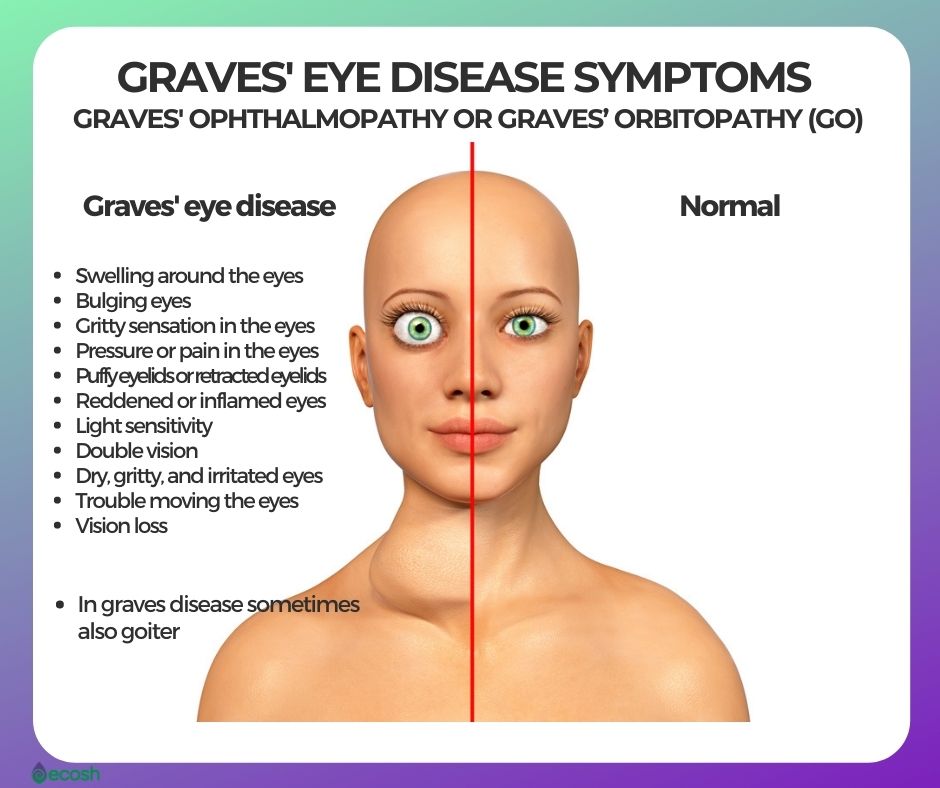
Ocular manifestations, known as Graves’ ophthalmopathy, include eye bulging, redness, and double vision. In severe cases, skin thickening on the lower legs, termed pretibial myxedema, may occur. The intensity of symptoms varies among individuals, with some presenting mildly and others severely.

Etiology

The etiology of Graves’ disease involves immune system dysfunction. The primary autoantibody implicated is thyroid-stimulating immunoglobulin, which disrupts normal thyroid regulation. Genetic factors, including specific human leukocyte antigen (HLA) types and other immune-regulatory genes, predispose individuals to the disease.

Environmental triggers such as smoking, infections, and high iodine intake have been linked to the onset of Graves’ disease. Hormonal fluctuations, particularly in women, are also believed to influence disease development and severity.

Pathophysiology

Graves’ disease results from the interplay between genetic susceptibility and environmental triggers, leading to autoimmune activation. The thyroid-stimulating immunoglobulin activates the TSH receptor on thyroid cells, causing unregulated thyroid hormone synthesis and release. Excess thyroid hormones amplify metabolic activity and enhance sympathetic nervous system sensitivity. Graves’ ophthalmopathy arises from the autoimmune attack on orbital tissues, leading to inflammation and fibrosis. Pretibial myxedema occurs due to the deposition of glycosaminoglycans in the dermis.

Diagnosis

Graves’ disease diagnosis combines clinical evaluation, laboratory tests, and imaging studies. Blood tests reveal elevated free thyroxine (T4) and triiodothyronine (T3) levels, with suppressed TSH levels. Thyroid-stimulating immunoglobulin or TSH receptor antibody tests confirm autoimmune activity.

A radioactive iodine uptake scan demonstrates diffuse thyroid gland overactivity. Ultrasound may identify thyroid enlargement and increased vascularity. A comprehensive assessment helps distinguish Graves’ disease from other causes of hyperthyroidism, such as toxic nodular goiter.

Treatment Regimens

Treatment options for Graves’ disease aim to control hyperthyroidism, alleviate symptoms, and address autoimmune activity. Antithyroid medications such as methimazole and propylthiouracil reduce thyroid hormone production. Beta-blockers like propranolol manage cardiovascular symptoms, including palpitations and tachycardia. Radioactive iodine therapy destroys overactive thyroid tissue, leading to a reduction in hormone production.

In some cases, thyroidectomy, the surgical removal of the thyroid gland, is performed. For Graves’ ophthalmopathy, glucocorticoids and orbital decompression surgery may be necessary in severe cases.

(Nursing Paper Example on Graves’ Disease)

Patient Education

Patient education is crucial for effective management of Graves’ disease. Individuals should understand the chronic nature of the condition and the importance of adhering to treatment plans. Regular follow-up visits are essential for monitoring thyroid hormone levels and adjusting therapy. Patients should be informed about potential side effects of treatments, such as hypothyroidism following radioactive iodine therapy.

Lifestyle modifications, including stress reduction, avoiding smoking, and maintaining a balanced diet, support overall health and may reduce symptom severity.

Research and Future Directions

Ongoing research on Graves’ disease focuses on understanding its genetic basis and immune mechanisms to develop targeted therapies. Advances in biologics targeting specific immune pathways, such as monoclonal antibodies, show promise in reducing autoimmune activity.

Studies exploring the role of gut microbiota in immune regulation may provide new insights into disease prevention and management. Improved diagnostic tools for early detection and risk assessment are also under investigation.

Conclusion

Graves’ disease is a complex autoimmune disorder with diverse clinical manifestations. Its pathogenesis involves genetic and environmental factors, leading to unregulated thyroid hormone production. Early diagnosis and tailored treatment strategies are essential for managing symptoms and preventing complications. Advances in research continue to enhance understanding and provide hope for improved therapeutic options in the future.

References

Davies, T. F., & Larsen, P. R. (2022). Thyroid hormone synthesis and secretion. New England Journal of Medicine. https://www.nejm.org
Smith, T. J., & Hegedüs, L. (2023). Graves’ disease: Pathophysiology and treatment approaches. Lancet Diabetes & Endocrinology. https://www.thelancet.com
Bahn, R. S. (2020). Graves’ ophthalmopathy. New England Journal of Medicine. https://www.nejm.org
Antonelli, A., Ferrari, S. M., & Fallahi, P. (2021). Environmental triggers in Graves’ disease. Autoimmunity Reviews. https://www.sciencedirect.com
Cooper, D. S., & Ross, D. S. (2023). Antithyroid drugs in the treatment of Graves’ disease. Journal of Clinical Endocrinology & Metabolism. https://academic.oup.com

 
Do you need a similar assignment done for you from scratch? Order now!
Use Discount Code "Newclient" for a 15% Discount!

Nursing Paper Example on Granulomatosis with Polyangiitis

Nursing Paper Example on Granulomatosis with Polyangiitis

Granulomatosis with polyangiitis (GPA), formerly known as Wegener’s granulomatosis, is a rare, systemic vasculitis affecting small to medium-sized blood vessels. It is characterized by necrotizing granulomas primarily in the respiratory tract and kidneys. GPA often presents with a wide range of symptoms, making diagnosis challenging. Early detection and treatment are crucial to preventing severe organ damage and improving prognosis. This autoimmune disease typically occurs in adults, with a slight predominance in males, and has no clear genetic or environmental cause.

Nursing Paper Example on Granulomatosis with Polyangiitis

Causes

The exact cause of GPA remains unknown. However, it is believed to result from an abnormal immune response triggered by environmental or infectious agents. Research suggests that bacterial or viral infections may initiate the autoimmune response.

Genetic predisposition may also play a role, particularly variants in human leukocyte antigen (HLA) genes that regulate immune function. Environmental factors such as exposure to silica and other occupational hazards have been loosely associated with GPA but require further study.

Signs and Symptoms

The clinical presentation of GPA is variable, but it typically involves the respiratory tract, kidneys, and other organ systems. Common symptoms include persistent nasal congestion, nosebleeds, and sinus infections due to inflammation in the upper respiratory tract. Lung involvement may lead to coughing, shortness of breath, and hemoptysis.  Kidney involvement often manifests as rapidly progressive glomerulonephritis, which can cause hematuria and proteinuria.

Generalized symptoms such as fever, weight loss, and fatigue are also common. Skin rashes, joint pain, and eye inflammation may occur in some patients, reflecting systemic vasculitis.

Etiology

GPA is an autoimmune disease primarily driven by the production of antineutrophil cytoplasmic antibodies (ANCAs). These autoantibodies target proteins in neutrophils, particularly proteinase-3, leading to neutrophil activation and subsequent vascular inflammation.

The underlying etiology involves complex interactions between genetic susceptibility, environmental triggers, and immune system dysregulation. Although the precise mechanism remains unclear, studies suggest that activated neutrophils release inflammatory mediators, causing endothelial injury and granuloma formation.

Pathophysiology

The pathophysiology of GPA involves immune-mediated inflammation of blood vessels and surrounding tissues. ANCAs play a central role by activating neutrophils, which adhere to vascular endothelium and release reactive oxygen species and proteolytic enzymes. This process damages blood vessel walls, leading to necrosis and granuloma formation.

Granulomas, a hallmark of GPA, consist of macrophages, multinucleated giant cells, and lymphocytes and are particularly prominent in the respiratory tract. Kidney involvement occurs when immune complexes deposit in glomeruli, causing glomerulonephritis. The systemic nature of GPA reflects widespread vascular inflammation.

(Nursing Paper Example on Granulomatosis with Polyangiitis)

Diagnosis

The diagnosis of GPA relies on clinical evaluation, laboratory tests, and imaging studies. Blood tests often reveal elevated inflammatory markers such as erythrocyte sedimentation rate (ESR) and C-reactive protein (CRP). The presence of ANCAs, specifically proteinase-3 ANCA, is highly suggestive of GPA but not definitive.

Imaging studies, including chest X-rays and computed tomography (CT), can identify granulomas and other abnormalities in the lungs and sinuses. Tissue biopsy remains the gold standard for diagnosis, with findings of necrotizing granulomas and vasculitis confirming the disease.

Treatment Regimens

Treatment for GPA aims to suppress inflammation, prevent disease progression, and manage organ damage. Initial therapy typically involves high-dose corticosteroids combined with immunosuppressive agents such as cyclophosphamide or rituximab. Corticosteroids rapidly reduce inflammation, while immunosuppressive drugs target the underlying autoimmune process.

For maintenance therapy, methotrexate or azathioprine is often used to reduce the risk of relapse. In severe cases, plasma exchange may be employed to remove ANCAs from the bloodstream. Patients with kidney involvement may require dialysis or, in rare cases, kidney transplantation.

Patient Education

Education is a critical component of GPA management. Patients should understand the chronic nature of the disease and the importance of adherence to treatment regimens to prevent relapses. Awareness of potential medication side effects, including infections and bone loss due to long-term corticosteroid use, is essential.

Regular follow-ups with healthcare providers are necessary to monitor disease activity and organ function. Patients should also be encouraged to maintain a healthy lifestyle, including smoking cessation, to reduce additional cardiovascular risks.

Research and Future Directions

Advances in understanding GPA are leading to new therapeutic approaches. Ongoing research focuses on identifying specific genetic markers and immune pathways involved in the disease. Monoclonal antibodies targeting inflammatory cytokines and immune cells, such as rituximab, are being studied for their efficacy and safety in long-term disease control.

Additionally, biomarkers for early detection and monitoring disease activity are under investigation. These developments hold promise for improving outcomes and reducing the burden of GPA.

Conclusion

Granulomatosis with polyangiitis is a complex autoimmune disease characterized by systemic vasculitis and granuloma formation. Although its exact cause is unknown, it is closely associated with ANCAs and immune dysregulation. Early diagnosis and aggressive treatment are vital to preventing irreversible organ damage and improving quality of life. Advances in immunotherapy and ongoing research provide hope for better management and outcomes for patients with this challenging condition.

References

Jennette, J. C., & Falk, R. J. (2020). Pathogenesis of ANCA-associated vasculitis: The role of autoantibodies and neutrophils. Annual Review of Pathology: Mechanisms of Disease. https://www.annualreviews.org
Falk, R. J., & Jennette, J. C. (2023). Clinical features and diagnosis of granulomatosis with polyangiitis. New England Journal of Medicine. https://www.nejm.org
Yates, M., Watts, R. A., & Scott, D. G. (2021). Advances in the treatment of ANCA-associated vasculitis. Lancet Rheumatology. https://www.thelancet.com
Niles, J. L., & Mejia-Vilet, J. M. (2023). Immunosuppressive therapy in granulomatosis with polyangiitis: Current strategies. Kidney International Reports. https://www.kidney-international.org
Luqmani, R. A., & Suppiah, R. (2020). Long-term management and prognosis of granulomatosis with polyangiitis. Rheumatology (Oxford). https://academic.oup.com/rheumatology

 
Do you need a similar assignment done for you from scratch? Order now!
Use Discount Code "Newclient" for a 15% Discount!

Nursing Paper Example on Guillain-Barré Syndrome

Nursing Paper Example on Guillain-Barré Syndrome

Guillain-Barré syndrome (GBS) is an acute autoimmune disorder that affects the peripheral nervous system, often triggered by an infection. It is characterized by the rapid onset of muscle weakness and paralysis, beginning in the lower extremities and potentially progressing to the upper body and respiratory muscles. Though rare, GBS is a serious condition that requires prompt diagnosis and treatment to prevent long-term disability or death. The condition is often self-limited, but the course can vary, with some individuals experiencing significant recovery while others may face lasting neurological deficits.

Nursing Paper Example on Guillain-Barré Syndrome

Causes

The primary trigger for GBS is an infection, with viral or bacterial pathogens being most commonly associated. The most frequently implicated virus is the cytomegalovirus, followed by Epstein-Barr virus, Zika virus, and the flu virus.

Bacterial infections, such as Campylobacter jejuni, are also a common precipitating factor. Vaccines, although rare, have also been linked to GBS, particularly after the administration of influenza or rabies vaccines. The exact mechanism of GBS involves molecular mimicry, where the immune system mistakenly attacks the myelin sheath of the nerves following an infection, leading to nerve damage.

Signs and Symptoms

GBS typically begins with a tingling sensation or weakness in the legs, often progressing to muscle paralysis. The symptoms can advance quickly, sometimes within hours or days, and affect the arms and facial muscles, leading to difficulty swallowing, speaking, and breathing. In severe cases, the paralysis can involve respiratory muscles, requiring mechanical ventilation.

Other signs include pain, particularly in the lower back, and autonomic dysfunction such as fluctuating blood pressure, heart rate abnormalities, and difficulties in temperature regulation. The severity and speed of symptom onset can vary from mild to life-threatening.

Etiology

The etiology of Guillain-Barré syndrome is autoimmune in nature. After an infection, the immune system produces antibodies that, in some cases, cross-react with the peripheral nervous system, leading to an inflammatory response. This response damages the myelin sheath that covers nerve fibers, impairing nerve signal transmission. The destruction of myelin results in the characteristic muscle weakness and sensory loss. The association between certain infections, particularly those caused by pathogens like Campylobacter, and the development of GBS underscores the importance of immune system dysfunction in its etiology.

Pathophysiology

Guillain-Barré syndrome is primarily a demyelinating condition, although some forms involve axonal damage. The immune system attacks peripheral nerves, initiating inflammation that damages the myelin sheath, which insulates nerve fibers. This damage leads to disrupted communication between nerves and muscles, resulting in the weakness, paralysis, and sensory disturbances that characterize the disorder.

In some variants of GBS, such as the acute motor axonal neuropathy, the axons themselves are damaged, causing more severe and longer-lasting disability. The pathophysiology involves both cellular immunity, particularly T cells, and humoral immunity, with antibodies playing a role in the damage to peripheral nerves.

Diagnosis

The diagnosis of Guillain-Barré syndrome is clinical and is supported by the patient’s history, symptoms, and diagnostic tests. The hallmark symptom is the progressive, symmetrical weakness starting in the legs. Diagnostic tests include nerve conduction studies, which reveal slowed nerve conduction due to demyelination, and cerebrospinal fluid analysis, which often shows elevated protein levels without a corresponding increase in white blood cells (albuminocytologic dissociation). Blood tests may help rule out other conditions, but no specific biomarker for GBS exists. In some cases, magnetic resonance imaging (MRI) can be used to assess the extent of nerve damage.

Treatment Regimens

The management of Guillain-Barré syndrome focuses on supportive care and immunotherapy. In the acute phase, hospitalization is usually required, especially if respiratory involvement is present. The main treatments are plasmapheresis (plasma exchange) and intravenous immunoglobulin (IVIg), both of which are used to modulate the immune response. Plasmapheresis involves removing harmful antibodies from the blood, while IVIg provides immune support by supplying healthy antibodies.

Pain management, physical therapy, and monitoring of autonomic functions are also key components of treatment. Early intervention is crucial for improving outcomes, although some patients may still experience prolonged weakness or residual effects.

Patient Education

Patient education is vital in the management of Guillain-Barré syndrome, both during the acute phase and throughout recovery. Patients should be informed about the potential for rapid deterioration of symptoms and the importance of early intervention. As recovery can be slow, patients and their families should be prepared for a gradual return to normal function, with some individuals experiencing long-term effects.

Rehabilitation therapies, including physical, occupational, and speech therapy, play an important role in recovery and should be initiated early. Support groups can help patients and their families manage the emotional challenges that often accompany the disease. Long-term monitoring may be necessary to address any ongoing complications.

Research and Future Directions

Ongoing research into Guillain-Barré syndrome is focused on better understanding its immune mechanisms, improving diagnostic techniques, and developing more effective treatments. Advances in immunotherapy, particularly monoclonal antibodies, are being investigated for their potential to more precisely target the autoimmune processes involved in GBS.

Researchers are also exploring the role of genetic predispositions and viral triggers in the development of the disease. The ultimate goal is to reduce the incidence of GBS, improve its prognosis, and provide better therapeutic options for patients with this potentially debilitating condition.

Conclusion

Guillain-Barré syndrome is a rare but potentially life-threatening condition that involves autoimmune damage to the peripheral nervous system. Though the disease has a variable course, with some individuals recovering fully and others facing lasting impairment, early diagnosis and prompt treatment significantly improve outcomes. Advances in immunotherapy and ongoing research provide hope for more effective treatments in the future. Awareness and education for both patients and healthcare providers are essential for managing this complex disease and ensuring the best possible recovery for those affected.

References

McGrogan, A., Leese, M., & Howell, J. (2023). Guillain-Barré syndrome: Epidemiology, clinical features, and diagnosis. Journal of Neurology, Neurosurgery, and Psychiatry. https://jnnp.bmj.com
Willison, H. J., & Yuki, N. (2023). Guillain-Barré syndrome: Mechanisms and management. The Lancet Neurology. https://www.thelancet.com/neurology
Van Doorn, P. A., & Hughes, R. A. C. (2023). Diagnosis and treatment of Guillain-Barré syndrome. British Medical Journal. https://www.bmj.com
Miller, R. G., & Haverkamp, M. H. (2023). Long-term outcomes in Guillain-Barré syndrome: A review of rehabilitation and management strategies. Journal of Neurologic Rehabilitation. https://www.journals.elsevier.com/journal-of-neurologic-rehabilitation
Willison, H. J., & Jacobs, B. C. (2023). Guillain-Barré syndrome and its infectious triggers. Journal of Clinical Investigation. https://www.jci.org

 
Do you need a similar assignment done for you from scratch? Order now!
Use Discount Code "Newclient" for a 15% Discount!

Nursing Paper Example on Glomerulonephritis

Nursing Paper Example on Glomerulonephritis

Glomerulonephritis refers to a group of kidney diseases characterized by inflammation of the glomeruli, the filtering units in the kidneys. This condition can occur as a primary kidney disease or secondary to systemic conditions like lupus or diabetes. The disease disrupts the kidneys’ ability to filter blood effectively, leading to complications such as proteinuria, hematuria, hypertension, and, in severe cases, kidney failure. The condition varies in severity and progression, requiring early detection and tailored treatment to prevent irreversible damage.

Nursing Paper Example on Glomerulonephritis

Causes

The causes of glomerulonephritis depend on its type. Primary glomerulonephritis occurs without an identifiable systemic disease, while secondary glomerulonephritis is associated with systemic conditions.

Common triggers include infections such as post-streptococcal infections, autoimmune diseases like systemic lupus erythematosus, and metabolic disorders such as diabetes mellitus.

Genetic predispositions and environmental factors, including toxins or drugs, may also play a role. The underlying pathogenesis involves immune system dysfunction, where immune complexes deposit in the glomeruli, triggering inflammation and damage.

Signs and Symptoms

The clinical presentation of glomerulonephritis varies. Early symptoms include swelling in the face, legs, or feet due to fluid retention, often accompanied by dark or pink-colored urine, indicating hematuria.

Patients may also experience frothy urine due to proteinuria. Hypertension and fatigue are common as the disease progresses.

In advanced cases, patients may develop uremia, characterized by nausea, confusion, and weight loss due to kidney failure. The variability in symptoms reflects the underlying cause and type of glomerulonephritis.

Etiology

The etiology of glomerulonephritis is linked to immune system dysregulation. In many cases, immune complexes formed in response to infections or autoantigens deposit in the glomeruli, leading to inflammation. For example, post-streptococcal glomerulonephritis follows a bacterial infection, while lupus nephritis is associated with autoantibody production. Genetic mutations affecting complement proteins can predispose individuals to glomerular injury, as seen in C3 glomerulopathy. Non-immune causes include hypertension and hyperglycemia, which damage the glomeruli over time.

Pathophysiology

Glomerulonephritis arises from immune-mediated or non-immune mechanisms. In immune-mediated types, immune complexes activate the complement system, resulting in recruitment of inflammatory cells that damage the glomerular structure. Non-immune forms involve direct injury to the glomerular cells, such as podocyte loss in diabetic nephropathy. This damage leads to proteinuria, reduced filtration capacity, and eventual glomerulosclerosis. Persistent inflammation and fibrosis contribute to chronic kidney disease, highlighting the importance of early intervention.

Diagnosis

Diagnosis begins with a thorough history, physical examination, and laboratory investigations. Urinalysis reveals hematuria and proteinuria, which are key indicators of glomerular damage.

Blood tests assess kidney function, including levels of serum creatinine and blood urea nitrogen. Complement levels and serologic tests help identify immune-mediated forms of the disease.

Imaging techniques, such as ultrasound, assess kidney size and structure. A kidney biopsy is often necessary for definitive diagnosis, providing insights into the type and extent of glomerular injury.

Treatment Regimens

Treatment for glomerulonephritis depends on the underlying cause and severity. For immune-mediated types, corticosteroids and immunosuppressive drugs like cyclophosphamide or rituximab are used to reduce inflammation. Infections causing glomerulonephritis are treated with antibiotics.

Angiotensin-converting enzyme inhibitors or angiotensin receptor blockers are prescribed to manage hypertension and proteinuria. In severe cases, plasmapheresis is employed to remove pathogenic antibodies from the blood. For patients with advanced kidney failure, dialysis or kidney transplantation may be necessary.

Patient Education

Patient education is critical for managing glomerulonephritis. Patients should understand the importance of adherence to prescribed medications to control inflammation and prevent disease progression. Dietary modifications, such as reducing salt and protein intake, help manage symptoms like swelling and hypertension.

Regular follow-ups are essential to monitor kidney function and adjust treatment as needed. Patients should be informed about the signs of disease exacerbation, such as worsening swelling or reduced urine output, to seek timely medical attention.

Research and Future Directions

Ongoing research aims to improve the understanding of glomerulonephritis and develop targeted therapies. Advances in genetic studies have identified novel mutations associated with the disease, opening avenues for personalized medicine. Biologic therapies targeting specific immune pathways, such as complement inhibitors, are being investigated for refractory cases. Improved biomarkers and imaging techniques are being developed to enhance early diagnosis and monitor treatment response. These efforts hold promise for better outcomes in patients with glomerulonephritis.

Conclusion

Glomerulonephritis encompasses a spectrum of kidney diseases with diverse causes, manifestations, and outcomes. Early recognition and a multidisciplinary approach are essential for effective management. Advances in diagnostics and treatment have improved patient outcomes, but challenges remain, particularly for severe or resistant cases. Continued research and patient education are crucial in combating the burden of this complex disease.

References

Jha, V., & Rathi, M. (2023). Glomerulonephritis: Overview and management. The Lancet. https://www.thelancet.com
Fervenza, F. C., & Appel, G. B. (2023). Advances in the treatment of glomerulonephritis. Kidney International. https://www.kidney-international.org
Bomback, A. S., & Canetta, P. A. (2023). Diagnosis of glomerular diseases: Role of biopsy and biomarkers. Clinical Journal of the American Society of Nephrology. https://cjasn.asnjournals.org
Turner, N., & Liyanage, T. (2023). Immune mechanisms in glomerular diseases. Nature Reviews Nephrology. https://www.nature.com/nrneph
Tang, S. C. W., & Lai, K. N. (2023). Innovations in glomerulonephritis treatment. Journal of Nephrology. https://www.jnephrology.org

 
Do you need a similar assignment done for you from scratch? Order now!
Use Discount Code "Newclient" for a 15% Discount!

Nursing Paper Example on Gaucher Disease

Nursing Paper Example on Gaucher Disease

Gaucher disease is a rare inherited lysosomal storage disorder caused by a deficiency in the enzyme beta-glucocerebrosidase. This enzyme’s dysfunction leads to the accumulation of a fatty substance called glucocerebroside in various cells and tissues, causing multi-organ dysfunction. The disease is most prevalent among Ashkenazi Jews, although it occurs worldwide. There are three primary subtypes of Gaucher disease, classified based on the presence and progression of neurological symptoms. Advances in therapeutic approaches have significantly enhanced disease management and patient outcomes, although challenges remain for more severe forms.

Nursing Paper Example on Gaucher Disease

Causes

The underlying cause of Gaucher disease is mutations in the GBA gene, which provides instructions for producing beta-glucocerebrosidase. This enzyme is essential for breaking down glucocerebroside into glucose and ceramide within lysosomes. When the enzyme is deficient or defective, glucocerebroside accumulates, particularly in macrophages, leading to the formation of Gaucher cells. These cells infiltrate organs such as the liver, spleen, and bone marrow, disrupting their function. The disease follows an autosomal recessive inheritance pattern, meaning two defective copies of the GBA gene—one from each parent—are required for the disease to manifest.

Signs and Symptoms

The symptoms of Gaucher disease vary by subtype. Type 1, the most common and non-neurological form, presents with hepatosplenomegaly, anemia, thrombocytopenia, bone pain, and fractures. Fatigue, growth delays, and a predisposition to infections are also observed. In type 2, the acute neuropathic form, symptoms manifest early in infancy and include severe neurological decline, spasticity, seizures, and difficulty swallowing. This form is typically fatal by age two. Type 3, the chronic neuropathic form, presents with systemic symptoms similar to type 1 but with progressive neurological impairment, such as ataxia, cognitive decline, and ocular abnormalities. Complications like pulmonary hypertension and an increased risk of malignancies, particularly multiple myeloma, are common across all forms.

Etiology

The etiology of Gaucher disease is directly linked to the biochemical and genetic defects caused by mutations in the GBA gene. These mutations hinder the normal degradation of glucocerebroside, leading to its pathological accumulation. The resultant Gaucher cells contribute to inflammation, organomegaly, and bone disease. Environmental and genetic factors, such as the specific type of mutation, influence the severity and type of Gaucher disease. Certain mutations, such as N370S, are associated with milder forms, whereas others like L444P are linked to severe neurological involvement.

Pathophysiology

The pathological hallmark of Gaucher disease is the presence of Gaucher cells, which are engorged macrophages containing undigested glucocerebroside. These cells infiltrate organs like the spleen, liver, bone marrow, and, in neuropathic types, the central nervous system. Their presence triggers inflammation and disrupts normal tissue architecture. In the bones, Gaucher cells interfere with vascularization and remodeling, leading to pain and fractures. In the nervous system, accumulated substrates disrupt lysosomal function, causing neuronal death and progressive neurodegeneration. The systemic inflammatory state induced by Gaucher cells further exacerbates organ dysfunction and contributes to disease complications.

Diagnosis

Gaucher disease diagnosis begins with clinical suspicion based on symptoms such as unexplained splenomegaly or bone pain. Enzyme assay testing is the gold standard, measuring beta-glucocerebrosidase activity in leukocytes or fibroblasts. Genetic testing identifies GBA mutations, confirming the diagnosis and assisting in subtype classification. Imaging studies, such as magnetic resonance imaging (MRI) or ultrasound, help evaluate organ involvement and monitor disease progression. Biomarkers like chitotriosidase and glucosylsphingosine levels are elevated in Gaucher disease and are valuable for tracking treatment response and disease activity.

Treatment Regimens

The management of Gaucher disease depends on its type and severity. Enzyme replacement therapy (ERT) is the cornerstone treatment for type 1 and some aspects of type 3 disease. Intravenous administration of recombinant beta-glucocerebrosidase, such as imiglucerase or velaglucerase, effectively reduces glucocerebroside accumulation, alleviating systemic symptoms. Substrate reduction therapy (SRT) with oral agents like eliglustat offers an alternative, especially for patients who cannot tolerate ERT. For neurological symptoms in type 3, interventions are largely supportive, as ERT cannot cross the blood-brain barrier. Palliative care is the mainstay for type 2, focusing on symptom management and quality of life. Additional therapies include blood transfusions for anemia, bisphosphonates for bone health, and pain management. Hematopoietic stem cell transplantation (HSCT) is considered in severe cases but carries significant risks.

Patient Education

Patient education plays a vital role in managing Gaucher disease. Patients and families must understand the genetic basis of the disease and the implications for family planning, including the importance of genetic counseling. Adherence to treatment regimens, whether ERT or SRT, is critical for symptom control and preventing complications. Patients should recognize early signs of disease progression, such as worsening fatigue or bone pain, to seek timely medical intervention. Lifestyle modifications, such as maintaining a balanced diet, regular exercise, and avoiding smoking, can help improve overall health and reduce disease complications.

Research and Future Directions

Ongoing research in Gaucher disease aims to address limitations in current therapies and uncover new treatment strategies. Gene therapy is being explored to provide a long-term cure by correcting the underlying genetic defect. Pharmacological chaperones, which stabilize misfolded beta-glucocerebrosidase, show promise in restoring enzyme function. Investigations into biomarkers and imaging techniques aim to improve disease monitoring and individualized treatment plans. Understanding the association between GBA mutations and Parkinson’s disease may provide insights into shared pathophysiological mechanisms, potentially benefiting both conditions.

Conclusion

Gaucher disease is a complex and multisystemic lysosomal storage disorder that poses significant challenges for patients and healthcare providers. While advancements in enzyme replacement and substrate reduction therapies have improved outcomes, severe neurological forms of the disease remain difficult to manage. Early diagnosis, personalized treatment strategies, and ongoing research are essential to optimize care for individuals with Gaucher disease. Continued education and multidisciplinary support are key to enhancing the quality of life for affected patients and their families.

References

Cox, T. M. (2023). Gaucher disease: Clinical features and current treatment. The Lancet. https://www.thelancet.com
Zimran, A., & Elstein, D. (2023). Enzyme replacement and substrate reduction in Gaucher disease. Orphanet Journal of Rare Diseases. https://ojrd.biomedcentral.com
Balwani, M., & Desnick, R. J. (2023). Genetic basis of Gaucher disease. Genetics in Medicine. https://geneticsinmedicine.org
Grabowski, G. A., & Mistry, P. K. (2023). Pathophysiology of Gaucher disease. Nature Reviews Endocrinology. https://nature.com/nrendocrin
de Fost, M., & Aerts, J. M. (2023). Advances in Gaucher biomarkers and therapies. Journal of Inherited Metabolic Disease. https://onlinelibrary.wiley.com

 
Do you need a similar assignment done for you from scratch? Order now!
Use Discount Code "Newclient" for a 15% Discount!

Nursing Paper Example on Focal Segmental Glomerulosclerosis

Nursing Paper Example on Focal Segmental Glomerulosclerosis

Focal segmental glomerulosclerosis (FSGS) is a kidney disorder characterized by scarring in some parts of the glomeruli, the filtration units of the kidneys. It is a significant cause of nephrotic syndrome in children and adults and a leading cause of end-stage renal disease (ESRD). The disease is classified as primary, secondary, or genetic, with diverse etiologies and clinical manifestations. Understanding its pathophysiology, diagnostic criteria, and treatment options is crucial for effective management and improving patient outcomes.

Nursing Paper Example on Focal Segmental Glomerulosclerosis

Causes

The causes of FSGS are broadly categorized into primary, secondary, and genetic forms. Primary FSGS occurs without an identifiable cause and is often associated with circulating permeability factors that damage the glomerular filtration barrier. Secondary FSGS arises from identifiable conditions, such as obesity, infections like HIV, drug toxicity (e.g., heroin or pamidronate), and adaptive responses to kidney damage or hyperfiltration. Genetic FSGS involves mutations in genes encoding podocyte or glomerular basement membrane proteins, leading to structural abnormalities and impaired kidney function. Understanding the underlying cause is essential for determining appropriate management strategies.

Signs and Symptoms

FSGS presents with a spectrum of clinical features depending on its type and severity. Common symptoms include proteinuria (high levels of protein in the urine), which may be nephrotic (greater than 3.5 grams per day) or non-nephrotic. Patients may also experience edema (swelling), particularly in the legs, ankles, and face, due to hypoalbuminemia from protein loss. Hypertension is frequently observed in FSGS, and some patients may develop hematuria (blood in the urine). Over time, progressive kidney damage can lead to reduced glomerular filtration rate (GFR) and chronic kidney disease (CKD), culminating in ESRD if left untreated.

Etiology

The etiology of FSGS is diverse, encompassing immune-mediated, genetic, and environmental factors. Primary FSGS is believed to result from circulating permeability factors that disrupt podocyte function, although the exact nature of these factors remains elusive. Genetic forms of FSGS are associated with mutations in genes such as NPHS1, NPHS2, and ACTN4, which affect podocyte structure and function. Secondary FSGS results from conditions causing glomerular hyperfiltration or direct injury, such as reflux nephropathy, sickle cell anemia, or chronic drug use. Identifying the etiology is critical, as it informs treatment decisions and prognosis.

Pathophysiology

The hallmark of FSGS is podocyte injury, leading to structural and functional abnormalities in the glomeruli. Podocytes, essential for maintaining the glomerular filtration barrier, are particularly vulnerable to injury from immune factors, mechanical stress, or genetic mutations. Once podocytes are damaged, they detach from the glomerular basement membrane, causing glomerulosclerosis (scarring). This process reduces the kidney’s ability to filter waste and maintain fluid balance. Over time, compensatory hyperfiltration in unaffected glomeruli exacerbates the damage, creating a vicious cycle that accelerates disease progression and contributes to CKD.

Diagnosis

Diagnosing FSGS involves clinical evaluation, laboratory testing, and kidney biopsy. Proteinuria, a defining feature, is assessed through a 24-hour urine protein test or spot urine protein-to-creatinine ratio. Blood tests may reveal hypoalbuminemia, hyperlipidemia, and elevated creatinine levels, indicating impaired kidney function. Imaging studies like ultrasound are useful for assessing kidney size and structure. A definitive diagnosis requires kidney biopsy, which identifies segmental scarring in some glomeruli. Immunofluorescence and electron microscopy can further classify FSGS subtypes and help differentiate it from other glomerular diseases.

Treatment Regimens

Treatment for FSGS aims to reduce proteinuria, slow disease progression, and address the underlying cause. Primary FSGS is typically managed with immunosuppressive therapy, including corticosteroids, calcineurin inhibitors (e.g., tacrolimus, cyclosporine), and rituximab. Secondary FSGS requires addressing the underlying condition, such as treating obesity, controlling hypertension, or discontinuing nephrotoxic drugs. Renin-angiotensin-aldosterone system (RAAS) blockers, such as angiotensin-converting enzyme (ACE) inhibitors or angiotensin receptor blockers (ARBs), are used to reduce proteinuria and preserve kidney function. In genetic FSGS, specific treatments depend on the genetic mutation involved, with supportive care playing a central role. Advanced cases may require dialysis or kidney transplantation, though recurrence in transplanted kidneys is a concern in primary FSGS.

Patient Education

Educating patients about FSGS is critical for effective disease management and improving quality of life. Patients should understand the importance of adhering to prescribed medications, dietary modifications (e.g., low-sodium and protein-restricted diets), and regular follow-up appointments to monitor kidney function. Lifestyle changes, such as maintaining a healthy weight and controlling blood pressure, are essential for slowing disease progression. For patients with secondary FSGS, addressing the underlying cause, such as reducing drug use or managing diabetes, is vital. Patients should also be aware of potential complications, including CKD and ESRD, and the need for timely intervention to prevent these outcomes.

Research and Future Directions

Current research on FSGS focuses on identifying the circulating factors implicated in primary FSGS, developing targeted therapies, and understanding the genetic basis of the disease. Advances in podocyte biology have provided insights into the mechanisms of podocyte injury and repair, opening new avenues for therapeutic interventions. Clinical trials are evaluating novel agents, such as endothelin receptor antagonists and novel immunomodulators, for reducing proteinuria and preserving kidney function. Precision medicine approaches, including genetic testing, are expected to improve the diagnosis and treatment of FSGS, offering personalized care for affected individuals.

Conclusion

Focal segmental glomerulosclerosis is a complex kidney disorder with diverse causes, manifestations, and outcomes. Early recognition, accurate diagnosis, and tailored treatment strategies are crucial for mitigating disease progression and preventing complications. Advances in understanding the pathophysiology of FSGS and ongoing research into novel therapies hold promise for improving outcomes for patients with this challenging condition. Patient education and lifestyle modifications remain integral components of comprehensive care, ensuring better disease management and quality of life.

References

D’Agati, V. D., & Kaskel, F. J. (2023). Focal segmental glomerulosclerosis: Pathophysiology and clinical features. Journal of the American Society of Nephrologyhttps://jasn.asnjournals.org

Chung, E. Y., & Wei, R. (2023). Current treatment approaches for FSGS. Clinical Journal of the American Society of Nephrology. https://cjasn.asnjournals.org

López-Gómez, J. M., & Morales, E. (2023). Genetic insights into focal segmental glomerulosclerosis. Kidney International. https://kidney-international.org

Rosenberg, A. Z., & Kopp, J. B. (2023). FSGS: Advances in diagnosis and therapy. Nature Reviews Nephrology. https://nature.com/nrneph

Meyrier, A. (2023). Therapeutic advances in primary and secondary FSGS. Nephrology Dialysis Transplantation. https://academic.oup.com/ndt

 
Do you need a similar assignment done for you from scratch? Order now!
Use Discount Code "Newclient" for a 15% Discount!

Nursing Paper Example on Fifth Disease

Nursing Paper Example on Fifth Disease

Fifth disease, also referred to as erythema infectiosum, is a viral illness commonly affecting children, though it can also occur in adults. It is caused by human parvovirus B19 and is primarily transmitted through respiratory droplets. The disease is often mild, with characteristic symptoms including fever, malaise, and a distinctive facial rash known as the “slapped cheek” appearance. Despite its generally benign nature, fifth disease can lead to significant complications in pregnant women, immunocompromised individuals, and those with underlying hematologic disorders.

Nursing Paper Example on Fifth Disease

Causes

Fifth disease is caused by human parvovirus B19, a single-stranded DNA virus from the Parvoviridae family. It is highly contagious and spreads through respiratory droplets during close contact with infected individuals. The virus can also be transmitted through blood products, organ transplantation, or from mother to fetus via vertical transmission. Outbreaks frequently occur in late winter and early spring, particularly in schools, daycare centers, and other settings involving children. Parvovirus B19 selectively infects red blood cell precursors, leading to the suppression of erythropoiesis, the production of red blood cells. This contributes to its clinical manifestations and complications in susceptible individuals.

Signs and Symptoms

The clinical presentation of fifth disease varies depending on the age and immune status of the patient. In children, the illness begins with nonspecific symptoms such as fever, headache, fatigue, sore throat, and mild cold-like symptoms. These prodromal symptoms precede the hallmark rash phase. The facial rash typically appears two to five days later, with bright red cheeks resembling a slapped cheek. This rash may spread to the trunk, arms, and legs, forming a lacy, reticulated pattern. In children, the rash is usually not itchy, but it may worsen with exposure to sunlight, heat, or exercise.

Adults often experience more systemic manifestations, including joint pain and swelling, particularly in the wrists, knees, and ankles. These symptoms, known as arthropathy, can persist for weeks or months. Complications arise in high-risk groups. For individuals with underlying hematologic conditions such as sickle cell disease or hereditary spherocytosis, parvovirus B19 infection can precipitate a transient aplastic crisis, a severe form of anemia. Pregnant women may develop complications, including fetal hydrops and miscarriage, if the infection occurs during pregnancy. Immunocompromised individuals are at risk for persistent anemia due to an inability to clear the virus effectively.

Etiology

The etiologic agent of fifth disease, parvovirus B19, primarily targets red blood cell precursors in the bone marrow. It gains entry by binding to the P antigen on the surface of these cells, allowing the virus to replicate. This replication disrupts erythropoiesis, causing anemia. While most individuals mount an effective immune response, clearing the virus within a few weeks, certain groups are more vulnerable to prolonged or severe infections. The immune response to parvovirus B19 is responsible for many of the symptoms seen in fifth disease, including the rash and joint pain caused by immune complex deposition.

Pathophysiology

Parvovirus B19 infection disrupts the normal production of red blood cells by directly infecting erythroid progenitor cells in the bone marrow. This leads to transient erythroid aplasia, which is generally well-tolerated in healthy individuals. However, in individuals with conditions requiring high red blood cell turnover, such as sickle cell anemia, the virus can cause an aplastic crisis, characterized by severe anemia and a drop in hemoglobin levels.

The immune system plays a dual role in fifth disease. On one hand, it clears the infection, but on the other, it contributes to the clinical manifestations. The distinctive rash and joint symptoms are mediated by the deposition of immune complexes in the skin and synovial tissues. During pregnancy, maternal infection with parvovirus B19 can cross the placenta, infecting the fetus and leading to severe complications, such as fetal anemia and hydrops fetalis, due to impaired red blood cell production and increased fluid accumulation.

Diagnosis

Fifth disease is typically diagnosed clinically based on the characteristic rash and symptom progression. Laboratory testing is reserved for cases where the diagnosis is uncertain or in high-risk individuals, such as pregnant women or immunocompromised patients. Serologic tests detecting parvovirus B19-specific IgM and IgG antibodies are commonly used to confirm recent or past infections. Polymerase chain reaction (PCR) testing for viral DNA is especially valuable in diagnosing infection in immunocompromised individuals or detecting fetal infection in pregnancy. Prenatal ultrasound may be employed to monitor for signs of fetal complications such as hydrops fetalis.

Treatment Regimens

Management of fifth disease is primarily supportive, as the illness is self-limiting in most individuals. Rest, hydration, and over-the-counter medications such as acetaminophen or ibuprofen can relieve fever and joint pain. Antihistamines may help if itching occurs. High-risk individuals, including those with aplastic crises, may require hospitalization and supportive care, including blood transfusions. Intravenous immunoglobulin (IVIG) therapy is indicated for immunocompromised patients with persistent anemia due to parvovirus B19 infection. Pregnant women with confirmed fetal complications may require intrauterine blood transfusions under specialist care.

Patient Education

Educating patients and caregivers is crucial for managing fifth disease and preventing its spread. Patients should be informed that the disease is most contagious during the prodromal phase, before the appearance of the rash. Children with the rash are generally no longer contagious and can return to school or daycare. Pregnant women exposed to parvovirus B19 should seek prompt medical evaluation to assess the risk of fetal complications. Emphasizing good hygiene practices, such as frequent handwashing and covering coughs, can help reduce transmission. High-risk individuals, such as those with sickle cell disease, should be educated about the symptoms of aplastic crisis and the importance of early medical intervention.

Prevention

Currently, there is no vaccine for parvovirus B19. Preventive measures focus on limiting the spread of the virus, particularly in settings such as schools and daycare centers. These include encouraging good hand hygiene, isolating infected individuals during the contagious phase, and avoiding contact with high-risk individuals. Pregnant women should avoid exposure to individuals with fifth disease during outbreaks.

Conclusion

Fifth disease is a common yet clinically significant viral illness, particularly for high-risk populations. While it is self-limiting in most cases, complications can arise in individuals with hematologic disorders, pregnant women, and immunocompromised patients. Early recognition and appropriate management are essential to mitigate its impact. As research progresses, the development of targeted antiviral therapies and vaccines may further improve outcomes, particularly for vulnerable populations. Public health measures and patient education remain critical components in reducing the burden of this disease.

References

Heegaard, E. D., & Brown, K. E. (2023). Human parvovirus B19: Epidemiology and clinical manifestations. Clinical Microbiology Reviewshttps://cmr.asm.org

Young, N. S., & Brown, K. E. (2023). Parvovirus B19 and human disease. Annual Review of Medicine. https://annualreviews.org

Jordan, J., et al. (2023). Clinical features and treatment of fifth disease. Pediatric Infectious Disease Journal. https://journals.lww.com/pidj

Anderson, L. J., et al. (2023). Diagnosis and management of parvovirus B19 infections. Journal of Infectious Diseaseshttps://academic.oup.com/jid

Cohen, B. J., & Buckley, M. M. (2023). Parvovirus B19 infection in pregnancy. Obstetrics and Gynecology Clinics of North Americahttps://www.obgyn.theclinics.com

 
Do you need a similar assignment done for you from scratch? Order now!
Use Discount Code "Newclient" for a 15% Discount!

Nursing Paper Example on Felty Syndrome

Nursing Paper Example on Felty Syndrome


Felty syndrome is a rare complication of rheumatoid arthritis, characterized by the triad of chronic inflammatory arthritis, splenomegaly, and neutropenia. It primarily affects adults with severe, long-standing rheumatoid arthritis, often associated with positive rheumatoid factor and anti-cyclic citrullinated peptide antibodies. First described in the early 20th century, Felty syndrome remains a significant clinical concern due to its complications, such as recurrent infections and hematologic abnormalities. Its rarity and complexity necessitate a thorough understanding for effective diagnosis and management.

Nursing Paper Example on Felty Syndrome

Causes

Felty syndrome is predominantly associated with advanced rheumatoid arthritis, an autoimmune disease characterized by systemic inflammation and joint destruction. The exact cause of Felty syndrome remains unknown, but genetic predisposition plays a key role. The strong association with HLA-DR4 and HLA-DR1 genetic alleles suggests that hereditary factors contribute to its development. Immune system dysregulation in rheumatoid arthritis, including the production of autoantibodies and immune complexes, exacerbates hematologic abnormalities. Environmental triggers, infections, and long-standing systemic inflammation are thought to further contribute to the syndrome’s onset.

Signs and Symptoms

Patients with Felty syndrome typically exhibit symptoms related to the underlying rheumatoid arthritis, including chronic joint pain, stiffness, and deformities.

Splenomegaly is often present and may cause upper abdominal discomfort or early satiety. Neutropenia, a hallmark of the condition, leads to an increased risk of recurrent infections, particularly respiratory and skin infections.

Other features include anemia, thrombocytopenia, leg ulcers, and occasionally hepatomegaly. Some individuals experience systemic symptoms like fever, fatigue, and weight loss, reflecting the chronic inflammatory nature of the syndrome.

Etiology

The etiology of Felty syndrome is multifactorial, involving a combination of genetic, immunologic, and environmental factors. The disease primarily develops in individuals with severe, erosive rheumatoid arthritis, often with positive rheumatoid factor and anti-cyclic citrullinated peptide antibodies. The strong genetic association with HLA-DR4 and related alleles underlines a hereditary predisposition. Chronic immune system activation leads to the production of autoantibodies targeting neutrophils, causing their destruction in the spleen and bone marrow. Environmental factors, such as infections, may act as triggers, exacerbating the disease process.

Pathophysiology

The pathophysiology of Felty syndrome revolves around immune-mediated destruction of neutrophils and other blood cells, along with splenomegaly. Chronic inflammation in rheumatoid arthritis induces the production of autoantibodies and immune complexes that bind to neutrophils, marking them for destruction by the immune system. This process occurs predominantly in the spleen and bone marrow, leading to neutropenia and splenomegaly. Over time, increased sequestration of blood cells in the spleen can result in pancytopenia, including anemia and thrombocytopenia. These mechanisms explain the susceptibility to infections and other hematologic complications in Felty syndrome.

Diagnosis

Diagnosing Felty syndrome involves correlating clinical, laboratory, and imaging findings. Clinically, it is suspected in patients with a history of severe rheumatoid arthritis who present with splenomegaly and recurrent infections. Laboratory tests typically reveal neutropenia, defined as an absolute neutrophil count below 2000/mm³, alongside anemia and thrombocytopenia. Rheumatoid factor and anti-cyclic citrullinated peptide antibodies are usually positive. Imaging studies, such as ultrasound or CT scans, confirm splenomegaly and assess for hepatomegaly. It is essential to rule out other causes of neutropenia, such as hematologic malignancies or drug-induced conditions.

Treatment Regimens

The management of Felty syndrome focuses on controlling rheumatoid arthritis and addressing its hematologic complications. Disease-modifying antirheumatic drugs (DMARDs), such as methotrexate and hydroxychloroquine, are the cornerstone of treatment, reducing inflammation and improving neutropenia. Biologic agents, including rituximab and tocilizumab, are used in refractory cases to target specific immune pathways.

Granulocyte-colony stimulating factor (G-CSF) may be employed to increase neutrophil production in severe neutropenia. Splenectomy is considered in patients with recurrent infections or refractory hematologic abnormalities. Prompt treatment of infections with antibiotics is critical for reducing morbidity and mortality.

Patient Education

Patient education plays a vital role in managing Felty syndrome. Patients should be informed about the importance of adhering to their prescribed treatments, including DMARDs and biologics, to control rheumatoid arthritis and prevent complications. Infection prevention strategies, such as maintaining good hygiene and seeking medical attention for fever or signs of infection, are essential. Regular monitoring through blood tests and medical check-ups helps detect and manage complications early. Encouraging a healthy lifestyle, including a balanced diet, regular exercise, and smoking cessation, can improve overall health and reduce disease burden.

Conclusion

Felty syndrome is a rare but serious condition associated with advanced rheumatoid arthritis. Its triad of chronic arthritis, splenomegaly, and neutropenia requires a multidisciplinary approach for effective management. Advances in understanding the disease’s pathogenesis have improved diagnostic accuracy and therapeutic options. Early recognition and tailored treatment can significantly enhance patient outcomes, reducing the risk of complications like infections and hematologic abnormalities. Continued research is needed to develop more targeted therapies and improve long-term prognosis for individuals with Felty syndrome.

References

Bykerk, V. P., Massarotti, E. M., & Costenbader, K. H. (2023). Managing complications of rheumatoid arthritis: Felty syndrome. Current Rheumatology Reports.
https://www.springer.com/journal/currentrheumrep

Goronzy, J. J., & Weyand, C. M. (2023). Pathophysiology of Felty syndrome: A review. Nature Reviews Rheumatology.
https://www.nature.com/nrrheumatology

Katsikis, P., et al. (2023). Neutropenia in Felty syndrome: Mechanisms and clinical implications. Seminars in Arthritis and Rheumatism.
https://www.semartrheumjournal.com

Smolen, J. S., Aletaha, D., & McInnes, I. B. (2023). Rheumatoid arthritis with extra-articular manifestations. The Lancet.
https://www.thelancet.com/journals/lancet

Vasoo, S. (2023). Splenomegaly and neutropenia in rheumatoid arthritis: The spectrum of Felty syndrome. Rheumatology International.
https://www.springer.com/journal/rheumintl

 
Do you need a similar assignment done for you from scratch? Order now!
Use Discount Code "Newclient" for a 15% Discount!

Nursing Paper Example on Familial Mediterranean Fever

Nursing Paper Example on Familial Mediterranean Fever

Familial Mediterranean fever (FMF) is a hereditary autoinflammatory disorder that predominantly affects populations from Mediterranean regions, such as Armenians, Turks, Arabs, and Sephardic Jews. FMF is characterized by recurrent episodes of fever, serositis, and inflammation, often leading to amyloidosis if untreated. It results from mutations in the MEFV gene, which encodes the pyrin protein involved in regulating inflammation. Understanding FMF’s genetic basis, clinical manifestations, and treatment options is essential for timely diagnosis and management.


Nursing Paper Example on Familial Mediterranean Fever

Causes

FMF is caused by mutations in the MEFV gene, which encodes the pyrin protein. Pyrin plays a critical role in the innate immune response by regulating the activation of inflammatory pathways. Mutations in the MEFV gene lead to uncontrolled activation of the inflammasome, a protein complex responsible for producing interleukin-1β (IL-1β), a potent pro-inflammatory cytokine. This hyperactivation results in recurrent inflammatory episodes characteristic of FMF. The disorder is inherited in an autosomal recessive manner, meaning two copies of the mutated gene are typically required for disease expression (Touitou et al., 2023).


Signs and Symptoms

The clinical presentation of FMF includes recurrent episodes of fever and inflammation lasting 12 to 72 hours. Common symptoms include:

Fever: Sudden, high-grade fever that resolves spontaneously.

Serositis: Inflammation of the serous membranes, causing abdominal pain (peritonitis), chest pain (pleuritis), and joint pain (arthritis).

Erysipelas-like erythema: Red, painful skin lesions, usually on the lower extremities.

Amyloidosis: Untreated FMF may lead to amyloid A protein deposition, particularly in the kidneys, causing renal failure.
Symptoms often begin in childhood, with severity and frequency varying between individuals (Livneh et al., 2023).


Etiology

FMF is most prevalent among specific ethnic groups with historically high carrier rates of the MEFV mutation. Environmental factors, such as infections or stress, may trigger episodes in genetically predisposed individuals. The disorder’s etiology reflects a complex interplay between genetic predisposition and environmental triggers (Ozen, 2023).


Pathophysiology

The hallmark of FMF is uncontrolled inflammation driven by mutations in the MEFV gene. Pyrin mutations disrupt the inflammasome’s regulation, leading to excessive production of IL-1β and other pro-inflammatory cytokines. This dysregulated inflammatory response causes the fever, serositis, and other symptoms characteristic of FMF. Over time, chronic inflammation may result in amyloid A protein deposition in organs, particularly the kidneys, causing secondary amyloidosis and its complications (Tunca et al., 2023).


Diagnosis

FMF diagnosis is primarily clinical, supported by genetic testing and family history. Diagnostic criteria include recurrent febrile episodes, typical serositis symptoms, and a favorable response to colchicine therapy. Genetic testing confirms MEFV mutations but may not identify all cases, as some patients lack identifiable mutations. Additional tests, such as elevated erythrocyte sedimentation rate (ESR), C-reactive protein (CRP), and serum amyloid A levels during attacks, support the diagnosis (Yalcinkaya et al., 2023).


Treatment Regimens

The cornerstone of FMF treatment is colchicine, an anti-inflammatory medication that prevents acute episodes and amyloidosis. Colchicine is effective in reducing attack frequency and severity in most patients. For colchicine-resistant cases, biologic agents targeting IL-1β, such as anakinra and canakinumab, are used. These therapies provide significant symptom relief and prevent complications. Patient adherence to long-term treatment is crucial to prevent amyloidosis. Symptomatic treatments, such as pain management, are also employed during acute episodes (Ben-Zvi & Livneh, 2023).


Complications

FMF’s most severe complication is secondary amyloidosis, caused by chronic inflammation leading to amyloid A protein deposition in organs. This condition primarily affects the kidneys, resulting in proteinuria and eventual renal failure. Other complications include infertility in women due to chronic pelvic inflammation and psychological stress related to recurrent attacks. Prompt treatment with colchicine significantly reduces the risk of these complications (Shohat et al., 2023).

(Nursing Paper Example on Familial Mediterranean Fever)


Patient Education

Educating patients with FMF is essential for effective disease management. Key topics include:

Treatment adherence: The importance of regular colchicine use to prevent attacks and complications.

Trigger identification: Recognizing and avoiding triggers such as infections and stress.

Monitoring: Regular follow-ups to assess disease activity and monitor renal function.

Genetic counseling: Informing at-risk individuals about inheritance patterns and the availability of genetic testing.

Empowering patients with knowledge about their condition enhances adherence to treatment and improves long-term outcomes (Ozen et al., 2023).


Conclusion

Familial Mediterranean fever is a genetic autoinflammatory disorder characterized by recurrent fevers, serositis, and risk of amyloidosis. Early diagnosis, primarily through clinical evaluation and genetic testing, enables timely treatment with colchicine, effectively reducing symptom burden and preventing complications. Patient education and long-term monitoring remain crucial components of FMF management, ensuring optimal quality of life for affected individuals. Continued research into the disease’s genetic and molecular mechanisms offers hope for improved diagnostic and therapeutic approaches in the future.


References

Ben-Zvi, I., & Livneh, A. (2023). Colchicine-resistant FMF: A review of treatment alternatives. Clinical Rheumatology. https://www.clinicalrheumatology.org

Livneh, A., Langevitz, P., & Zemer, D. (2023). FMF in children and adults: Similarities and differences. Journal of Rheumatology. https://www.jrheumatology.org

Ozen, S. (2023). Familial Mediterranean Fever: Pathogenesis and current treatment. Best Practice & Research: Clinical Rheumatology. https://www.bpcrheumatology.com

Shohat, M., & Halpern, G. J. (2023). Familial Mediterranean Fever: A genetic perspective. American Journal of Human Genetics. https://www.ajhg.org

Touitou, I., & Tunca, M. (2023). The molecular basis of FMF. Autoinflammatory Diseases. https://www.autoinflammatorydiseasesjournal.com

 
Do you need a similar assignment done for you from scratch? Order now!
Use Discount Code "Newclient" for a 15% Discount!

Nursing Paper Example on Factor V Leiden Thrombophilia

Nursing Paper Example on Factor V Leiden Thrombophilia

Factor V Leiden thrombophilia is a genetic condition characterized by an increased risk of blood clot formation due to a mutation in the Factor V gene. This mutation leads to a form of Factor V that is resistant to inactivation by activated protein C, a protein responsible for regulating blood clotting. As a result, individuals with this condition are predisposed to venous thromboembolism, a condition encompassing deep vein thrombosis and pulmonary embolism. Factor V Leiden is the most common inherited thrombophilia, especially in Caucasian populations, and can manifest in heterozygous or homozygous forms. While many individuals remain asymptomatic, others experience life-threatening complications, emphasizing the importance of understanding this condition comprehensively.

Nursing Paper Example on Factor V Leiden Thrombophilia

Causes

Factor V Leiden thrombophilia results from a single point mutation in the Factor V gene, specifically a substitution of arginine with glutamine at position 506. This mutation alters the protein’s structure, impairing the ability of activated protein C to cleave and inactivate Factor V. Consequently, the mutated protein persists in the blood, contributing to an exaggerated clotting response.

The condition is inherited in an autosomal dominant manner, meaning a person needs only one mutated copy of the gene to have an increased risk of thrombosis. Homozygous individuals, carrying two copies of the mutated gene, face a significantly higher risk compared to heterozygous individuals.

External factors such as immobilization, surgery, pregnancy, and hormonal therapies further increase the risk of thrombotic events in affected individuals (Dahlbäck, 2023).

Signs and Symptoms

The clinical presentation of Factor V Leiden thrombophilia varies widely, ranging from asymptomatic cases to recurrent thrombotic events. The most common manifestation is venous thromboembolism, which often presents as deep vein thrombosis or pulmonary embolism. Deep vein thrombosis typically causes swelling, pain, redness, and warmth in the affected limb, most commonly the lower extremities. Pulmonary embolism occurs when a clot from the veins dislodges and travels to the lungs, leading to symptoms such as chest pain, shortness of breath, rapid heart rate, and, in severe cases, sudden death.

Women with Factor V Leiden thrombophilia may experience complications during pregnancy, including preeclampsia, miscarriage, and placental abruption. Despite these potential manifestations, a significant number of individuals remain symptom-free throughout their lives (Mayo Clinic, 2023).

Etiology

The etiology of Factor V Leiden thrombophilia lies in a genetic mutation that disrupts the natural anticoagulation mechanisms of the blood. While the mutation itself is sufficient to increase thrombotic risk, additional genetic and environmental factors influence disease expression.

Environmental triggers, including prolonged immobility, surgery, or trauma, often precipitate clot formation in genetically predisposed individuals. Similarly, hormonal factors such as oral contraceptive use or hormone replacement therapy further enhance the risk, particularly in women. The interaction between these genetic and environmental factors determines the likelihood and severity of thrombotic events (National Institutes of Health, 2023).

Pathophysiology

The pathophysiology of Factor V Leiden thrombophilia centers on resistance to activated protein C, a crucial anticoagulant. Activated protein C normally inactivates Factor Va, a protein essential for the clotting cascade. In individuals with Factor V Leiden, the mutated Factor V protein is resistant to degradation, resulting in prolonged and excessive thrombin generation. This dysregulation predisposes individuals to form clots within veins, especially under conditions of reduced blood flow or endothelial injury.

Unlike other forms of thrombophilia, Factor V Leiden primarily affects venous rather than arterial circulation. This selective impact explains the predominance of deep vein thrombosis and pulmonary embolism among affected individuals (Eikelboom & Weitz, 2023).

Diagnosis

The diagnosis of Factor V Leiden thrombophilia involves clinical evaluation and genetic testing. Affected individuals are often identified after experiencing an unprovoked venous thromboembolism or recurrent thrombotic episodes. Genetic testing is the gold standard for confirming the mutation in the Factor V gene.

Functional assays, such as activated protein C resistance tests, are also employed to screen for the condition. Testing is typically recommended for individuals with a personal or family history of thrombosis, especially if the event occurs at a young age or in unusual locations. Pregnant women and individuals undergoing high-risk surgeries may also be screened to assess thrombotic risk (American College of Medical Genetics, 2023).

Treatment Regimens

Treatment strategies for Factor V Leiden thrombophilia focus on managing and preventing thrombotic events. Acute thrombotic episodes are treated with anticoagulants such as heparin or warfarin. Long-term anticoagulation therapy is reserved for individuals with recurrent events, homozygous mutations, or additional risk factors.

For asymptomatic individuals, treatment is typically unnecessary unless they are exposed to high-risk situations, such as surgery or prolonged immobilization. Preventive measures, including prophylactic anticoagulation, are crucial during high-risk periods, particularly in pregnancy or post-surgical recovery. Lifestyle modifications, such as maintaining a healthy weight and staying active, also play an essential role in reducing thrombotic risk (Genetics Home Reference, 2023).

Complications

Complications of Factor V Leiden thrombophilia arise from recurrent thrombotic episodes and their sequelae. Chronic venous insufficiency can develop after repeated deep vein thrombosis, causing leg swelling, pain, and skin changes. Pulmonary embolism may lead to pulmonary hypertension, a serious condition characterized by increased pressure in the pulmonary arteries.

In pregnant women, the condition increases the risk of adverse outcomes, including miscarriage, preeclampsia, and intrauterine growth restriction. Rarely, massive venous thromboembolism can result in life-threatening outcomes, including stroke and multi-organ failure (National Institutes of Health, 2023).

Patient Education

Education is a cornerstone of managing Factor V Leiden thrombophilia. Patients should understand their genetic predisposition and the importance of identifying potential risk factors, such as prolonged immobility or hormonal therapies. Women should be counseled on the increased risks associated with pregnancy and hormonal contraceptive use. Patients with a history of thrombotic events should adhere to prescribed anticoagulant therapy and attend regular follow-ups.

Emphasis on maintaining a healthy lifestyle, including regular exercise and smoking cessation, can significantly reduce the risk of clot formation. For those undergoing high-risk procedures, preventive anticoagulation strategies should be discussed with healthcare providers (American Academy of Family Physicians, 2023).

Conclusion

Factor V Leiden thrombophilia is a genetic condition that significantly increases the risk of venous thromboembolism due to resistance to activated protein C. While many individuals remain asymptomatic, others experience serious complications requiring prompt diagnosis and management. Understanding the genetic and environmental factors that contribute to the condition is crucial for effective prevention and treatment. With appropriate medical intervention, lifestyle modifications, and patient education, individuals with Factor V Leiden thrombophilia can lead healthy lives while minimizing thrombotic risks.

References

Dahlbäck, B. (2023). Advances in understanding the mechanisms of thrombophilia in Factor V Leiden. Thrombosis Research. https://www.thrombosisresearch.com

National Institutes of Health. (2023). Factor V Leiden thrombophilia. https://www.ncbi.nlm.nih.gov

Mayo Clinic. (2023). Factor V Leiden. https://www.mayoclinic.org

Eikelboom, J. W., & Weitz, J. I. (2023). Anticoagulation in inherited thrombophilia: Current approaches. Journal of Thrombosis and Haemostasis. https://www.journalofthrombosisandhaemostasis.org

Genetics Home Reference. (2023). Factor V Leiden mutation. https://ghr.nlm.nih.gov

 
Do you need a similar assignment done for you from scratch? Order now!
Use Discount Code "Newclient" for a 15% Discount!