Nursing Paper Example on Familial Mediterranean Fever

Nursing Paper Example on Familial Mediterranean Fever

Familial Mediterranean fever (FMF) is a hereditary autoinflammatory disorder that predominantly affects populations from Mediterranean regions, such as Armenians, Turks, Arabs, and Sephardic Jews. FMF is characterized by recurrent episodes of fever, serositis, and inflammation, often leading to amyloidosis if untreated. It results from mutations in the MEFV gene, which encodes the pyrin protein involved in regulating inflammation. Understanding FMF’s genetic basis, clinical manifestations, and treatment options is essential for timely diagnosis and management.


Nursing Paper Example on Familial Mediterranean Fever

Causes

FMF is caused by mutations in the MEFV gene, which encodes the pyrin protein. Pyrin plays a critical role in the innate immune response by regulating the activation of inflammatory pathways. Mutations in the MEFV gene lead to uncontrolled activation of the inflammasome, a protein complex responsible for producing interleukin-1β (IL-1β), a potent pro-inflammatory cytokine. This hyperactivation results in recurrent inflammatory episodes characteristic of FMF. The disorder is inherited in an autosomal recessive manner, meaning two copies of the mutated gene are typically required for disease expression (Touitou et al., 2023).


Signs and Symptoms

The clinical presentation of FMF includes recurrent episodes of fever and inflammation lasting 12 to 72 hours. Common symptoms include:

Fever: Sudden, high-grade fever that resolves spontaneously.

Serositis: Inflammation of the serous membranes, causing abdominal pain (peritonitis), chest pain (pleuritis), and joint pain (arthritis).

Erysipelas-like erythema: Red, painful skin lesions, usually on the lower extremities.

Amyloidosis: Untreated FMF may lead to amyloid A protein deposition, particularly in the kidneys, causing renal failure.
Symptoms often begin in childhood, with severity and frequency varying between individuals (Livneh et al., 2023).


Etiology

FMF is most prevalent among specific ethnic groups with historically high carrier rates of the MEFV mutation. Environmental factors, such as infections or stress, may trigger episodes in genetically predisposed individuals. The disorder’s etiology reflects a complex interplay between genetic predisposition and environmental triggers (Ozen, 2023).


Pathophysiology

The hallmark of FMF is uncontrolled inflammation driven by mutations in the MEFV gene. Pyrin mutations disrupt the inflammasome’s regulation, leading to excessive production of IL-1β and other pro-inflammatory cytokines. This dysregulated inflammatory response causes the fever, serositis, and other symptoms characteristic of FMF. Over time, chronic inflammation may result in amyloid A protein deposition in organs, particularly the kidneys, causing secondary amyloidosis and its complications (Tunca et al., 2023).


Diagnosis

FMF diagnosis is primarily clinical, supported by genetic testing and family history. Diagnostic criteria include recurrent febrile episodes, typical serositis symptoms, and a favorable response to colchicine therapy. Genetic testing confirms MEFV mutations but may not identify all cases, as some patients lack identifiable mutations. Additional tests, such as elevated erythrocyte sedimentation rate (ESR), C-reactive protein (CRP), and serum amyloid A levels during attacks, support the diagnosis (Yalcinkaya et al., 2023).


Treatment Regimens

The cornerstone of FMF treatment is colchicine, an anti-inflammatory medication that prevents acute episodes and amyloidosis. Colchicine is effective in reducing attack frequency and severity in most patients. For colchicine-resistant cases, biologic agents targeting IL-1β, such as anakinra and canakinumab, are used. These therapies provide significant symptom relief and prevent complications. Patient adherence to long-term treatment is crucial to prevent amyloidosis. Symptomatic treatments, such as pain management, are also employed during acute episodes (Ben-Zvi & Livneh, 2023).


Complications

FMF’s most severe complication is secondary amyloidosis, caused by chronic inflammation leading to amyloid A protein deposition in organs. This condition primarily affects the kidneys, resulting in proteinuria and eventual renal failure. Other complications include infertility in women due to chronic pelvic inflammation and psychological stress related to recurrent attacks. Prompt treatment with colchicine significantly reduces the risk of these complications (Shohat et al., 2023).

(Nursing Paper Example on Familial Mediterranean Fever)


Patient Education

Educating patients with FMF is essential for effective disease management. Key topics include:

Treatment adherence: The importance of regular colchicine use to prevent attacks and complications.

Trigger identification: Recognizing and avoiding triggers such as infections and stress.

Monitoring: Regular follow-ups to assess disease activity and monitor renal function.

Genetic counseling: Informing at-risk individuals about inheritance patterns and the availability of genetic testing.

Empowering patients with knowledge about their condition enhances adherence to treatment and improves long-term outcomes (Ozen et al., 2023).


Conclusion

Familial Mediterranean fever is a genetic autoinflammatory disorder characterized by recurrent fevers, serositis, and risk of amyloidosis. Early diagnosis, primarily through clinical evaluation and genetic testing, enables timely treatment with colchicine, effectively reducing symptom burden and preventing complications. Patient education and long-term monitoring remain crucial components of FMF management, ensuring optimal quality of life for affected individuals. Continued research into the disease’s genetic and molecular mechanisms offers hope for improved diagnostic and therapeutic approaches in the future.


References

Ben-Zvi, I., & Livneh, A. (2023). Colchicine-resistant FMF: A review of treatment alternatives. Clinical Rheumatology. https://www.clinicalrheumatology.org

Livneh, A., Langevitz, P., & Zemer, D. (2023). FMF in children and adults: Similarities and differences. Journal of Rheumatology. https://www.jrheumatology.org

Ozen, S. (2023). Familial Mediterranean Fever: Pathogenesis and current treatment. Best Practice & Research: Clinical Rheumatology. https://www.bpcrheumatology.com

Shohat, M., & Halpern, G. J. (2023). Familial Mediterranean Fever: A genetic perspective. American Journal of Human Genetics. https://www.ajhg.org

Touitou, I., & Tunca, M. (2023). The molecular basis of FMF. Autoinflammatory Diseases. https://www.autoinflammatorydiseasesjournal.com

 
Do you need a similar assignment done for you from scratch? Order now!
Use Discount Code "Newclient" for a 15% Discount!

Nursing Paper Example on Factor V Leiden Thrombophilia

Nursing Paper Example on Factor V Leiden Thrombophilia

Factor V Leiden thrombophilia is a genetic condition characterized by an increased risk of blood clot formation due to a mutation in the Factor V gene. This mutation leads to a form of Factor V that is resistant to inactivation by activated protein C, a protein responsible for regulating blood clotting. As a result, individuals with this condition are predisposed to venous thromboembolism, a condition encompassing deep vein thrombosis and pulmonary embolism. Factor V Leiden is the most common inherited thrombophilia, especially in Caucasian populations, and can manifest in heterozygous or homozygous forms. While many individuals remain asymptomatic, others experience life-threatening complications, emphasizing the importance of understanding this condition comprehensively.

Nursing Paper Example on Factor V Leiden Thrombophilia

Causes

Factor V Leiden thrombophilia results from a single point mutation in the Factor V gene, specifically a substitution of arginine with glutamine at position 506. This mutation alters the protein’s structure, impairing the ability of activated protein C to cleave and inactivate Factor V. Consequently, the mutated protein persists in the blood, contributing to an exaggerated clotting response.

The condition is inherited in an autosomal dominant manner, meaning a person needs only one mutated copy of the gene to have an increased risk of thrombosis. Homozygous individuals, carrying two copies of the mutated gene, face a significantly higher risk compared to heterozygous individuals.

External factors such as immobilization, surgery, pregnancy, and hormonal therapies further increase the risk of thrombotic events in affected individuals (Dahlbäck, 2023).

Signs and Symptoms

The clinical presentation of Factor V Leiden thrombophilia varies widely, ranging from asymptomatic cases to recurrent thrombotic events. The most common manifestation is venous thromboembolism, which often presents as deep vein thrombosis or pulmonary embolism. Deep vein thrombosis typically causes swelling, pain, redness, and warmth in the affected limb, most commonly the lower extremities. Pulmonary embolism occurs when a clot from the veins dislodges and travels to the lungs, leading to symptoms such as chest pain, shortness of breath, rapid heart rate, and, in severe cases, sudden death.

Women with Factor V Leiden thrombophilia may experience complications during pregnancy, including preeclampsia, miscarriage, and placental abruption. Despite these potential manifestations, a significant number of individuals remain symptom-free throughout their lives (Mayo Clinic, 2023).

Etiology

The etiology of Factor V Leiden thrombophilia lies in a genetic mutation that disrupts the natural anticoagulation mechanisms of the blood. While the mutation itself is sufficient to increase thrombotic risk, additional genetic and environmental factors influence disease expression.

Environmental triggers, including prolonged immobility, surgery, or trauma, often precipitate clot formation in genetically predisposed individuals. Similarly, hormonal factors such as oral contraceptive use or hormone replacement therapy further enhance the risk, particularly in women. The interaction between these genetic and environmental factors determines the likelihood and severity of thrombotic events (National Institutes of Health, 2023).

Pathophysiology

The pathophysiology of Factor V Leiden thrombophilia centers on resistance to activated protein C, a crucial anticoagulant. Activated protein C normally inactivates Factor Va, a protein essential for the clotting cascade. In individuals with Factor V Leiden, the mutated Factor V protein is resistant to degradation, resulting in prolonged and excessive thrombin generation. This dysregulation predisposes individuals to form clots within veins, especially under conditions of reduced blood flow or endothelial injury.

Unlike other forms of thrombophilia, Factor V Leiden primarily affects venous rather than arterial circulation. This selective impact explains the predominance of deep vein thrombosis and pulmonary embolism among affected individuals (Eikelboom & Weitz, 2023).

Diagnosis

The diagnosis of Factor V Leiden thrombophilia involves clinical evaluation and genetic testing. Affected individuals are often identified after experiencing an unprovoked venous thromboembolism or recurrent thrombotic episodes. Genetic testing is the gold standard for confirming the mutation in the Factor V gene.

Functional assays, such as activated protein C resistance tests, are also employed to screen for the condition. Testing is typically recommended for individuals with a personal or family history of thrombosis, especially if the event occurs at a young age or in unusual locations. Pregnant women and individuals undergoing high-risk surgeries may also be screened to assess thrombotic risk (American College of Medical Genetics, 2023).

Treatment Regimens

Treatment strategies for Factor V Leiden thrombophilia focus on managing and preventing thrombotic events. Acute thrombotic episodes are treated with anticoagulants such as heparin or warfarin. Long-term anticoagulation therapy is reserved for individuals with recurrent events, homozygous mutations, or additional risk factors.

For asymptomatic individuals, treatment is typically unnecessary unless they are exposed to high-risk situations, such as surgery or prolonged immobilization. Preventive measures, including prophylactic anticoagulation, are crucial during high-risk periods, particularly in pregnancy or post-surgical recovery. Lifestyle modifications, such as maintaining a healthy weight and staying active, also play an essential role in reducing thrombotic risk (Genetics Home Reference, 2023).

Complications

Complications of Factor V Leiden thrombophilia arise from recurrent thrombotic episodes and their sequelae. Chronic venous insufficiency can develop after repeated deep vein thrombosis, causing leg swelling, pain, and skin changes. Pulmonary embolism may lead to pulmonary hypertension, a serious condition characterized by increased pressure in the pulmonary arteries.

In pregnant women, the condition increases the risk of adverse outcomes, including miscarriage, preeclampsia, and intrauterine growth restriction. Rarely, massive venous thromboembolism can result in life-threatening outcomes, including stroke and multi-organ failure (National Institutes of Health, 2023).

Patient Education

Education is a cornerstone of managing Factor V Leiden thrombophilia. Patients should understand their genetic predisposition and the importance of identifying potential risk factors, such as prolonged immobility or hormonal therapies. Women should be counseled on the increased risks associated with pregnancy and hormonal contraceptive use. Patients with a history of thrombotic events should adhere to prescribed anticoagulant therapy and attend regular follow-ups.

Emphasis on maintaining a healthy lifestyle, including regular exercise and smoking cessation, can significantly reduce the risk of clot formation. For those undergoing high-risk procedures, preventive anticoagulation strategies should be discussed with healthcare providers (American Academy of Family Physicians, 2023).

Conclusion

Factor V Leiden thrombophilia is a genetic condition that significantly increases the risk of venous thromboembolism due to resistance to activated protein C. While many individuals remain asymptomatic, others experience serious complications requiring prompt diagnosis and management. Understanding the genetic and environmental factors that contribute to the condition is crucial for effective prevention and treatment. With appropriate medical intervention, lifestyle modifications, and patient education, individuals with Factor V Leiden thrombophilia can lead healthy lives while minimizing thrombotic risks.

References

Dahlbäck, B. (2023). Advances in understanding the mechanisms of thrombophilia in Factor V Leiden. Thrombosis Research. https://www.thrombosisresearch.com

National Institutes of Health. (2023). Factor V Leiden thrombophilia. https://www.ncbi.nlm.nih.gov

Mayo Clinic. (2023). Factor V Leiden. https://www.mayoclinic.org

Eikelboom, J. W., & Weitz, J. I. (2023). Anticoagulation in inherited thrombophilia: Current approaches. Journal of Thrombosis and Haemostasis. https://www.journalofthrombosisandhaemostasis.org

Genetics Home Reference. (2023). Factor V Leiden mutation. https://ghr.nlm.nih.gov

 
Do you need a similar assignment done for you from scratch? Order now!
Use Discount Code "Newclient" for a 15% Discount!

Nursing Paper Example on Erythema Multiforme

Nursing Paper Example on Erythema Multiforme

(Nursing Paper Example on Erythema Multiforme) Erythema multiforme (EM) is an acute, immune-mediated skin disorder characterized by the appearance of target-like lesions. It primarily affects the skin and mucous membranes, but in severe cases, it can involve other organs. The condition often arises as a response to infections, most notably the herpes simplex virus, but can also be triggered by medications or, in rare cases, other systemic diseases. Erythema multiforme is classified into two main categories: minor and major, based on the extent of involvement and severity of the symptoms. While erythema multiforme minor is typically self-limiting and resolves with treatment, erythema multiforme major can be more severe, requiring prompt medical attention to prevent complications such as organ involvement.

Nursing Paper Example on Erythema Multiforme

Causes

The primary cause of erythema multiforme is an immune reaction to various triggers. The most common trigger is infection, particularly the herpes simplex virus (HSV), which accounts for a significant number of cases. HSV infection often leads to reactivation, which can result in erythema multiforme.

In addition to herpes, other infections, such as mycoplasma pneumonia, upper respiratory tract infections, and fungal infections, have also been implicated as potential triggers. Medications can also induce erythema multiforme, with antibiotics, nonsteroidal anti-inflammatory drugs (NSAIDs), and anticonvulsants being common culprits.

In rare instances, the condition may arise due to autoimmune diseases or malignancies. The specific immune mechanism behind erythema multiforme involves the activation of T lymphocytes, which causes inflammation and the development of characteristic skin lesions.

Signs and Symptoms

Erythema multiforme typically presents with distinctive skin lesions that appear suddenly, often starting as small red or purple spots. These spots evolve into larger, target-like lesions, with a central dark area surrounded by pale or erythematous rings. These lesions can range in size and often occur on the extremities, particularly the hands and feet, as well as the face, neck, and trunk. In some cases, the lesions may also involve mucous membranes, such as the mouth, eyes, and genitals, leading to pain and ulceration. The condition is often accompanied by systemic symptoms, including fever, fatigue, headache, and malaise. Erythema multiforme minor typically resolves with conservative treatment, while erythema multiforme major can cause more severe symptoms, including difficulty swallowing, eye irritation, and difficulty breathing if the airway is affected.

Etiology

The etiology of erythema multiforme is predominantly linked to an abnormal immune response triggered by infections, medications, or other environmental factors. The herpes simplex virus is the most frequently implicated pathogen, particularly in recurrent cases.

Other viral infections, such as influenza and varicella-zoster virus, may also contribute to the development of the condition. Additionally, bacterial infections, particularly mycoplasma pneumonia, are known to trigger erythema multiforme in some cases. Medications, such as antibiotics (penicillin, sulfonamides), anticonvulsants (phenytoin), and NSAIDs, have also been recognized as triggers.

In rare cases, autoimmune conditions or malignancies can contribute to the development of erythema multiforme. The immune response involves the activation of cytotoxic T cells, which target and damage the skin and mucosal tissues, leading to the formation of the characteristic lesions.

Pathophysiology

The pathophysiology of erythema multiforme involves an immune-mediated process in which the body’s T cells become activated in response to a triggering event. These activated T cells release inflammatory cytokines, leading to the recruitment of additional immune cells, such as macrophages and neutrophils, to the site of injury. This inflammatory response results in the development of the characteristic erythematous lesions, which have a central area of necrosis surrounded by a ring of edema and erythema.

In more severe cases, erythema multiforme major can lead to mucosal involvement, particularly in the eyes, mouth, and genitals, causing ulcerations and pain. The immune response is primarily directed at the epidermis and dermis, which explains the skin and mucosal manifestations. The immune cells cause direct cytotoxic damage to keratinocytes, leading to cell death and inflammation.

DSM-5 Diagnosis

Erythema multiforme does not have a specific diagnosis listed in the Diagnostic and Statistical Manual of Mental Disorders (DSM-5) because it is a dermatologic and immunologic disorder rather than a psychiatric condition. Diagnosis is primarily clinical, based on the appearance of the skin lesions and a detailed patient history.

Laboratory tests and skin biopsy may be used to rule out other conditions and confirm the diagnosis. A skin biopsy typically reveals perivascular inflammation, necrosis of the epidermis, and a mild dermal infiltrate of lymphocytes.

Serologic tests for herpes simplex virus or mycoplasma pneumoniae may also be conducted if an infectious etiology is suspected. The condition is categorized as erythema multiforme minor or major based on the extent of skin involvement and mucosal involvement.

Treatment Regimens

Treatment for erythema multiforme focuses on addressing the underlying cause and managing symptoms. For cases triggered by infections, antiviral or antibiotic medications may be prescribed to treat the primary infection, particularly in cases of herpes simplex virus reactivation.

In severe cases, systemic corticosteroids may be used to reduce inflammation and control symptoms. Topical corticosteroids or emollients can also be applied to the affected skin to help alleviate discomfort and promote healing.

If erythema multiforme is medication-induced, discontinuing the offending drug is crucial. In severe cases of erythema multiforme major, hospitalization may be required for supportive care, including pain management, fluid replacement, and monitoring for complications. Mucosal involvement may necessitate specialized treatments such as mouthwashes or eye drops to reduce irritation and prevent secondary infections.

Complications

Complications of erythema multiforme can occur, particularly in the more severe form, erythema multiforme major. Mucosal involvement is a significant concern, as it can cause painful ulcers in the mouth, eyes, and genital regions, making swallowing, vision, and urination difficult.

In rare cases, respiratory compromise may occur if the upper airway becomes affected. Severe skin damage and scarring may result from extensive lesions, and secondary bacterial infections can develop due to the breakdown of the skin barrier. Prolonged or recurrent episodes of erythema multiforme, especially in individuals with HSV, may lead to chronic skin changes and increased morbidity. In very rare cases, systemic involvement or complications related to the underlying infection or medication may occur.

Prevention

Prevention of erythema multiforme centers on minimizing the risk of triggers. Individuals with a history of herpes simplex virus infection should take preventive measures to avoid reactivation, such as antiviral medications during outbreaks or at times of increased risk. For those with drug-induced erythema multiforme, avoiding the causative medication is crucial.

In general, maintaining good hygiene practices, avoiding known infections, and early treatment of infections can help reduce the risk of developing erythema multiforme. For individuals with recurrent episodes, regular follow-up with a healthcare provider and the use of prophylactic treatments may help prevent flare-ups.

Patient Education

Patient education for individuals with erythema multiforme should focus on the importance of identifying triggers, such as infections or medications. Patients should be educated about recognizing the symptoms of the condition, including the appearance of target-like lesions, and seek medical attention promptly if they experience new or worsening symptoms.

For patients with herpes simplex virus-related erythema multiforme, education on managing viral outbreaks and preventing reactivation is important. Patients should also be informed about the importance of adherence to prescribed treatments and regular follow-ups. Those with severe mucosal involvement should be educated about pain management strategies, including the use of mouthwashes and eye drops, and how to care for affected areas to prevent secondary infections.

Conclusion

Erythema multiforme is an immune-mediated skin disorder that typically results from infections, medications, or other triggers. It presents with characteristic target-like lesions and can involve the skin and mucous membranes. While erythema multiforme minor usually resolves with treatment, erythema multiforme major can lead to significant complications, including mucosal ulcerations and respiratory distress. Early diagnosis and treatment are crucial in managing the condition and preventing complications. Prevention strategies, such as avoiding triggers and managing underlying infections, can help reduce the incidence of erythema multiforme. With proper management and education, individuals with erythema multiforme can achieve good outcomes and minimize the impact of the disease.

References

Mayo Clinic. (2023). Erythema multiforme. https://www.mayoclinic.org/diseases-conditions/erythema-multiforme

National Institutes of Health. (2023). Erythema multiforme overview. https://www.ncbi.nlm.nih.gov

American Academy of Dermatology. (2023). Erythema multiforme: Symptoms, causes, and treatment. https://www.aad.org/dermatology/erythema-multiforme

 
Do you need a similar assignment done for you from scratch? Order now!
Use Discount Code "Newclient" for a 15% Discount!

Nursing Paper Example on Ergotism

Nursing Paper Example on Ergotism

(Nursing Paper Example on Ergotism) Ergotism is a poisoning caused by consuming grains contaminated with the fungus Claviceps purpurea, which produces ergot alkaloids. The condition has been known since ancient times, often resulting in widespread outbreaks during times of famine or when ergot-contaminated rye was used in baking. The disease manifests in two forms: convulsive and gangrenous. While convulsive ergotism primarily affects the nervous system, leading to seizures and hallucinations, gangrenous ergotism causes vasoconstriction, leading to necrosis and the gangrene of limbs. Despite modern improvements in agriculture and food safety, ergotism still poses a risk in areas with inadequate regulation and control over grain contamination.

Nursing Paper Example on Ergotism

Causes

Ergotism results from consuming food contaminated with the ergot fungus, Claviceps purpurea, which grows primarily on rye, wheat, and barley. The fungus produces alkaloids, particularly ergotamine, which are highly toxic when ingested. These alkaloids interfere with blood flow and can cause severe neurological and vascular effects. The disease has been more common in regions where rye is a staple food, particularly during periods of poor harvest when the fungus grows more abundantly. Ergotism outbreaks typically occur when people unknowingly consume bread or porridge made from contaminated grain. Modern agricultural practices have significantly reduced the occurrence of ergotism, but contamination still poses a threat in areas with limited access to food safety regulation.

Signs and Symptoms

Ergotism manifests in two forms: convulsive and gangrenous. Convulsive ergotism is characterized by neurological symptoms such as seizures, spasms, hallucinations, and delirium, caused by the toxic effects of ergot alkaloids on the central nervous system. It often starts with gastrointestinal symptoms, such as nausea, vomiting, and diarrhea. Gangrenous ergotism, on the other hand, occurs when the alkaloids cause severe vasoconstriction, restricting blood flow to limbs and leading to necrosis and gangrene. Affected individuals may experience pain, coldness, and a loss of sensation in the affected areas, typically starting in the fingers and toes before progressing to larger parts of the body. The lack of blood flow can result in irreversible tissue damage, leading to limb amputations in severe cases.

Etiology

The etiology of ergotism is directly linked to the ingestion of ergot-contaminated grains, primarily rye. The Claviceps purpurea fungus infects the flowering parts of the rye plant, replacing the grain with a hard, dark sclerotium that contains high levels of alkaloids. When consumed, these alkaloids interfere with various physiological processes, particularly the regulation of blood flow and neurotransmission. The primary toxins responsible for ergotism include ergotamine, ergonovine, and lysergic acid amides. These compounds cause vasoconstriction by stimulating serotonin and dopamine receptors in the smooth muscle, leading to symptoms of gangrene and convulsions. The disease is more likely to occur during times of grain shortage or poor harvest conditions, which promote the growth of the fungus on the grains.

Pathophysiology

The pathophysiology of ergotism revolves around the toxic effects of ergot alkaloids on the body’s vascular and nervous systems. The alkaloids cause vasoconstriction by binding to serotonin, dopamine, and adrenergic receptors on the smooth muscles of blood vessels. This action restricts blood flow, particularly to the extremities, leading to gangrene in severe cases. In the central nervous system, ergot alkaloids interfere with neurotransmission, leading to convulsions, hallucinations, and altered mental states. The altered blood flow can also result in ischemia, leading to tissue death and necrosis in affected limbs. In severe cases, the damage caused by reduced circulation and ischemia may require surgical intervention, such as amputation, to prevent the spread of gangrene.

DSM-5 Diagnosis

Ergotism does not have a specific diagnostic code in the Diagnostic and Statistical Manual of Mental Disorders (DSM-5) since it is a toxicological condition rather than a mental health disorder. However, the diagnosis is typically clinical, based on the patient’s history of ergot exposure and the characteristic symptoms of convulsive or gangrenous ergotism. Laboratory tests can confirm the presence of ergot alkaloids in urine or blood samples, although these are not routinely performed unless there is suspicion of poisoning. Diagnosis is usually based on symptoms, patient history, and the exclusion of other potential causes of the observed neurological or vascular symptoms.

Treatment Regimens

The treatment of ergotism depends on the form of the disease. In the case of convulsive ergotism, treatment focuses on controlling seizures and alleviating neurological symptoms. Benzodiazepines or anticonvulsant medications may be administered to control seizures, while sedatives or antipsychotic drugs may be used to address hallucinations and agitation. For gangrenous ergotism, the primary treatment is aimed at improving blood flow to the affected limbs, which may involve the administration of vasodilators or intravenous fluids. In severe cases, surgical intervention, including amputation, may be necessary to prevent the spread of gangrene. Early recognition and treatment are key to minimizing long-term damage and improving patient outcomes.

Complications

The complications of ergotism are significant, particularly in severe cases. Gangrenous ergotism can result in irreversible tissue damage, leading to amputations of fingers, toes, or even entire limbs. In extreme cases, the reduction of blood flow can cause multi-organ failure. Convulsive ergotism can also lead to long-term neurological effects, including cognitive impairment, memory loss, and persistent mental health symptoms. The prolonged use of ergot-based medications, such as ergotamine for migraine treatment, can also result in chronic poisoning, leading to recurrent episodes of ergotism. Psychological symptoms, such as paranoia, depression, and anxiety, may persist long after the acute effects of the poisoning have resolved.

(Nursing Paper Example on Ergotism)

Prevention

The prevention of ergotism primarily involves proper agricultural practices and food safety measures. Grains, particularly rye, should be monitored for contamination by the Claviceps purpurea fungus, especially during periods of high rainfall or when conditions are conducive to fungal growth. The use of ergot-resistant grain strains, improved harvesting techniques, and regular inspection of grain supplies can help reduce the risk of contamination. In regions where rye is a staple food, educating the population about the symptoms of ergotism and the risks of consuming contaminated food can help prevent outbreaks. Additionally, modern pharmacological practices involve using ergotamine only under strict medical supervision to avoid the accumulation of toxins in the body.

Patient Education

Patient education for individuals at risk of ergotism centers on the importance of food safety and recognizing the symptoms of poisoning. It is crucial to educate people on the risks of consuming bread or other food products made from contaminated rye. Patients who are prescribed ergotamine or other ergot-derived medications for conditions such as migraines should be aware of the potential risks of overuse and the symptoms of ergot toxicity. Educating the public about the importance of proper grain storage, including preventing the growth of Claviceps purpurea, can help reduce the occurrence of ergotism. In addition, patients should be encouraged to seek prompt medical attention if they experience symptoms of ergotism, particularly if they suspect they have consumed contaminated food.

Conclusion

Ergotism, though rare in modern times, remains a significant historical and toxicological concern. The condition is caused by consuming food contaminated with ergot alkaloids, produced by the Claviceps purpurea fungus. It manifests in two forms: convulsive, affecting the nervous system, and gangrenous, causing vasoconstriction and tissue death. Early recognition and treatment are crucial for minimizing long-term effects and improving patient outcomes. Prevention strategies focus on proper agricultural practices, food safety, and awareness. While rare, ergotism serves as a reminder of the importance of food security and the potential dangers of improperly managed agricultural products.

References

Mayo Clinic. (2023). Ergotism. https://www.mayoclinic.org/diseases-conditions/ergotism

Centers for Disease Control and Prevention. (2023). Ergotism: Public health concerns. https://www.cdc.gov/ergotism

National Library of Medicine. (2023). Ergot alkaloids and their effects on human health. https://www.ncbi.nlm.nih.gov/ergotism

 
Do you need a similar assignment done for you from scratch? Order now!
Use Discount Code "Newclient" for a 15% Discount!

Nursing Paper Example on Epidermolysis Bullosa

Nursing Paper Example on Epidermolysis Bullosa

Epidermolysis bullosa (EB) is a rare group of inherited skin disorders characterized by blister formation and skin fragility. The condition arises from mutations in genes responsible for the proteins that bind skin layers together. This leads to skin that is easily damaged by friction or trauma, causing blisters and sores. The severity of EB can vary significantly, with some forms presenting mild skin fragility and others causing widespread blistering, scarring, and systemic complications. EB can affect not only the skin but also mucous membranes, including those of the mouth, eyes, and gastrointestinal tract. Individuals with EB face challenges that extend beyond the physical symptoms, as the condition can impact quality of life and require ongoing care to manage wounds and prevent complications.

Nursing Paper Example on Epidermolysis Bullosa

Causes

Epidermolysis bullosa is primarily caused by genetic mutations in genes that produce structural proteins involved in the attachment of skin layers. These include mutations in the COL7A1 gene responsible for type VII collagen, which plays a critical role in anchoring the skin’s layers. Inherited as either an autosomal dominant or recessive trait, the different forms of EB are classified based on the type of gene mutation and the layer of skin affected. In simplex EB, the mutations primarily affect the basal cells of the epidermis, while dystrophic EB involves mutations in type VII collagen. Junctional EB results from mutations in genes encoding proteins that form the basement membrane zone. There are also rare forms, such as Kindler syndrome, caused by mutations in the FERMT1 gene.

The severity of the condition correlates with the nature of the mutation. Autosomal dominant inheritance typically results in a less severe form, while autosomal recessive inheritance often leads to more severe manifestations, such as extensive blistering and scarring.

Signs and Symptoms

The primary feature of epidermolysis bullosa is the formation of blisters after even minimal trauma. The severity and location of blisters depend on the type of EB. In epidermolysis bullosa simplex, blisters often form on the hands, feet, and knees, with minimal scarring. Dystrophic EB is characterized by deeper blisters, which heal with scarring, leading to contractures and deformities. In junctional EB, the blisters may appear in the mouth and other mucosal surfaces, with a tendency for severe systemic complications. Blistering can occur spontaneously or as a result of friction, such as rubbing or pressure. The affected skin appears fragile, and wounds may heal slowly or with increased scarring. In more severe cases, complications such as infections, skin cancer, and difficulty with swallowing or breathing can occur.

The chronic nature of the disease leads to ongoing issues with wound care, pain management, and infections. In some forms of EB, such as dystrophic EB, patients may develop contractures, deformities, and shortened limbs due to extensive scarring. These physical symptoms are often compounded by psychological impacts, including social and emotional stress, due to the visible nature of the condition and the need for continuous medical attention.

Etiology

The etiology of epidermolysis bullosa is rooted in genetic mutations that disrupt the skin’s ability to maintain its integrity. These mutations typically affect structural proteins that are involved in maintaining the cohesion of the epidermis and dermis. In epidermolysis bullosa simplex, mutations often involve keratins, proteins that provide strength and resilience to the skin. In dystrophic EB, the genetic mutations affect type VII collagen, a protein that forms anchoring fibrils that secure the epidermis to the dermis. This instability leads to mechanical fragility in the skin.

The onset and severity of symptoms depend on the specific gene mutation and its inheritance pattern. In junctional EB, mutations in laminin or collagen XVII lead to defects in the basement membrane, disrupting the connection between the epidermis and dermis. In Kindler syndrome, a form that involves both blistering and photosensitivity, mutations in the FERMT1 gene impair the skin’s response to UV light, contributing to both mechanical fragility and photosensitivity.

Pathophysiology

The pathophysiology of epidermolysis bullosa involves a breakdown in the structural integrity of the skin layers due to defective adhesion molecules or structural proteins. In the case of epidermolysis bullosa simplex, defects in keratins, which provide mechanical strength to skin cells, make the epidermis prone to mechanical injury. The loss of keratin leads to the formation of fluid-filled blisters, which rupture easily.

In dystrophic EB, mutations in type VII collagen disrupt the structural framework between the epidermis and dermis. This weakens the anchoring fibrils that hold these layers together, making the skin prone to separation. When trauma occurs, the layers of skin pull apart, forming large blisters that often lead to scarring. Junctional EB results from defects in the proteins of the basement membrane zone, including laminins and collagen XVII, which form the interface between the epidermis and dermis. These defects contribute to blistering, not just at the skin surface but also within mucosal areas such as the mouth and eyes.

The severity of the disease depends on whether the mutations cause partial or complete loss of function in the affected proteins. In Kindler syndrome, defective FERMT1 impairs cell-cell and cell-matrix interactions, leading to increased skin fragility and photosensitivity.

DSM-5 Diagnosis

While there is no specific diagnostic criterion for epidermolysis bullosa in the DSM-5, diagnosis typically relies on clinical presentation and genetic testing. A skin biopsy may be performed to determine the type of EB by examining the ultrastructure of the skin and identifying the presence of blisters in the appropriate skin layers. Immunofluorescence studies can also identify specific proteins affected by mutations. Genetic testing is the most definitive method for diagnosing EB, allowing for identification of the specific gene mutation and confirming the type of EB.

Treatment Regimens

The primary approach to treating epidermolysis bullosa involves symptom management and preventing complications. Because EB is a genetic disorder, there is currently no cure, and treatment focuses on wound care, pain relief, and infection prevention. Regular wound care, including the use of non-stick bandages and moisturizers, is essential for managing blisters. Infection control is a critical aspect of care, as open wounds are susceptible to bacterial infections. Topical antibiotics and oral antibiotics may be used to treat infections.

In more severe cases, surgical intervention may be necessary to release contractures or repair deformities caused by scarring. Stem cell therapy has shown promise in experimental treatments, where stem cells may help regenerate healthy skin. Genetic therapies are being researched as potential future treatments, though they are not yet widely available.

Complications

Complications from epidermolysis bullosa can be severe, depending on the type and severity of the condition. Chronic wounds can lead to infections, which may spread to deeper tissues or even the bloodstream, causing sepsis. In dystrophic EB, scarring can result in contractures, deformities, and loss of function in affected limbs. Individuals with EB are also at higher risk of developing skin cancer, particularly squamous cell carcinoma, due to chronic UV exposure and ongoing skin damage.

In cases of junctional EB, the involvement of mucosal surfaces can lead to serious complications, such as difficulty swallowing, respiratory problems, and corneal scarring, which can cause vision loss. Chronic anemia, malnutrition, and growth retardation may also result from the difficulty in maintaining proper nutrition due to oral and esophageal blistering.

Prevention

Preventing epidermolysis bullosa is not possible because it is a genetic condition. However, individuals with a family history of EB may benefit from genetic counseling to understand the risks of passing the condition to offspring. Early diagnosis through genetic testing can help families understand the specific type of EB and the severity of the disease, which may inform treatment options.

While the condition itself cannot be prevented, preventing complications is a crucial aspect of management. Protective measures, such as avoiding trauma to the skin and using gentle skin care products, can help minimize blister formation. Regular follow-up with healthcare providers is essential to manage wounds, infections, and other complications.

Patient Education

Education is a key component of managing epidermolysis bullosa. Patients and caregivers should be taught proper wound care techniques, including cleaning and dressing blisters to prevent infection. Using soft fabrics, wearing padded shoes, and avoiding friction are critical for reducing skin trauma. Patients should be informed about the importance of protecting their skin from sun exposure to reduce the risk of skin cancer.

Family members should be educated on the psychological impacts of living with a chronic, visible condition. Support groups and counseling may provide emotional support and coping strategies for both patients and caregivers. Nutrition management is also essential, particularly in cases where oral involvement makes eating difficult.

Conclusion

Epidermolysis bullosa is a group of inherited disorders that cause skin fragility and blistering, leading to chronic pain, infections, and potential deformities. While the condition cannot be cured, early diagnosis, careful management of symptoms, and prevention of complications can improve the quality of life for individuals affected by EB. Continued research into genetic therapies and advanced wound care techniques holds promise for the future treatment of this challenging condition.


References

Epidermolysis Bullosa Research Partnership. (2023). Epidermolysis bullosa overview. https://www.ebresearch.org/overview

Mayo Clinic. (2023). Epidermolysis bullosa. https://www.mayoclinic.org/diseases-conditions/epidermolysis-bullosa

National Institute of Arthritis and Musculoskeletal and Skin Diseases. (2023). Epidermolysis bullosa. https://www.niams.nih.gov/health-topics/epidermolysis-bullosa

The Ehlers-Danlos Society. (2023). Epidermolysis bullosahttps://www.ehlers-danlos.com/epidermolysis-bullosa

 
Do you need a similar assignment done for you from scratch? Order now!
Use Discount Code "Newclient" for a 15% Discount!

Nursing Paper Example on Ehlers-Danlos Syndrome

Nursing Paper Example on Ehlers-Danlos Syndrome

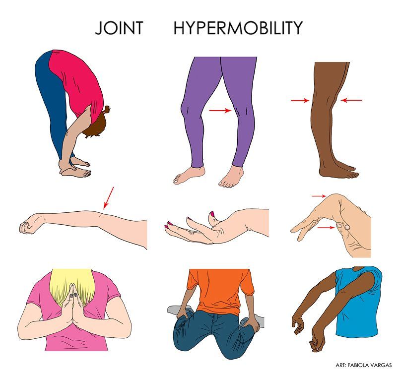
(Nursing Paper Example on Ehlers-Danlos Syndrome) Ehlers-Danlos Syndrome (EDS) is a group of rare, inherited connective tissue disorders that affect the skin, joints, and blood vessels. It is characterized by a range of symptoms, including hypermobility of joints, skin hyperextensibility, and tissue fragility, which vary in severity depending on the subtype. EDS is primarily caused by genetic mutations affecting the structure or synthesis of collagen, a vital protein in connective tissue. These mutations lead to compromised tissue integrity and function, resulting in complications such as chronic pain, joint instability, and, in severe cases, vascular rupture. While there are 13 recognized subtypes of EDS, each linked to distinct genetic abnormalities, the syndrome’s diagnosis and management remain challenging due to its diverse presentation.

Nursing Paper Example on Ehlers-Danlos Syndrome

Causes of Ehlers-Danlos Syndrome

Ehlers-Danlos Syndrome (EDS) is caused by genetic mutations that affect connective tissue components, primarily collagen. Collagen is a crucial protein that provides strength and elasticity to tissues such as skin, ligaments, and blood vessels. Mutations that impair collagen production or structure lead to weakened connective tissues, which underlie the manifestations of EDS.

Genetic Mutations and Subtypes
There are 13 recognized subtypes of EDS, each linked to specific genetic mutations. For example, classical EDS results from mutations in the COL5A1 and COL5A2 genes, which encode type V collagen. These mutations lead to fragile and hyperextensible skin. In vascular EDS, mutations in the COL3A1 gene disrupt the synthesis of type III collagen, which compromises blood vessel integrity and increases the risk of vascular rupture. Other subtypes, such as hypermobile EDS, lack a clearly defined genetic mutation, although recent studies suggest potential links to the TNXB gene.

Hereditary Patterns
The mode of inheritance varies by subtype. Autosomal dominant inheritance is observed in most forms, including classical and vascular EDS, meaning an affected individual has a 50% chance of passing the condition to their offspring. Autosomal recessive inheritance is less common and is seen in subtypes like dermatosparaxis EDS, requiring both parents to carry a copy of the mutated gene.

Environmental and Epigenetic Influences
While EDS is predominantly genetic, environmental and epigenetic factors may influence the severity of symptoms. Factors such as mechanical stress on joints, repetitive strain, or nutritional deficiencies can exacerbate symptoms but do not directly cause the condition.


Signs and Symptoms of Ehlers-Danlos Syndrome

The signs and symptoms of Ehlers-Danlos Syndrome (EDS) can vary widely depending on the subtype, but they primarily affect the skin, joints, and blood vessels. Common symptoms include hypermobility of joints, fragile skin, and a tendency to bruise easily.

Skin Involvement
One of the hallmark signs of EDS is hyperelasticity, or the ability of the skin to stretch beyond normal limits. Skin in individuals with EDS may be soft, velvety, and fragile, leading to frequent bruising and scarring. These individuals often develop atrophic scars that are thin, narrow, and stretch easily. In classical EDS, these skin changes are more pronounced, with a higher risk of wound complications and delayed wound healing.

Joint Hypermobility
Joint hypermobility is another defining feature, particularly in the hypermobile subtype of EDS. Affected individuals may experience loose or unstable joints that easily dislocate or subluxate. This hypermobility can lead to chronic joint pain, instability, and an increased risk of injury. Joint hypermobility often affects the fingers, knees, elbows, and shoulders.

Vascular Complications
In more severe forms of EDS, such as vascular EDS, blood vessels, internal organs, and tissues are more fragile. This increases the risk of spontaneous ruptures, particularly in the arteries, which can be life-threatening. Individuals with vascular EDS may experience internal bleeding, organ rupture, or arterial dissection, which can cause severe complications.

Other Symptoms
Additional symptoms may include muscle weakness, scoliosis, and early onset of osteoarthritis due to joint instability. Individuals with EDS often experience chronic fatigue, and in some cases, there may be a higher susceptibility to developing cardiac issues like mitral valve prolapse.

These symptoms vary in severity and can significantly impact daily functioning, making early diagnosis and intervention important for managing the condition.


Pathophysiology of Ehlers-Danlos Syndrome

Ehlers-Danlos Syndrome (EDS) is primarily caused by genetic mutations that affect the production or processing of collagen, a key structural protein in the body. Collagen provides strength and elasticity to connective tissues, and its defects result in the hallmark features of EDS, such as hyper-elastic skin and joint hypermobility.

Collagen Deficiency or Abnormalities
Collagen plays a crucial role in the structural integrity of skin, tendons, ligaments, and blood vessels. In EDS, mutations in genes that encode collagen molecules, or in genes responsible for collagen processing enzymes, lead to weakened collagen fibers. In classical EDS, for instance, mutations affect type I or type III collagen, making the collagen fibers weaker and more prone to stretching. In vascular EDS, mutations in the COL3A1 gene affect type III collagen, leading to fragile blood vessels and internal organs, which increases the risk of life-threatening ruptures.

Impact on Connective Tissue
Collagen abnormalities disrupt the normal function of connective tissues, impairing their ability to support and protect internal structures. As a result, individuals with EDS experience increased skin fragility, joint hypermobility, and soft tissue that is easily injured. The instability of collagenous structures also contributes to the characteristic pain, instability, and frequent dislocations seen in affected individuals.

Increased Risk of Vascular and Organ Rupture
In severe forms like vascular EDS, the defective collagen in the blood vessels leads to their fragility, making them more susceptible to rupture. This defect also affects internal organs such as the intestines and the uterus, increasing the risk of spontaneous organ rupture and internal bleeding. The pathophysiology of EDS thus underscores the essential role of collagen in maintaining tissue integrity, with disruptions leading to widespread systemic issues.

(Nursing Paper Example on Ehlers-Danlos Syndrome)


Diagnosis of Ehlers-Danlos Syndrome

Diagnosing Ehlers-Danlos Syndrome (EDS) involves a combination of clinical assessment, genetic testing, and family history analysis. Since there are several types of EDS, each with different genetic mutations, it is essential to accurately identify the specific subtype in order to provide the best management strategy.

Clinical Assessment
The diagnosis of EDS often begins with a detailed clinical evaluation, which includes a review of the patient’s symptoms and family medical history. Key diagnostic features include hyperelastic skin, joint hypermobility, and a history of frequent dislocations or joint instability. A physical examination is conducted to assess skin texture, joint flexibility, and the presence of characteristic bruising or scarring. The Beighton score, a system used to measure joint hypermobility, may be used as part of this assessment.

Genetic Testing
Genetic testing plays a crucial role in confirming the diagnosis and identifying the specific mutation responsible for the disorder. Whole-exome sequencing or targeted genetic tests are used to identify mutations in genes related to collagen synthesis and processing, such as COL1A1, COL1A2, COL3A1, and others, depending on the type of EDS suspected. These tests can help differentiate between the various subtypes of EDS and rule out other connective tissue disorders.

Family History and Genetic Counseling
Because EDS is inherited in an autosomal dominant or recessive pattern, understanding the family history is essential. If there is a history of EDS or related connective tissue disorders, it strengthens the suspicion of the condition. Genetic counseling may be offered to the family, especially when a specific mutation is identified, to discuss inheritance patterns, recurrence risks, and prenatal testing options for future pregnancies.

Skin Biopsy and Imaging
In some cases, a skin biopsy or imaging studies may be performed to assess the integrity of connective tissues or to rule out other causes of similar symptoms, such as other collagenopathies or connective tissue disorders.


Treatment Regimens for Ehlers-Danlos Syndrome

There is no cure for Ehlers-Danlos Syndrome (EDS), so treatment focuses on managing symptoms, preventing complications, and improving quality of life. The treatment regimen varies based on the subtype of EDS and the specific symptoms the patient experiences.

Multidisciplinary Approach
A multidisciplinary team of healthcare providers, including geneticists, dermatologists, orthopedic specialists, and physical therapists, is often involved in managing EDS. This team approach ensures that all aspects of the condition are addressed, including skin care, joint stability, and cardiovascular health.

Joint and Skin Care
Due to joint hypermobility, individuals with EDS are at higher risk for joint dislocations and sprains. Physical therapy plays a critical role in strengthening muscles around the joints to reduce the risk of injury. Bracing or splinting may be used to provide additional support and stability to hypermobile joints. Patients are also advised to avoid high-impact activities that could lead to joint trauma. Skin care is also important, as patients may experience hyperelasticity and fragile skin prone to bruising and scarring. Gentle skin care routines and protective measures, such as avoiding excessive friction and trauma, can help minimize skin injuries.

Pain Management
Chronic pain is a common symptom of EDS, especially in patients with frequent joint dislocations and soft tissue damage. Non-steroidal anti-inflammatory drugs (NSAIDs), acetaminophen, and other analgesics may be used to manage pain. In some cases, patients may benefit from corticosteroid injections or other pain-relieving treatments.

Cardiovascular Management
For patients with vascular EDS, regular monitoring of cardiovascular health is essential. Blood pressure management and surveillance for arterial abnormalities, such as aneurysms, are critical components of treatment. Lifestyle modifications, such as avoiding smoking and maintaining a healthy weight, are encouraged to reduce cardiovascular risk.

Surgical Interventions
In some cases, surgical procedures may be necessary to repair joint or vascular damage. However, surgery should be approached with caution due to the increased risk of complications such as wound healing issues and bleeding. Careful planning and coordination with the healthcare team are essential to minimize risks.


Complications of Ehlers-Danlos Syndrome

Ehlers-Danlos Syndrome (EDS) can lead to a variety of complications, some of which are potentially life-threatening. These complications arise due to the connective tissue abnormalities that affect various body systems, including the skin, joints, and blood vessels. The severity and range of complications depend on the specific subtype of EDS.

Joint and Musculoskeletal Issues
The most common complications of EDS involve the musculoskeletal system, especially due to joint hypermobility and skin hyperelasticity. Individuals with EDS are prone to frequent joint dislocations, sprains, and fractures. Chronic pain from musculoskeletal problems is another significant issue, as damaged tissues and joints can lead to long-term discomfort. In some cases, osteoarthritis may develop earlier than usual due to joint instability.

Vascular Complications
For those with vascular EDS, one of the most serious complications is the risk of arterial rupture or aneurysms. The fragile blood vessels are more susceptible to damage, and even minor trauma can lead to life-threatening internal bleeding. These individuals may also experience spontaneous rupture of internal organs, such as the intestines or uterus, which can be fatal without prompt medical attention.

Skin Complications
Individuals with EDS often have highly fragile skin, which can lead to frequent bruising, scarring, and delayed wound healing. Due to the skin’s hyperelasticity, wounds may stretch or tear easily, and even minor cuts or abrasions can result in significant scarring. In severe cases, skin fragility can lead to spontaneous skin rupture, requiring careful wound management.

Cardiovascular and Internal Organ Damage
In addition to vascular complications, individuals with EDS may experience internal organ damage due to the fragility of connective tissues. This can lead to complications such as lung or gastrointestinal perforations, which require immediate medical intervention to prevent life-threatening consequences.

Neurological Issues
EDS can also affect the nervous system. Individuals may experience chronic headaches, nerve compression, or even issues with proprioception, where the body loses the ability to sense its position in space. These neurological complications can significantly impact daily functioning and overall quality of life.


Prevention of Complications

Ehlers-Danlos Syndrome (EDS) is a genetic disorder, and as such, prevention strategies are limited. Since the condition is inherited, the focus of prevention lies in early detection, managing symptoms, and minimizing the risk of complications. While there is no known cure for EDS, certain measures can help manage the condition and improve quality of life.

Genetic Counseling
For individuals with a family history of EDS or those at risk of passing the condition to their offspring, genetic counseling is crucial. This service helps couples understand the inheritance patterns of EDS and the likelihood of passing the condition to their children. Genetic testing can also confirm a diagnosis in those with a family history, allowing for early intervention and management.

Early Diagnosis and Regular Monitoring
Early diagnosis of EDS can significantly improve outcomes by allowing individuals to start management plans early. Regular monitoring by healthcare providers specializing in EDS can help detect complications early. Cardiovascular and musculoskeletal issues, such as joint dislocations or artery rupture, can be addressed proactively, reducing the risks of severe outcomes.

Joint Protection and Physical Therapy
Joint protection is an essential preventative measure for individuals with EDS, particularly for those with hypermobile types. Physical therapy can help strengthen the muscles around joints, reducing the risk of dislocations and preventing musculoskeletal injuries. Occupational therapy may also be beneficial for managing activities of daily living and ensuring that individuals with EDS protect their joints during routine tasks.

Skin Care and Wound Management
Since people with EDS have fragile skin, good skin care is essential. Regular moisturizing, protection from excessive sunlight, and avoiding harsh chemicals can help maintain skin integrity. For wound care, prompt medical attention and proper techniques to handle minor cuts and bruises can prevent infections and minimize scarring.

Lifestyle Adjustments
Lifestyle adjustments, such as avoiding high-impact sports or activities that place excessive strain on the joints, can prevent further musculoskeletal damage. In addition, individuals with vascular EDS should avoid trauma or injury, as their blood vessels are highly susceptible to rupture. Wearing protective gear and taking precautions during physical activities can help reduce the risk of accidents.

By adopting these prevention strategies, individuals with EDS can minimize complications, manage symptoms more effectively, and improve their overall quality of life. However, ongoing research may lead to new prevention strategies and treatments in the future.


Patient Education

Patient education plays a vital role in managing Ehlers-Danlos Syndrome (EDS) and improving the quality of life for individuals affected by the condition. As EDS is a lifelong disorder, educating patients and their families about the nature of the disease, its symptoms, and management strategies is essential for both physical and emotional well-being.

Understanding EDS and Its Types
Patients should be educated about the specific type of EDS they have, as there are various subtypes, each with different manifestations and risks. Understanding the nature of the disease, including the genetic causes and inheritance patterns, helps individuals manage expectations and make informed decisions about family planning. This knowledge also empowers patients to take an active role in managing their health.

Symptom Awareness and Early Intervention
Patients should be encouraged to recognize common symptoms such as joint hypermobility, skin fragility, and easy bruising. Early detection of complications, such as joint dislocations, cardiovascular issues, or skin injuries, can reduce the risk of severe outcomes. Patients should be aware of warning signs and seek medical attention promptly if complications arise, particularly those related to the heart and blood vessels.

Joint Protection and Physical Therapy
Education on joint protection is crucial, especially for individuals with the hypermobility type of EDS. Patients should learn safe techniques for daily activities to avoid joint dislocations or strains. Physical therapy, including strengthening exercises, can help stabilize joints and improve muscle tone, reducing the likelihood of musculoskeletal injuries. Occupational therapy can also teach adaptive strategies for managing activities of daily living.

Skin Care and Wound Management
Because individuals with EDS have fragile skin, educating patients on proper skin care is important. This includes moisturizing regularly, using mild soaps, and protecting the skin from sun damage. Instructing patients on effective wound care techniques, such as cleaning and bandaging minor cuts, can help prevent infections and reduce scarring.

Psychosocial Support and Coping Strategies
EDS can be physically and emotionally challenging, so providing patients with information on coping strategies is essential. Encouraging them to seek psychological support or counseling may help them manage the mental strain of living with a chronic condition. Support groups and online communities can also offer a valuable source of emotional support, as sharing experiences with others facing similar challenges can reduce feelings of isolation.

By providing comprehensive education about EDS, healthcare professionals can empower patients to manage the condition proactively and improve their overall quality of life.


Prognosis

The prognosis of Ehlers-Danlos Syndrome (EDS) largely depends on the specific type of EDS a patient has, as well as the severity of symptoms and any associated complications. Since EDS is a genetic disorder affecting connective tissues, it is a lifelong condition, but the course of the disease varies greatly between individuals.

Impact of EDS Type on Prognosis
The different subtypes of EDS influence prognosis in distinct ways. For example, individuals with the hypermobility type (hEDS) often have a relatively normal life expectancy and can lead independent lives with proper management, though they may experience chronic pain, joint instability, and frequent dislocations. In contrast, individuals with vascular EDS (vEDS), caused by mutations affecting blood vessels, may have a significantly reduced life expectancy due to the risk of arterial rupture and internal bleeding. Early diagnosis and management can improve survival in these cases, but the risks associated with vascular complications remain high.

Management of Symptoms and Complications
The prognosis for patients with EDS can improve with early diagnosis, proper symptom management, and a tailored care plan. Regular monitoring for potential complications, such as heart issues or musculoskeletal problems, can prevent severe outcomes. With physical therapy, joint protection strategies, and proper wound care, many individuals with milder forms of EDS can live relatively normal lives. However, severe forms of the condition may require more intensive care, including surgical intervention and ongoing support from multidisciplinary teams.

Quality of Life Considerations
Although life expectancy may not be significantly impacted in most cases, the quality of life can be affected by chronic pain, mobility challenges, and the emotional toll of dealing with a lifelong condition. Ongoing psychological support, community engagement, and proper education on disease management can greatly improve the well-being of patients and enhance their coping mechanisms.

Conclusion

Ehlers-Danlos syndrome is a complex group of connective tissue disorders characterized by hypermobility, skin hyper-elasticity, and increased susceptibility to bruising and injury. The causes of this condition are often genetic, with various mutations in collagen and other connective tissue-related genes leading to different forms of the syndrome. Its symptoms can range from mild to severe, affecting the skin, joints, and blood vessels. Pathophysiologically, the impaired collagen production and structure contribute to tissue fragility, leading to the common clinical manifestations. Diagnosis relies heavily on clinical evaluation, family history, and genetic testing, while management focuses on symptom relief and preventing complications such as joint dislocations or vascular issues. Although no cure exists, early intervention and appropriate treatment can improve quality of life and reduce the risk of severe complications. Patient education and regular monitoring are crucial for managing the condition effectively, ensuring patients lead active, healthy lives.


References

Mayo Clinic. (2023). Ehlers-Danlos syndrome. Mayo Clinic. https://www.mayoclinic.org/diseases-conditions/ehlers-danlos-syndrome

National Library of Medicine. (2023). Ehlers-Danlos syndrome overview. National Institutes of Health. https://www.ncbi.nlm.nih.gov

Genetics Home Reference. (2023). Ehlers-Danlos syndromes. U.S. National Library of Medicine. https://ghr.nlm.nih.gov

Ehlers-Danlos Society. (2023). What is EDS? The Ehlers-Danlos Society. https://www.ehlers-danlos.com

 
Do you need a similar assignment done for you from scratch? Order now!
Use Discount Code "Newclient" for a 15% Discount!

Nursing Paper Example on Ecthyma

Nursing Paper Example on Ecthyma

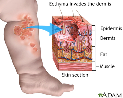
(Nursing Paper Example on Ecthyma) Ecthyma is a deeper skin infection characterized by ulcerative lesions that penetrate the epidermis and reach the dermis. Often regarded as a severe form of impetigo, ecthyma is most commonly caused by bacterial infections and typically affects individuals with poor hygiene or compromised immune systems. This paper provides a comprehensive overview of ecthyma, covering its causes, clinical presentation, pathophysiology, diagnostic criteria, treatment options, and preventive measures.


Nursing Paper Example on Ecthyma

Causes of Ecthyma

Ecthyma is primarily caused by bacterial infections. The main causative agents are:

  • Streptococcus pyogenes:

A beta-hemolytic bacterium often implicated in skin infections.

  • Staphylococcus aureus:

Frequently associated with ecthyma, especially strains resistant to methicillin (MRSA).

  • Secondary Factors:

Poor hygiene.

Trauma to the skin that provides entry points for bacteria.

Coexisting skin conditions such as eczema or insect bites.

Individuals with immune deficiencies, malnutrition, or systemic illnesses such as diabetes are at higher risk.


Signs and Symptoms of Ecthyma

Ecthyma lesions typically present as:

  • Initial Lesions:

Small, erythematous, and pustular sores that progress into vesicles.

  • Ulceration:

Vesicles rupture, forming round or oval ulcers with thick, adherent crusts.

Crusts are often yellowish-gray with erythematous, indurated margins.

  • Pain and Tenderness:

Lesions may cause discomfort and are tender to the touch.

  • Location:

Commonly affects the lower extremities but can appear on any part of the body.

If untreated, ecthyma may cause systemic symptoms, such as fever or lymphadenopathy, due to bacterial spread.


Pathophysiology of Ecthyma

Ecthyma begins with superficial bacterial colonization of damaged skin. If untreated, the infection spreads deeper into the dermis. Key factors include:

Host Immune Response: Inflammatory reactions to bacterial toxins result in tissue damage.

Enzymatic Breakdown: Bacterial enzymes, such as hyaluronidase, degrade dermal structures, facilitating deeper penetration.

Crust Formation: Exudates from necrotic tissues form the characteristic crust.

In severe or prolonged cases, secondary infections may develop, leading to cellulitis or systemic complications.


Diagnosis of Ecthyma

Diagnosing ecthyma involves a combination of clinical examination and laboratory investigations. Key diagnostic steps include:

Clinical Examination: Identification of ulcerative lesions with characteristic crusts.

Microbial Culture: Swabs from lesions help isolate causative bacteria and determine antibiotic sensitivity.

Histopathology: Rarely performed but may reveal dermal inflammation and necrosis.

Differential Diagnosis: Conditions such as impetigo, pyoderma gangrenosum, and cutaneous anthrax must be ruled out.

A thorough patient history, including hygiene practices and comorbidities, aids in diagnosis.


Treatment Regimens for Ecthyma

The primary goals of treatment are to eradicate the infection, promote healing, and prevent complications.

Topical Therapies

Antibacterial Ointments: Mupirocin or fusidic acid applied directly to lesions after cleaning.

Wound Care: Soaking the affected area with saline and debriding crusts promotes recovery.

Systemic Antibiotics

Indicated for extensive lesions or systemic symptoms.

First-Line Agents: Oral antibiotics such as penicillin or cephalexin targeting Streptococcus pyogenes.

MRSA Coverage: Clindamycin, doxycycline, or trimethoprim-sulfamethoxazole for suspected resistant strains.

Supportive Care

Maintaining hygiene and addressing underlying conditions such as diabetes or malnutrition is essential.


Complications of Ecthyma

If untreated, ecthyma can lead to severe complications, including:

Cellulitis: Bacterial spread into deeper tissues.

Post-Streptococcal Glomerulonephritis: Rare but serious immune-mediated kidney damage following Streptococcus pyogenes infections.

Scarring: Lesions may leave permanent scars due to dermal involvement.

Systemic Infections: Bacteremia and septicemia in immunocompromised individuals.

Prompt treatment minimizes the risk of these complications.


Prevention of Ecthyma

Preventive measures focus on reducing bacterial colonization and maintaining skin integrity. These include:

Personal Hygiene: Regular bathing and appropriate wound care.

Injury Management: Immediate cleaning and dressing of cuts or abrasions.

Addressing Underlying Conditions: Managing systemic illnesses like diabetes.

Prophylactic Measures: In high-risk groups, antibacterial soaps or preventive antibiotics may be recommended.

Education on hygiene practices is critical, especially in communities with limited resources.


Prognosis

With timely and appropriate treatment, ecthyma generally resolves without long-term consequences. However, severe or neglected cases may result in scarring or systemic complications.


Patient Education

Patients should be educated about:

Recognizing early signs of ecthyma to seek prompt treatment.

Maintaining hygiene and avoiding practices that damage the skin, such as excessive scratching.

Adhering to prescribed treatments and follow-ups to ensure complete resolution of the infection.

Support from healthcare providers, including detailed explanations and demonstrations of wound care, fosters adherence and better outcomes.


Conclusion

Ecthyma is a significant skin infection that can cause considerable discomfort and complications if untreated. Early diagnosis, targeted antibiotic therapy, and proper wound care are crucial for successful management. Preventive measures and patient education play vital roles in reducing the incidence of ecthyma, particularly in vulnerable populations.


References

American Academy of Dermatology Association. (2023). Ecthyma: Causes, symptoms, and treatments.
https://www.aad.org

Centers for Disease Control and Prevention (CDC). (2023). Group A Streptococcal (GAS) diseases.
https://www.cdc.gov

National Library of Medicine. (2023). Ecthyma: A bacterial skin infection.
https://www.ncbi.nlm.nih.gov

Mayo Clinic. (2023). Ecthyma: Diagnosis and management.
https://www.mayoclinic.org

 
Do you need a similar assignment done for you from scratch? Order now!
Use Discount Code "Newclient" for a 15% Discount!

Nursing Paper Example on Dysautonomia

Nursing Paper Example on Dysautonomia

Dysautonomia refers to a group of disorders that affect the autonomic nervous system, responsible for controlling involuntary bodily functions such as heart rate, blood pressure, digestion, and temperature regulation. The condition can manifest in various forms and degrees of severity, ranging from mild to life-altering.


Nursing Paper Example on Dysautonomia

Causes of Dysautonomia

The causes of dysautonomia depend on the specific type of the disorder. Some cases arise due to genetic predisposition, while others develop secondary to other conditions or triggers. Key causes include:

  • Primary Dysautonomia:

Genetic mutations, such as those seen in familial dysautonomia (Riley-Day syndrome).

Autoimmune dysfunction, as observed in autoimmune autonomic ganglionopathy.

  • Secondary Dysautonomia:

Chronic illnesses such as diabetes, Parkinson’s disease, or multiple sclerosis.

Physical trauma or spinal cord injuries.

Viral infections, including Epstein-Barr virus and COVID-19.

Exposure to toxins or certain medications.

The underlying mechanism typically involves damage, degeneration, or dysfunction of the autonomic nerves or their receptors (National Institute of Neurological Disorders and Stroke [NINDS], 2023).


Signs and Symptoms of Dysautonomia

Dysautonomia can present with a broad spectrum of symptoms that vary based on the affected autonomic functions. Common signs include:

  • Cardiovascular Symptoms:

Orthostatic hypotension: A sudden drop in blood pressure upon standing.

Tachycardia or bradycardia.

  • Gastrointestinal Symptoms:

Gastroparesis: Delayed stomach emptying leading to nausea, bloating, and vomiting.

Diarrhea or constipation.

  • Neurological Symptoms:

Dizziness or fainting (syncope).

Headaches and fatigue.

  • Other Symptoms:

Heat intolerance and abnormal sweating.

Bladder dysfunction.

Difficulty regulating body temperature.

The severity and combination of symptoms can vary significantly among individuals.


Types of Dysautonomia

Several forms of dysautonomia exist, each with unique characteristics. Common types include:

  • Postural Orthostatic Tachycardia Syndrome (POTS):

Affects mostly young women and causes an abnormal increase in heart rate upon standing.

  • Multiple System Atrophy (MSA):

A rare neurodegenerative disorder with autonomic failure and motor dysfunction.

  • Neurocardiogenic Syncope:

Leads to fainting episodes triggered by stress, pain, or prolonged standing.

  • Familial Dysautonomia (Riley-Day Syndrome):

A genetic condition primarily affecting individuals of Ashkenazi Jewish descent.

  • Autoimmune Autonomic Ganglionopathy:

An autoimmune condition where antibodies attack autonomic nerve receptors.

Each type has distinct features and may require tailored treatment.


Pathophysiology of Dysautonomia

The autonomic nervous system is divided into the sympathetic and parasympathetic branches. Dysautonomia arises from the imbalance or failure of these systems, which may involve:

  • Nerve Degeneration:

Loss of nerve fibers in the autonomic ganglia or peripheral nerves, as seen in MSA.

  • Immune-Mediated Damage:

Antibody attacks on acetylcholine receptors, leading to autonomic dysfunction.

  • Abnormal Reflexes:

Dysregulated baroreceptor reflexes cause blood pressure instability.

  • Metabolic Impairment:

Poor glucose metabolism in diabetes-induced autonomic neuropathy.

These abnormalities lead to systemic dysregulation of multiple physiological functions.


Diagnosis of Dysautonomia

Diagnosing dysautonomia is challenging due to its diverse presentation. Common diagnostic methods include:

  • Clinical History:

Detailed evaluation of symptoms and their progression.

  • Autonomic Testing:

Tilt table test to assess blood pressure and heart rate responses.

Sweat tests to evaluate sudomotor function.

Heart rate variability and Valsalva maneuver.

  • Blood Tests:

To rule out secondary causes like infections, autoimmune disorders, or metabolic issues.

  • Imaging:

MRI or CT scans to identify structural abnormalities in the brain or spinal cord.

Collaborative care involving neurologists, cardiologists, and other specialists is often required for an accurate diagnosis.


Treatment Regimens for Dysautonomia

Treatment for dysautonomia is individualized, focusing on symptom management and addressing the underlying cause.

Lifestyle Modifications

Increased salt and fluid intake to improve blood volume and prevent orthostatic hypotension.

Compression garments to enhance venous return.

Regular exercise tailored to the patient’s capacity.

Medications

  • Cardiovascular Drugs:

Midodrine for low blood pressure.

Beta-blockers for heart rate control in POTS.

  • Neurological Medications:

Gabapentin or pregabalin for neuropathic pain.

Immunosuppressants for autoimmune causes.

  • Gastrointestinal Treatments:

Prokinetics like metoclopramide for gastroparesis.

Laxatives or antidiarrheal agents as needed.

Physical Therapy

Recumbent or water-based exercises to improve cardiovascular fitness without triggering symptoms.

Advanced cases, such as MSA, may require palliative care to address progressive disability.


Complications Associated with Dysautonomia

Untreated dysautonomia can lead to significant complications, including:

Chronic fatigue and reduced quality of life.

Recurrent syncope, increasing the risk of falls and injuries.

Progressive organ dysfunction in neurodegenerative forms.

Prompt diagnosis and treatment are essential to mitigate these risks.


Prognosis

The prognosis for dysautonomia varies based on the underlying cause. While conditions like POTS often respond well to treatment, neurodegenerative forms like MSA have a poorer outlook due to their progressive nature.


Patient Education

Education is vital for effective dysautonomia management. Patients should:

Understand their condition and triggers for symptom exacerbation.

Follow prescribed treatment plans and attend regular follow-ups.

Maintain a healthy lifestyle and monitor hydration and nutrition.

Support groups and resources from organizations like Dysautonomia International can provide additional support.


Conclusion

Dysautonomia encompasses a range of disorders with diverse causes, symptoms, and outcomes. Comprehensive management involves accurate diagnosis, symptom control, and addressing the underlying etiology. With appropriate care, many individuals with dysautonomia can achieve improved function and quality of life.


References

Dysautonomia International. (2023). What is Dysautonomia?
https://www.dysautonomiainternational.org

National Institute of Neurological Disorders and Stroke (NINDS). (2023). Dysautonomia Information Page.
https://www.ninds.nih.gov

Mayo Clinic. (2023). Dysautonomia: Symptoms and Causes.
https://www.mayoclinic.org

Cleveland Clinic. (2023). Dysautonomia: Types, Symptoms, Causes, Diagnosis & Treatment.
https://my.clevelandclinic.org

 
Do you need a similar assignment done for you from scratch? Order now!
Use Discount Code "Newclient" for a 15% Discount!

Nursing Paper Example on Dupuytren’s Contracture

Nursing Paper Example on Dupuytren’s Contracture

(Nursing Paper Example on Dupuytren’s Contracture) Dupuytren’s contracture, also known as palmar fibromatosis, is a progressive condition affecting the connective tissue in the palm of the hand. This condition leads to thickening and tightening of the fascia, causing one or more fingers to bend permanently toward the palm. It can significantly impact hand function, particularly grip strength and dexterity.


Nursing Paper Example on Dupuytren's Contracture

Causes of Dupuytren’s Contracture

The exact cause of Dupuytren’s contracture remains unclear, but research suggests it results from a combination of genetic, environmental, and lifestyle factors. Key contributing factors include:

  • Genetics: A strong hereditary component has been noted, with a higher prevalence among individuals of Northern European descent.
  • Gender: Men are more commonly affected than women.
  • Age: The condition usually develops after the age of 50.
  • Lifestyle Factors: Smoking and alcohol consumption are associated with an increased risk.
  • Medical Conditions: It is more common in individuals with diabetes and those undergoing treatment for epilepsy.

These factors may trigger changes in the connective tissue, leading to the abnormal production of collagen and subsequent fibrotic nodules and cords in the palm (National Health Service [NHS], 2023).


Signs and Symptoms of Dupuytren’s Contracture

Dupuytren’s contracture often progresses gradually over several years. Common signs and symptoms include:

  1. Palmar Nodules: Small, firm lumps that form in the palm, often near the base of the ring or little finger.
  2. Thickened Cords: Bands of fibrous tissue develop, extending from the nodules to the fingers.
  3. Finger Flexion: The fingers, especially the ring and little fingers, gradually bend toward the palm.
  4. Loss of Function: As the contracture progresses, affected individuals may struggle to fully straighten their fingers, affecting grip strength and manual dexterity.

The condition is usually painless, but some individuals may experience tenderness around the nodules in the early stages.


Pathophysiology of Dupuytren’s Contracture

Dupuytren’s contracture is characterized by fibroproliferative changes in the palmar fascia. The following mechanisms contribute to the condition:

  • Collagen Overproduction: Type III collagen, which is normally present during wound healing, accumulates excessively in the fascia, forming nodules and cords.
  • Myofibroblast Activity: Myofibroblasts, specialized cells involved in tissue repair, become overactive and contract, leading to thickening and shortening of the connective tissue.
  • Vascular and Immune Dysregulation: Altered blood flow and immune responses in the affected tissue may further exacerbate fibrosis (Mayo Clinic, 2023).

Diagnosis of Dupuytren’s Contracture

Dupuytren’s contracture is typically diagnosed through a clinical examination. Key diagnostic steps include:

  1. Physical Examination: The physician palpates the palm and fingers to assess the presence of nodules, cords, and contractures.
  2. Functional Assessment: Patients may be asked to place their hand flat on a table. Inability to fully straighten the fingers is a hallmark feature.
  3. Differential Diagnosis: Other conditions, such as trigger finger and tenosynovitis, are ruled out.

Imaging studies, such as ultrasound or magnetic resonance imaging (MRI), are rarely needed but may be used to evaluate the extent of fibrosis in complex cases.


Treatment Regimens for Dupuytren’s Contracture

Treatment options for Dupuytren’s contracture depend on the severity and functional impact of the condition. These include non-invasive and surgical approaches.

Non-Surgical Treatments

  • Watchful Waiting: Mild cases with minimal functional impairment may only require observation.
  • Steroid Injections: Corticosteroids can reduce inflammation and slow the progression of nodules.
  • Collagenase Injections: Enzymatic injections, such as collagenase clostridium histolyticum, break down the fibrous cords, improving finger extension.

Surgical Treatments

  • Needle Aponeurotomy: A minimally invasive procedure where a needle is used to divide the thickened cords.
  • Fasciectomy: Partial or total removal of the affected fascia to release the contracture.
  • Dermofasciectomy: Removal of the fascia along with the overlying skin, often requiring skin grafting to prevent recurrence.

Rehabilitation

Post-procedure rehabilitation, including physical therapy and splinting, is crucial to restore range of motion and prevent recurrence.


Complications Associated with Dupuytren’s Contracture

Without treatment, Dupuytren’s contracture can lead to severe hand deformity and loss of function. Even after treatment, complications may arise, including:

  • Recurrence of contractures.
  • Nerve or blood vessel damage during surgery.
  • Stiffness and reduced range of motion due to scar formation.

Prognosis

The prognosis for Dupuytren’s contracture varies. Early intervention and treatment generally lead to better outcomes, but recurrence is common. Surgical procedures are more likely to yield lasting improvements compared to non-surgical methods.


Patient Education

Education is vital for managing Dupuytren’s contracture. Patients should:

  • Understand the progressive nature of the disease.
  • Avoid hand trauma and repetitive strain to minimize aggravation.
  • Seek medical evaluation promptly if contractures worsen.
  • Follow postoperative care instructions, including physical therapy and splinting.

Support groups and patient organizations can also provide resources and assistance.


Conclusion

Dupuytren’s contracture is a chronic connective tissue disorder with significant functional implications. While its exact cause remains unclear, advancements in non-surgical and surgical treatments have improved management options. Early diagnosis, tailored interventions, and rehabilitation are essential for preserving hand function and quality of life.


References

Mayo Clinic. (2023). Dupuytren’s Contracture: Symptoms and Causes. https://www.mayoclinic.org

National Health Service (NHS). (2023). Dupuytren’s Contracture. https://www.nhs.uk

American Academy of Orthopaedic Surgeons. (2023). Dupuytren’s Contracture: Overview. https://www.aaos.org

MedlinePlus. (2023). Dupuytren’s Disease. https://medlineplus.gov

 
Do you need a similar assignment done for you from scratch? Order now!
Use Discount Code "Newclient" for a 15% Discount!

Nursing Paper Example on Down Syndrome

Nursing Paper Example on Down Syndrome

Down syndrome, also known as trisomy 21, is a genetic disorder caused by the presence of an extra copy of chromosome 21. This condition leads to intellectual disability, distinctive physical features, and an increased risk of certain medical conditions. Down syndrome is one of the most common chromosomal disorders and has significant implications for physical, cognitive, and social development.


Nursing Paper Example on Down Syndrome

Causes of Down Syndrome

Down syndrome is caused by abnormal cell division involving chromosome 21, which leads to an extra partial or full chromosome. The three main types of Down syndrome are:

  1. Trisomy 21: This is the most common type, accounting for 95% of cases. It occurs when every cell in the body has three copies of chromosome 21 instead of the usual two.
  2. Translocation: In about 4% of cases, a portion of chromosome 21 becomes attached (translocated) to another chromosome. This type can occur before or after conception.
  3. Mosaicism: This rare form, present in about 1% of cases, occurs when some cells have the usual two copies of chromosome 21, while others have three. Mosaic Down syndrome tends to result in fewer symptoms.

The exact reason for the abnormal division is unknown, but maternal age is a significant risk factor. Women aged 35 and older have a higher likelihood of having a child with Down syndrome (Centers for Disease Control and Prevention [CDC], 2023).


Signs and Symptoms of Down Syndrome

Individuals with Down syndrome exhibit a range of physical, cognitive, and developmental characteristics. These include:

Physical Features

  • Flattened facial profile
  • Upward slanting eyes with epicanthal folds
  • Short neck
  • Small ears and mouth
  • Broad, short hands with a single palmar crease
  • Poor muscle tone (hypotonia) during infancy
  • Short stature

Cognitive and Developmental Features

  • Intellectual disability, often mild to moderate
  • Delayed speech and language development
  • Delayed motor milestones, such as walking and sitting

Medical Conditions

  • Congenital heart defects (present in about 50% of cases)
  • Hearing loss
  • Vision problems, including cataracts and strabismus
  • Thyroid disorders, particularly hypothyroidism
  • Increased risk of leukemia and autoimmune diseases
  • Sleep apnea due to structural abnormalities and hypotonia
  • Gastrointestinal abnormalities, such as duodenal atresia or Hirschsprung’s disease

The severity of symptoms varies widely, and some individuals with Down syndrome may experience fewer complications than others.


Pathophysiology of Down Syndrome

The presence of an extra chromosome 21 leads to an overexpression of its genes, disrupting normal development and function. The genes on chromosome 21 are involved in various biological processes, including:

Neurodevelopment: Overexpression of specific genes contributes to brain structure changes, resulting in intellectual disability and learning challenges.

Cellular Processes: Dysregulation in signaling pathways impacts immune function, cardiovascular health, and metabolism.

Aging: Chromosome 21 overexpression is associated with premature aging and an increased risk of Alzheimer’s disease.

The pathophysiological mechanisms are complex and involve both genetic and epigenetic factors (National Institutes of Health [NIH], 2023).


Diagnosis of Down Syndrome

Prenatal Diagnosis

Prenatal testing helps detect Down syndrome before birth and includes:

  1. Screening Tests:
    • Blood tests measuring biomarkers, such as human chorionic gonadotropin (hCG) and alpha-fetoprotein (AFP).
    • Ultrasound to identify physical markers like nuchal translucency.
  2. Diagnostic Tests:
    • Amniocentesis: Extracts amniotic fluid for chromosomal analysis.
    • Chorionic Villus Sampling (CVS): Analyzes placental tissue for genetic abnormalities.
    • Non-Invasive Prenatal Testing (NIPT): Examines fetal DNA in the mother’s blood.

Postnatal Diagnosis

After birth, Down syndrome is confirmed using:

Physical Examination: Identification of characteristic physical traits.

Karyotyping: A blood test to analyze the number and structure of chromosomes, confirming trisomy 21.


Management of Down Syndrome

There is no cure for Down syndrome, but early intervention and multidisciplinary care can enhance quality of life.

Medical Management

Congenital Heart Defects: Regular cardiology evaluations; surgical correction if necessary.

Hearing and Vision Care: Routine screenings; use of hearing aids and corrective lenses when required.

Thyroid Disorders: Routine thyroid function tests; thyroid hormone replacement therapy if needed.

Developmental and Educational Support

Early intervention programs for physical, occupational, and speech therapy.

Special education tailored to the individual’s cognitive abilities.

Support for inclusion in mainstream classrooms to promote social development.

Behavioral and Psychological Support

Counseling and behavioral therapy for managing anxiety, depression, and other emotional challenges.

Support groups for families to provide emotional and practical assistance.

Regular Health Monitoring

Routine screenings for common complications like sleep apnea, leukemia, and gastrointestinal issues.

Immunizations to protect against infections, as individuals with Down syndrome may have weaker immune systems.


Prognosis and Quality of Life

The life expectancy of individuals with Down syndrome has significantly improved, reaching an average of 60 years due to advancements in medical care. Many individuals lead fulfilling lives, participate in social activities, and contribute to their communities.

Supportive families, access to healthcare, and opportunities for education and employment play vital roles in improving quality of life.


Complications Associated with Down Syndrome

If not managed effectively, Down syndrome can lead to several complications:

Severe Congenital Heart Disease: Can result in heart failure or other life-threatening conditions.

Early-Onset Alzheimer’s Disease: Common in individuals aged 40 and older.

Chronic Health Conditions: Recurrent infections, diabetes, and obesity are more prevalent.

Psychosocial Challenges: Stigmatization and lack of societal inclusion can impact mental health.


Prevention of Down Syndrome

There is no way to prevent Down syndrome, but certain measures can help:

  • Genetic counseling for families with a history of Down syndrome.
  • Advanced maternal age pregnancies benefit from prenatal testing to inform decision-making.

Conclusion

Down syndrome remains a complex condition with significant physical, cognitive, and social challenges. However, early diagnosis, tailored interventions, and comprehensive healthcare can significantly improve outcomes. As society continues to advance in medical care and social inclusion, individuals with Down syndrome have more opportunities to thrive and contribute meaningfully.


References

Centers for Disease Control and Prevention. (2023). Facts about Down Syndrome. https://www.cdc.gov

Johns Hopkins Medicine. (2023). Down Syndrome: Overview and Care. https://www.hopkinsmedicine.org

Mayo Clinic. (2023). Down Syndrome: Symptoms and Causes. https://www.mayoclinic.org

National Institutes of Health. (2023). Understanding Down Syndrome. https://www.nih.gov

World Health Organization. (2023). Genetic Disorders – Down Syndrome. https://www.who.int

 
Do you need a similar assignment done for you from scratch? Order now!
Use Discount Code "Newclient" for a 15% Discount!