Nursing Paper Example on Acromegaly

Nursing Paper Example on Acromegaly

Acromegaly is a rare but serious hormonal disorder caused by an overproduction of growth hormone (GH). It typically results from a benign tumor (pituitary adenoma) in the pituitary gland, which is responsible for secreting excessive amounts of GH. The condition is most commonly diagnosed in middle-aged adults and can lead to significant health complications if left untreated. In this paper, we will explore the causes, signs and symptoms, etiology, pathophysiology, DSM-5 diagnosis, treatment regimens, and patient education related to acromegaly. Understanding this disease is crucial for early detection and effective management to prevent severe complications.
Nursing Paper Example on Acromegaly

Causes of Acromegaly

The primary cause of acromegaly is the presence of a pituitary adenoma, a non-cancerous tumor in the pituitary gland. The tumor secretes an abnormally high level of growth hormone, leading to the overproduction of insulin-like growth factor 1 (IGF-1) by the liver. The excessive IGF-1 promotes abnormal growth of bones and tissues throughout the body. Although pituitary adenomas are the most common cause, acromegaly can also result from other conditions, such as ectopic production of growth hormone-releasing hormone (GHRH), which stimulates the pituitary gland to release excess growth hormone.

Signs and Symptoms

The signs and symptoms of acromegaly develop gradually and can be subtle at first, making early diagnosis challenging. The most common symptoms include:

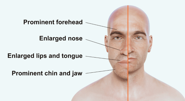
  • Facial Changes: Enlargement of facial features such as the nose, jaw, lips, and tongue. Patients may experience a coarsening of the facial features, including a protruding jaw (prognathism) and a broadened nose.
  • Hand and Foot Enlargement: Increased size of the hands and feet, often noticed when patients find that their rings or shoes no longer fit.
  • Joint Pain and Arthropathy: Due to the abnormal growth of bones, acromegaly often leads to joint pain, osteoarthritis, and carpal tunnel syndrome.
  • Skin Changes: Thickening and increased sweating of the skin, as well as the development of skin tags.
  • Vision Problems: As the pituitary adenoma grows, it can compress the optic nerves, leading to visual disturbances, such as peripheral vision loss.
  • Sleep Apnea: Due to the enlargement of soft tissues in the throat and tongue, patients may develop obstructive sleep apnea.

Etiology

The underlying etiology of acromegaly is almost always related to the development of a pituitary adenoma. These adenomas are classified as either microadenomas (less than 10 mm in diameter) or macroadenomas (greater than 10 mm in diameter). The majority of these adenomas are somatotroph adenomas, which specifically secrete growth hormone. In rare cases, acromegaly may be caused by ectopic GH or GHRH production, most often by tumors in the lungs, pancreas, or gastrointestinal tract.

Several genetic conditions have also been linked to acromegaly, including:

  • Multiple Endocrine Neoplasia Type 1 (MEN1): A hereditary disorder that increases the risk of developing pituitary adenomas and other endocrine tumors.
  • Carney Complex: A genetic disorder associated with pituitary adenomas, among other tumors.

Pathophysiology

Acromegaly is characterized by the overproduction of growth hormone, which leads to an increase in IGF-1 levels. The elevated IGF-1 stimulates the growth of bones and soft tissues, especially in areas such as the face, hands, and feet. The high levels of GH and IGF-1 also have metabolic effects, leading to insulin resistance, hypertension, and dyslipidemia.

The excess growth hormone can also cause tissue hypertrophy and increased cell division, contributing to the development of various complications, including:

  • Cardiovascular Disease: Acromegaly is associated with an increased risk of cardiovascular conditions such as hypertension, heart failure, and arrhythmias. The excess GH can lead to the thickening of the heart muscle (hypertrophic cardiomyopathy), which can impair cardiac function.
  • Sleep Apnea: As mentioned, the enlargement of tissues in the upper airway can result in obstructive sleep apnea, further exacerbating cardiovascular complications.
  • Diabetes: Growth hormone’s antagonistic effect on insulin can lead to insulin resistance and, eventually, type 2 diabetes.

DSM-5 Diagnosis

Acromegaly is not specifically listed in the Diagnostic and Statistical Manual of Mental Disorders, Fifth Edition (DSM-5), as it is not primarily a psychiatric condition. However, diagnosis is made based on clinical signs, biochemical tests, and imaging studies.

The diagnostic approach for acromegaly involves the following:

  1. Biochemical Testing: The first step is to measure the serum levels of IGF-1, as it provides a reliable indication of growth hormone excess. An oral glucose tolerance test (OGTT) is then conducted, in which a failure to suppress GH levels after glucose administration supports the diagnosis of acromegaly.
  2. Imaging: Once biochemical evidence of acromegaly is confirmed, an MRI of the pituitary gland is performed to identify the presence of a pituitary adenoma.
  3. Visual Field Testing: Given the risk of optic nerve compression from a pituitary tumor, visual field testing may be conducted to assess any vision abnormalities.

Treatment Regimens

The treatment of acromegaly aims to normalize GH and IGF-1 levels, alleviate symptoms, and prevent complications. The main treatment options include:

  • Surgical Treatment: The primary treatment for acromegaly is the surgical removal of the pituitary adenoma. The goal is to achieve a complete resection of the tumor. However, surgery is not always curative, especially if the adenoma is large or located near critical structures like the optic nerves.
  • Medical Treatment: If surgery is unsuccessful or not feasible, medical therapy may be used. The following medications are commonly employed:
    • Somatostatin Analogs (Octreotide, Lanreotide): These drugs inhibit GH secretion and reduce IGF-1 levels.
    • Growth Hormone Receptor Antagonists (Pegvisomant): Pegvisomant blocks the effects of GH at its receptor and lowers IGF-1 levels.
    • Dopamine Agonists (Cabergoline): In some cases, dopamine agonists can help reduce tumor size and lower GH production.
  • Radiotherapy: In cases where surgery and medication are not sufficient, radiotherapy may be used to shrink the tumor over time. This is typically reserved for patients with persistent disease after other treatments.

Patient Education

Patient education is a vital component of managing acromegaly. Key educational points for patients include:

  • Understanding the Disease: Patients should be informed about the nature of acromegaly, including its symptoms, complications, and the importance of early diagnosis and treatment.
  • Treatment Adherence: It is crucial for patients to adhere to prescribed treatment regimens, whether surgical, medical, or a combination of both. Non-adherence can lead to persistent disease and further complications.
  • Monitoring and Follow-Up: Regular monitoring of GH and IGF-1 levels is necessary to assess treatment efficacy. Patients should also be monitored for complications such as cardiovascular disease, diabetes, and sleep apnea.
  • Lifestyle Modifications: Patients may benefit from lifestyle changes, such as weight management, regular exercise, and a healthy diet, to manage associated conditions like hypertension and diabetes.

Conclusion

Acromegaly is a rare endocrine disorder that can have significant, long-term effects on a patient’s health if not properly managed. Early diagnosis and treatment are essential for reducing the risk of severe complications. Medical advancements, including surgical and pharmacological interventions, have greatly improved the prognosis for individuals with acromegaly. Patient education is also a critical component of managing the disease, ensuring adherence to treatment, and preventing further health issues.

References

Fagin, J. A., & Kastelein, J. J. (2019). Acromegaly. The Lancet Diabetes & Endocrinology, 7(6), 450-460. https://doi.org/10.1016/S2213-8587(19)30087-4

Melmed, S. (2016). Acromegaly. New England Journal of Medicine, 375(17), 1664-1673. https://doi.org/10.1056/NEJMra1602113

Colao, A., & Di Somma, C. (2017). Acromegaly: Pathophysiology and clinical management. Endocrinology and Metabolism Clinics of North America, 46(4), 685-704. https://doi.org/10.1016/j.ecl.2017.07.001

Giustina, A., & Chanson, P. (2017). Acromegaly: Pathophysiology and clinical management. The Lancet Diabetes & Endocrinology, 5(7), 561-575. https://doi.org/10.1016/S2213-8587(17)30119-9

 
Do you need a similar assignment done for you from scratch? Order now!
Use Discount Code "Newclient" for a 15% Discount!

Nursing Paper Example on Zika Fever

Nursing Paper Example on Zika Fever

(Nursing Paper Example on Zika Fever) Zika fever, caused by the Zika virus, is a mosquito-borne illness that became a significant global concern during outbreaks in the 21st century. Its association with severe congenital anomalies and neurological complications underlines its importance as a public health issue. This paper provides an in-depth look into the causes, symptoms, transmission, pathophysiology, diagnosis, treatment, prevention, complications, and global health implications of Zika fever.


Nursing Paper Example on Zika Fever

Causes and Transmission of Zika Fever

Causative Agent

The Zika virus, an arbovirus from the genus Flavivirus within the family Flaviviridae, is the etiological agent of Zika fever. It was first identified in rhesus monkeys in Uganda’s Zika Forest in 1947 and later in humans in 1952. Two primary strains exist: the African lineage and the Asian lineage, the latter linked to the 2015–2016 global outbreaks (Centers for Disease Control and Prevention [CDC], 2023).

Transmission Pathways

The primary mode of Zika virus transmission is via Aedes mosquito bites (Aedes aegypti and Aedes albopictus). These mosquitoes are most active during the day. Other routes of transmission include:

  1. Maternal-Fetal Transmission: The virus can cross the placenta and infect the fetus, leading to congenital Zika syndrome.
  2. Sexual Transmission: The virus can be present in semen, vaginal fluids, and saliva, spreading through sexual contact.
  3. Blood Transfusion: Rare cases of Zika virus transmission through contaminated blood have been reported.
  4. Occupational Exposure: Accidental exposure in laboratory and healthcare settings is another route (World Health Organization [WHO], 2023).

Understanding these transmission methods is essential for controlling the spread of Zika fever.


Signs and Symptoms of Zika Fever

Zika fever often presents with mild symptoms, and many individuals remain asymptomatic. When symptomatic, the illness has an incubation period of 3–14 days and typically includes:

  • Low-Grade Fever: Usually below 38.5°C.
  • Maculopapular Rash: Rash starting on the face and spreading to the rest of the body.
  • Arthralgia: Joint pain, particularly in smaller joints of the hands and feet.
  • Conjunctivitis: Non-purulent redness of the eyes.
  • Fatigue, Myalgia, and Headache: Other common but non-specific symptoms.

Symptoms generally last for 2–7 days. Severe illness is rare but may occur in vulnerable populations, including pregnant women and individuals with compromised immune systems (Johns Hopkins Medicine, 2023).

(Nursing Paper Example on Zika Fever)


Pathophysiology of Zika Fever

The Zika virus enters the human body through the bite of an infected mosquito. It binds to receptors such as AXL on host cells, allowing it to invade keratinocytes, dendritic cells, and fibroblasts. Once inside, the virus replicates, triggering an inflammatory response and cell death.

In pregnant women, the virus can cross the placental barrier, infecting fetal neural progenitor cells. This disrupts neurodevelopment, causing conditions such as microcephaly. The exact mechanisms are still under investigation, but studies suggest viral-induced apoptosis and disruption of neural cell function (Rasmussen et al., 2016).

The virus also stimulates an immune response that, in some cases, may lead to autoimmune complications such as Guillain-Barré syndrome, characterized by peripheral nervous system damage.


Diagnosis of Zika Fever

Diagnosing Zika fever involves clinical evaluation and laboratory testing:

  1. Molecular Testing: Reverse transcription polymerase chain reaction (RT-PCR) is the gold standard for detecting Zika virus RNA in blood, urine, or saliva. It is most effective within the first week of symptom onset.
  2. Serological Testing: Enzyme-linked immunosorbent assay (ELISA) detects IgM and IgG antibodies. However, cross-reactivity with related flaviviruses, such as dengue, complicates diagnosis.
  3. Prenatal Ultrasound: Pregnant women suspected of Zika infection undergo fetal ultrasounds to detect anomalies associated with congenital Zika syndrome, such as microcephaly.
  4. Imaging: In neurological cases, brain imaging (MRI or CT) may reveal complications like calcifications or brain abnormalities in newborns (CDC, 2023).

Efforts to improve diagnostic accuracy are ongoing, particularly in resource-limited settings.


Treatment and Management of Zika Fever

There is no specific antiviral treatment for Zika fever. Management focuses on symptomatic relief:

  • Pain Management: Acetaminophen is recommended for fever and joint pain, but nonsteroidal anti-inflammatory drugs are avoided until dengue is ruled out due to the risk of bleeding complications.
  • Hydration and Rest: Patients are encouraged to drink fluids and rest to alleviate symptoms.
  • Monitoring in Pregnancy: Pregnant women require close monitoring for fetal anomalies, including regular ultrasounds.

Hospitalization is rarely needed except in severe cases or complications.


Prevention of Zika Fever

Preventing Zika fever requires targeted measures to reduce mosquito exposure and halt virus transmission:

  • Mosquito Control: Strategies include using insect repellents, eliminating standing water, and employing larvicides.
  • Protective Measures: Wearing long-sleeved clothing and using bed nets reduce the risk of mosquito bites.
  • Travel Warnings: Pregnant women are advised to avoid traveling to areas with active Zika virus transmission.
  • Safe Sexual Practices: Condom use is encouraged to prevent sexual transmission.
  • Vaccine Development: Although no vaccine is currently approved, ongoing research aims to develop safe and effective immunization options (WHO, 2023).

Public health campaigns play a vital role in educating communities about these preventive measures.


Complications of Zika Fever

Congenital Zika Syndrome

This syndrome results from vertical transmission of the virus during pregnancy and includes:

  • Microcephaly: Abnormal brain development leading to a reduced head size.
  • Neurological Abnormalities: Hearing and vision impairments, seizures, and intellectual disabilities.

Neurological Complications in Adults

Zika virus has been linked to Guillain-Barré syndrome, characterized by rapid-onset muscle weakness and paralysis. Other rare complications include meningoencephalitis and myelitis.


Public Health Impact of Zika Fever

The Zika virus outbreak of 2015–2016 revealed the virus’s potential to cause widespread health emergencies. In regions like the Americas and Southeast Asia, the epidemic led to significant socioeconomic consequences, including healthcare system strain and tourism revenue losses. The emergence of congenital Zika syndrome has further underscored the importance of global collaboration in controlling vector-borne diseases (Rasmussen et al., 2016).

Surveillance programs, public awareness campaigns, and investment in vaccine research are critical for mitigating the long-term effects of Zika fever.


Conclusion

Zika fever remains a global health concern due to its ability to cause severe congenital and neurological complications. Efforts to combat the disease focus on improving diagnosis, advancing research on vaccines, and implementing effective mosquito control strategies. A coordinated global response is crucial to reducing the impact of future Zika outbreaks.


References

Centers for Disease Control and Prevention. (2023). Zika virus. https://www.cdc.gov/zika

Johns Hopkins Medicine. (2023). Zika virus infection. https://www.hopkinsmedicine.org

Rasmussen, S. A., Jamieson, D. J., Honein, M. A., & Petersen, L. R. (2016). Zika virus and birth defects—reviewing the evidence for causality. New England Journal of Medicine, 374(20), 1981-1987. https://doi.org/10.1056/NEJMsr1604338

World Health Organization. (2023). Zika virus. https://www.who.int

 
Do you need a similar assignment done for you from scratch? Order now!
Use Discount Code "Newclient" for a 15% Discount!

Nursing Paper Example on Yersiniosis

Nursing Paper Example on Yersiniosis

(Nursing Paper Example on Yersiniosis) Yersiniosis is an infectious disease primarily caused by Yersinia enterocolitica, a bacterium often associated with gastroenteritis. While the condition generally results in mild gastrointestinal symptoms, it can occasionally lead to severe complications, especially in young children, immunocompromised individuals, and those with underlying health conditions. This paper examines the causes, symptoms, pathophysiology, diagnosis, treatment, prevention, and potential complications associated with yersiniosis.

Nursing Paper Example on Yersiniosis

Causes and Transmission of Yersiniosis

Causative Agent

Yersiniosis is primarily caused by Yersinia enterocolitica, a gram-negative coccobacillus bacterium. Less commonly, Yersinia pseudotuberculosis is also responsible for some cases. These bacteria can survive in cold temperatures, allowing them to thrive in refrigerated foods and other cool environments. Yersinia enterocolitica is often found in raw or undercooked pork, though it can also be present in contaminated water, milk, and a variety of other foods (Centers for Disease Control and Prevention [CDC], 2023).

Transmission Pathways

Yersiniosis is typically transmitted via the fecal-oral route through consumption of contaminated food or water. Common sources of infection include:

  • Raw or Undercooked Pork Products: Yersinia bacteria are often present in pork, with chitterlings (pork intestines) and other undercooked pork products posing a higher risk.
  • Unpasteurized Milk and Dairy Products: Dairy products that are not pasteurized may harbor the bacteria.
  • Contaminated Water or Vegetables: Ingestion of water or vegetables contaminated with Yersinia can also lead to infection, particularly in areas with poor sanitation.

In rare cases, direct contact with an infected animal or person can spread the bacteria. The infection is more common in colder climates, as the bacteria can survive and even multiply at low temperatures (Food Safety and Inspection Service, 2023).

Signs and Symptoms of Yersiniosis

The incubation period for yersiniosis ranges from four to seven days after exposure to the bacteria. Symptoms often resemble other gastrointestinal infections but can vary in severity.

  • Gastrointestinal Symptoms: The majority of patients experience diarrhea, abdominal pain (especially in the lower right quadrant), nausea, and vomiting. Fever may also be present. In children, yersiniosis may mimic appendicitis, leading to unnecessary surgical interventions.
  • Other Symptoms in Severe Cases: In rare cases, individuals may develop pharyngitis or experience symptoms beyond the gastrointestinal tract, such as skin rash, joint pain, or erythema nodosum—a painful red rash typically on the legs. Systemic symptoms are more common in immunocompromised individuals.

Symptoms generally last one to three weeks but can persist longer in individuals with weakened immune systems (Johns Hopkins Medicine, 2023).

Pathophysiology of Yersiniosis

Upon ingestion, Yersinia enterocolitica travels to the small intestine and adheres to intestinal epithelial cells. The bacteria utilize a set of virulence factors, including adhesins and invasins, to facilitate cellular entry and evade immune responses. Inside the cells, Yersinia can survive and replicate, triggering a host inflammatory response that leads to mucosal damage.

The body’s immune response to Yersinia involves both innate and adaptive immunity. However, certain virulence mechanisms enable the bacteria to resist phagocytosis and immune recognition, which allows it to persist in the host. This persistence can lead to chronic symptoms in some cases, and in rare situations, bacteremia (the spread of bacteria to the bloodstream), resulting in systemic infection (Carniel & Nogrady, 2021).

Diagnosis of Yersiniosis

Diagnosing yersiniosis can be challenging due to the similarity of its symptoms to other gastrointestinal conditions, including appendicitis and Crohn’s disease. The following diagnostic methods are used:

  • Stool Culture: Stool samples are collected and cultured to identify Yersinia enterocolitica. Due to the bacterium’s slower growth rate, stool cultures may take several days for definitive results.
  • Polymerase Chain Reaction (PCR): PCR testing for Yersinia DNA is a more rapid and sensitive method that can confirm infection. It is particularly useful during outbreaks or when other family members are also symptomatic.
  • Blood Culture (in severe cases): In cases with systemic symptoms or immunocompromised patients, blood cultures may be necessary to check for bacteremia.

Laboratory identification of Yersinia requires special growth media, as it does not thrive in routine stool culture conditions. This necessitates specific laboratory protocols to confirm yersiniosis (CDC, 2023).

Treatment and Management of Yersiniosis

Most cases of yersiniosis are self-limiting and do not require specific treatment beyond supportive care. Treatment options depend on symptom severity and the patient’s health status.

  • Hydration and Symptomatic Relief: Supportive care, including rehydration and electrolyte replacement, is essential for managing dehydration due to diarrhea. Over-the-counter pain relievers may be used for abdominal pain, but caution is needed with nonsteroidal anti-inflammatory drugs to prevent potential gastrointestinal complications.
  • Antibiotic Therapy (in severe cases): In cases with severe symptoms or systemic involvement, antibiotics such as ciprofloxacin, doxycycline, or trimethoprim-sulfamethoxazole may be prescribed. Antibiotics are typically reserved for immunocompromised individuals or those with prolonged symptoms to prevent the development of antibiotic resistance.

Children, elderly adults, and those with weakened immune systems should be closely monitored to prevent complications and to provide early intervention if symptoms worsen (Food Safety and Inspection Service, 2023).

Prevention of Yersiniosis

Preventive measures are essential to minimize the risk of yersiniosis, especially in areas or communities with higher incidence rates. Prevention strategies include:

  • Proper Food Handling and Cooking: Ensuring pork is thoroughly cooked is crucial to kill any potential Yersinia bacteria. The CDC recommends cooking pork to an internal temperature of 145°F (62.8°C).
  • Avoiding Unpasteurized Milk: Drinking pasteurized milk and dairy products reduces the risk of exposure to Yersinia and other pathogens.
  • Good Hygiene Practices: Washing hands after handling raw meat or visiting farms with animals can prevent cross-contamination. Food preparation areas should be sanitized after handling raw meat to prevent the spread of bacteria.

Public health education on proper food handling and personal hygiene is vital in reducing the spread of yersiniosis, particularly in colder regions where infection rates are higher (CDC, 2023).

Complications and Prognosis of Yersiniosis

While yersiniosis is typically a mild disease, complications can arise in rare cases, especially in immunocompromised individuals. These complications include:

  • Reactive Arthritis: Reactive arthritis, a condition that causes joint pain and swelling, can develop weeks after the initial infection. It is more common in adults than in children.
  • Erythema Nodosum: This inflammatory skin condition, presenting as painful red nodules on the shins, occurs in response to yersiniosis and other bacterial infections. It generally resolves on its own but can be painful.
  • Systemic Involvement: In cases with bloodstream infection (bacteremia), Yersinia can spread to other organs, leading to systemic complications such as endocarditis or sepsis. These are rare but serious complications.

Most individuals recover fully from yersiniosis, though recovery time may vary. With proper medical care and preventive measures, the prognosis for yersiniosis remains favorable, especially when appropriate hygiene practices are followed.

(Nursing Paper Example on Yersiniosis)

Conclusion

Yersiniosis is a bacterial infection caused by Yersinia enterocolitica, primarily affecting the gastrointestinal system. Although the infection is typically mild and self-limiting, it can cause severe complications, particularly in at-risk populations. Diagnosis relies on stool culture or PCR testing, while treatment focuses on supportive care and, in severe cases, antibiotics. Preventive measures such as safe food handling and thorough cooking of pork products are essential to minimize infection risk. Continued public health efforts and awareness can reduce the prevalence and impact of yersiniosis, especially in colder regions.

References

Centers for Disease Control and Prevention. (2023). Yersiniosis. https://www.cdc.gov/yersinia

Carniel, E., & Nogrady, N. (2021). The pathophysiology of Yersinia enterocolitica infections. Journal of Bacteriology and Pathogenesis, 12(3), 241-249. https://doi.org/10.1128/jbp2021031241

Food Safety and Inspection Service. (2023). Yersinia enterocolitica in pork products: Guidelines for safe food handling. https://www.fsis.usda.gov

 
Do you need a similar assignment done for you from scratch? Order now!
Use Discount Code "Newclient" for a 15% Discount!

Nursing Paper Example on Yellow Fever

Nursing Paper Example on Yellow Fever

(Nursing Paper Example on Yellow Fever) Yellow fever is a viral hemorrhagic disease transmitted primarily by Aedes and Haemagogus mosquitoes. Known for its high mortality rate and potential to cause outbreaks, yellow fever remains a significant public health concern in tropical regions of Africa and South America. This overview examines yellow fever’s etiology, symptoms, transmission, pathophysiology, diagnosis, treatment, prevention, and complications.

Nursing Paper Example on Yellow Fever

Causes and Transmission of Yellow Fever

Yellow Fever Virus

Yellow fever is caused by the yellow fever virus, an RNA virus belonging to the Flavivirus genus and the Flaviviridae family. This genus also includes other viruses such as dengue, Zika, and West Nile viruses. Yellow fever virus is an arbovirus, meaning it is transmitted through arthropods, particularly mosquitoes, to humans. It originated in Africa and spread through travel and trade, especially during the colonial period, contributing to global outbreaks (Centers for Disease Control and Prevention [CDC], 2023).

Transmission Cycle

Yellow fever spreads through the bite of infected mosquitoes, primarily the Aedes aegypti species in urban settings and Haemagogus species in jungle or forested areas. Transmission cycles include:

  • Sylvatic Cycle: In this cycle, yellow fever is transmitted between non-human primates (monkeys) and mosquito species in jungle environments. Human infection occurs when individuals enter these habitats and get bitten by infected mosquitoes.
  • Intermediate Cycle: This occurs in areas where humans and primates live in proximity, often in rural or village settings. Mosquitoes infected by primates can spread the virus to humans, leading to localized outbreaks.
  • Urban Cycle: In highly populated urban areas, Aedes aegypti mosquitoes can spread the virus from human to human, leading to significant outbreaks. This cycle is especially concerning due to its potential for rapid spread (World Health Organization [WHO], 2023).

Signs and Symptoms of Yellow Fever

The incubation period of yellow fever is generally three to six days. Symptoms range from mild to severe and can progress through different stages.

  • Early Stage (Acute Phase): Initial symptoms include sudden onset of fever, chills, headache, back pain, muscle pain, nausea, vomiting, and weakness. These symptoms typically last three to four days.
  • Toxic Phase: About 15% of cases progress to the toxic phase, characterized by more severe symptoms, including high fever, jaundice (yellowing of the skin and eyes), abdominal pain, vomiting (sometimes with blood), bleeding from the nose, mouth, eyes, or stomach, and organ failure, particularly affecting the liver and kidneys. This phase is highly fatal, with a mortality rate of up to 50% in severe cases (Johns Hopkins Medicine, 2023).

Pathophysiology of Yellow Fever

Upon infection, the yellow fever virus targets hepatocytes (liver cells) and Kupffer cells (liver macrophages), leading to direct viral damage and immune-mediated damage to the liver. The viral replication in liver cells results in apoptosis (cell death), which contributes to the development of jaundice, a hallmark sign of yellow fever. Furthermore, the virus affects endothelial cells lining blood vessels, leading to hemorrhagic symptoms. A high viral load in the blood exacerbates the body’s immune response, causing a cytokine storm, which can lead to multi-organ failure in severe cases (McNeill et al., 2021).

Diagnosis of Yellow Fever

Laboratory Testing

Diagnosis of yellow fever is essential during outbreaks. Diagnostic methods include:

  • Serology (IgM and IgG antibodies): Detection of specific antibodies against yellow fever virus in the blood helps confirm recent or past infection. Serological tests like enzyme-linked immunosorbent assays (ELISA) detect IgM antibodies, which typically appear within seven to 10 days after infection.
  • Reverse Transcriptase Polymerase Chain Reaction (RT-PCR): RT-PCR detects the virus’s genetic material in blood samples. This test is most effective in the early stages of infection when the viral load is high.
  • Liver Biopsy (in postmortem cases): Histopathology of liver tissues can reveal characteristic signs of yellow fever infection, such as midzonal necrosis without inflammation, which is pathognomonic for yellow fever (WHO, 2023).

(Nursing Paper Example on Yellow Fever)

Treatment and Management of Yellow Fever

Yellow fever has no specific antiviral treatment. Management focuses on supportive care and alleviating symptoms.

  • Hydration and Electrolyte Balance: Fluids and electrolytes are administered to maintain hydration and prevent dehydration, especially in patients experiencing high fever and vomiting.
  • Pain and Fever Management: Paracetamol is commonly used to manage pain and fever, as aspirin and nonsteroidal anti-inflammatory drugs (NSAIDs) are avoided due to bleeding risks.
  • Close Monitoring for Organ Failure: For severe cases, monitoring liver and kidney function is essential, and advanced medical support may be necessary, including renal replacement therapy if kidney failure occurs.

In cases of severe bleeding, blood transfusions may be required. Isolation of patients is also recommended to prevent further spread of the virus within healthcare settings (CDC, 2023).

Prevention of Yellow Fever

Yellow Fever Vaccine

Vaccination is the most effective method of preventing yellow fever. The yellow fever vaccine is a live-attenuated vaccine that provides lifelong immunity in most individuals after a single dose. Immunization campaigns in endemic regions have reduced yellow fever incidence significantly.

The yellow fever vaccine is recommended for:

  • Individuals aged nine months or older in endemic regions.
  • Travelers to areas where yellow fever is endemic, as some countries require proof of vaccination for entry.
  • Outbreak response and control measures during epidemics to create herd immunity in affected areas.

Vector Control Measures

To reduce mosquito populations, vector control strategies such as insecticide spraying, removing stagnant water sources, and encouraging the use of mosquito repellents and bed nets are recommended. These measures aim to minimize human exposure to mosquitoes, especially in areas where vaccination coverage may be low or outbreaks are active (WHO, 2023).

Complications and Prognosis of Yellow Fever

Yellow fever can lead to life-threatening complications, especially in cases that progress to the toxic phase. Complications may include:

  • Liver Failure and Jaundice: Liver failure is a severe consequence of yellow fever, as the virus directly targets liver cells. Jaundice is a prominent clinical feature that indicates liver dysfunction.
  • Kidney Failure: Renal impairment is common in severe cases, often necessitating dialysis or renal replacement therapy.
  • Hemorrhage and Shock: Due to vascular damage and coagulopathy, patients may experience significant bleeding, leading to shock.
  • Multi-Organ Failure and Death: Multi-organ dysfunction due to widespread viral replication and immune response can be fatal. The case fatality rate among individuals in the toxic phase can reach up to 50% (Johns Hopkins Medicine, 2023).

The prognosis for yellow fever varies depending on the severity of the infection and access to supportive care. While mild cases often resolve with proper management, severe cases carry a high risk of mortality without adequate medical support.

Conclusion

Yellow fever remains a severe and sometimes fatal disease with no specific antiviral treatment. Prevention through vaccination and vector control is essential in endemic areas and for travelers. Surveillance, rapid diagnosis, and supportive care play critical roles in managing outbreaks and reducing mortality. Continued efforts toward vaccination coverage, coupled with vector control, are essential to control yellow fever’s public health impact.

References

Centers for Disease Control and Prevention. (2023). Yellow fever. https://www.cdc.gov/yellowfever

Johns Hopkins Medicine. (2023). Yellow fever. https://www.hopkinsmedicine.org/health/conditions-and-diseases/yellow-fever

McNeill, J., Peters, A., & Walker, D. (2021). Pathophysiology of yellow fever and clinical implications. Tropical Medicine and Infectious Disease, 6(2), 98-105. https://doi.org/10.3390/tropmed6020098

World Health Organization. (2023). Yellow fever: Key facts and prevention. https://www.who.int

 
Do you need a similar assignment done for you from scratch? Order now!
Use Discount Code "Newclient" for a 15% Discount!

Nursing Paper Example on Warts

Nursing Paper Example on Warts

Warts, or verrucae, are benign skin growths caused by the human papillomavirus (HPV). These skin lesions are common worldwide and can affect individuals of any age. Warts can occur on various parts of the body and have different clinical presentations depending on the HPV strain involved. While they are generally harmless, warts can cause discomfort, and some types may spread easily through contact, especially among immunocompromised individuals.

Nursing Paper Example on Warts

Causes and Transmission of Warts

Human Papillomavirus (HPV)

The primary cause of warts is the human papillomavirus (HPV), a DNA virus from the Papillomaviridae family. More than 100 HPV types exist, and around 30 types are responsible for skin warts. HPV infects the top layer of skin through small abrasions or cuts, leading to rapid cell proliferation that causes the visible wart. HPV types that cause skin warts differ from those causing genital warts or other HPV-related cancers, which helps clarify their relatively benign nature (Centers for Disease Control and Prevention [CDC], 2023).

Transmission

HPV spreads through direct skin-to-skin contact or indirectly through contact with surfaces contaminated with the virus. Common sites of transmission include communal areas, such as public showers, gyms, and pools. The virus can also spread through autoinoculation, where a person transfers the virus from one part of their body to another, especially by scratching or shaving affected areas. Immunocompromised individuals are more susceptible to HPV infection and may experience recurrent warts (National Institutes of Health [NIH], 2023).

Types of Warts and Their Symptoms

Warts can be classified into several types based on their location and appearance, with each type associated with specific HPV strains.

  • Common Warts (Verruca Vulgaris): Typically caused by HPV types 2 and 4, these warts appear as rough, raised lesions with a cauliflower-like texture. They usually form on the hands and fingers, especially in areas prone to injury.
  • Plantar Warts (Verruca Plantaris): These warts, caused by HPV types 1, 2, and 4, develop on the soles of the feet and can grow inward due to pressure. They may cause discomfort while walking and often appear with a hard, thickened surface and tiny black dots, representing clotted blood vessels.
  • Flat Warts (Verruca Plana): Caused by HPV types 3, 10, and 28, flat warts are smooth, slightly elevated lesions often found on the face, neck, hands, and arms. These warts are smaller than other types and are more common among children and young adults.
  • Filiform Warts: Characterized by finger-like projections, filiform warts are typically found on the face, around the mouth, nose, or eyes. HPV types 1, 2, 4, and 27 commonly cause filiform warts.
  • Genital Warts: Caused by HPV types 6 and 11, genital warts are transmitted through sexual contact. They are soft, flesh-colored growths that can appear on the genital and anal areas. Although not generally dangerous, they require specialized treatment due to their location and potential risk of spread (American Academy of Dermatology [AAD], 2023).

Pathophysiology of Warts

Upon entering the body through minor skin breaks, HPV infects the basal epithelial cells and promotes rapid cellular proliferation. The virus replicates in the nuclei of infected cells, leading to the formation of hyperkeratotic (thickened) and papillomatous (warty) growths on the skin. HPV can evade the immune response, which may contribute to the persistence of warts and their ability to spread. The body’s immune system may eventually recognize and destroy the virus, leading to spontaneous resolution in some cases (Johnson et al., 2021).

Diagnosis of Warts

Clinical Examination

Diagnosis of warts is typically clinical, based on visual examination and identification of characteristic features. The following diagnostic techniques may also be used to confirm wart presence:

  • Dermatoscopy: This non-invasive technique uses a dermatoscope to visualize the wart’s surface, aiding in differentiation from other skin lesions.
  • Biopsy: In rare cases, especially if the wart does not respond to treatment or presents unusual characteristics, a biopsy may be conducted to rule out malignancy. Histopathological examination typically reveals hyperkeratosis, papillomatosis, and viral cytopathic effects (such as koilocytosis) indicative of HPV infection (American Academy of Dermatology [AAD], 2023).

Treatment and Management of Warts

Treatment for warts depends on the type, location, and symptoms of the lesions. While some warts may resolve spontaneously, treatment is often pursued to relieve discomfort, prevent spread, and improve cosmetic appearance.

  • Topical Salicylic Acid: This over-the-counter treatment is commonly used for warts on the hands, feet, and other areas. Salicylic acid exfoliates the outer layers of the wart, gradually reducing its size. Regular application and adherence are key to its effectiveness.
  • Cryotherapy: A healthcare provider applies liquid nitrogen to freeze the wart, causing cellular destruction. Multiple sessions are often required, and side effects can include temporary pain, blistering, and scarring.
  • Cantharidin: Derived from beetles, cantharidin causes blistering of the treated wart, leading it to separate from the skin. This treatment is typically administered by a dermatologist, as it requires precise application.
  • Immunotherapy: For recalcitrant warts, immunotherapy using agents like imiquimod or contact sensitizers like diphencyprone (DPCP) helps stimulate the immune response to attack the HPV-infected cells.
  • Laser Treatment and Electrosurgery: For large or stubborn warts, laser therapy (such as pulsed-dye lasers) and electrosurgery can be effective in destroying wart tissue, though these methods carry a higher risk of scarring (National Institutes of Health [NIH], 2023).

Prevention of Warts

Prevention focuses on reducing HPV exposure and strengthening the immune system’s ability to fight infection. Effective preventive measures include:

  • Good Hygiene Practices: Regular handwashing, especially after touching warts, reduces the risk of spreading HPV to other parts of the body or to others.
  • Protective Footwear: Wearing flip-flops or sandals in communal showers and pools reduces exposure to plantar wart-causing HPV strains.
  • Avoiding Skin Trauma: Minimizing cuts and abrasions on the skin decreases entry points for the virus, lowering the risk of wart formation.
  • HPV Vaccination: While primarily aimed at preventing genital HPV strains linked to cancer, the HPV vaccine may also reduce the risk of common warts, although this effect requires further study (Centers for Disease Control and Prevention [CDC], 2023).

Prognosis and Complications

Warts are generally benign and may resolve without intervention over months to years. However, complications can arise, particularly in individuals with compromised immune systems or persistent, recurrent warts. Possible complications include:

  • Pain and Discomfort: Plantar warts may cause pain during walking, impacting mobility and quality of life.
  • Secondary Infections: Warts can become secondarily infected, especially if scratched or injured.
  • Scarring and Pigmentation Changes: Some treatments, particularly invasive ones, may result in scarring or pigmentation alterations at the site (Johnson et al., 2021).

Conclusion

Warts are a widespread dermatological condition caused by HPV infection. While generally harmless, warts can be distressing due to their appearance and potential for spread. With various treatment options available, managing warts involves a combination of self-care, medical intervention, and preventive practices to mitigate their impact. Although spontaneous resolution is common, persistent or symptomatic cases benefit from professional evaluation and targeted treatment strategies.

References

American Academy of Dermatology. (2023). Warts: Diagnosis and treatment. https://www.aad.org

Centers for Disease Control and Prevention. (2023). Human papillomavirus (HPV) and warts: Causes, symptoms, and prevention. https://www.cdc.gov

Johnson, S. M., Roberts, J., & Smith, R. (2021). HPV-related skin lesions: A clinical overview. Journal of Dermatological Treatment, 32(3), 245-250. https://doi.org/10.1080/09546634.2020.1858629

National Institutes of Health. (2023). Warts: Overview and management. MedlinePlus. https://medlineplus.gov

 
Do you need a similar assignment done for you from scratch? Order now!
Use Discount Code "Newclient" for a 15% Discount!

Nursing Paper Example on Warkany Syndrome

Nursing Paper Example on Warkany Syndrome

(Nursing Paper Example on Warkany Syndrome) Warkany syndrome, also known as Trisomy 8 Mosaicism Syndrome, is a rare chromosomal disorder involving the presence of an extra chromosome 8 in some or all of the body’s cells. First described by Dr. Joseph Warkany, the syndrome has a variable presentation, with symptoms ranging from mild to severe physical abnormalities and developmental delays. Although Trisomy 8 can occur in a complete or mosaic form, the mosaic type is more common and compatible with life, as the complete form often results in miscarriage.

Nursing Paper Example on Warkany Syndrome

Genetic Basis and Etiology of Warkany Syndrome

Chromosomal Abnormality

Warkany syndrome is caused by trisomy of chromosome 8, meaning that instead of having two copies, affected cells have three copies of this chromosome. The syndrome typically presents as mosaic trisomy, where only a portion of the cells contain the extra chromosome, while others have a normal chromosomal arrangement. This genetic mosaicism arises during early embryonic cell division, leading to a mix of normal and trisomic cells in the body (Brooks et al., 2020).

Risk Factors

Although the exact cause of the chromosomal anomaly in Warkany syndrome is unknown, certain risk factors may increase the likelihood of chromosomal abnormalities, including maternal age. Advanced maternal age has been associated with an increased risk of chromosomal disorders in offspring, although it is less directly correlated with mosaic trisomy conditions like Warkany syndrome (National Organization for Rare Disorders, 2023).

Clinical Presentation and Signs and Symptoms

The manifestations of Warkany syndrome are variable and depend on the proportion and distribution of trisomic cells across different tissues and organs. However, some common clinical features include:

  • Distinctive Facial Features: These may include deep-set eyes, prominent forehead, and large, low-set ears.
  • Skeletal Abnormalities: Individuals often display musculoskeletal abnormalities, such as joint contractures, scoliosis, or spinal deformities.
  • Intellectual and Developmental Delays: Mild to moderate intellectual disability is common, along with delays in speech and motor skill development.
  • Hand and Foot Abnormalities: Features such as clinodactyly (curved fingers), broad thumbs, and small or webbed toes are often seen.
  • Cardiovascular and Renal Anomalies: Structural heart defects and kidney abnormalities are reported, adding to the complexity of the syndrome’s management.
  • Hypotonia: Reduced muscle tone, or hypotonia, is common, contributing to delayed motor milestones such as sitting and walking (National Institutes of Health, 2023).

Diagnosis of Warkany Syndrome

Prenatal Diagnosis

Prenatal diagnosis of Warkany syndrome is challenging due to the mosaic nature of the condition. Trisomy 8 mosaicism can sometimes be detected during prenatal testing, including:

  1. Amniocentesis: This procedure involves sampling amniotic fluid to analyze fetal cells for chromosomal abnormalities.
  2. Chorionic Villus Sampling (CVS): CVS samples placental tissue, which may also reveal mosaic trisomy 8 if present. However, CVS can miss mosaic cases if the extra chromosome is not present in the sampled cells.

Postnatal Diagnosis

Once born, the diagnosis of Warkany syndrome involves a combination of clinical assessment and genetic testing:

  1. Karyotyping: Chromosomal analysis of blood cells can reveal trisomy 8 mosaicism. However, because only some cells contain the extra chromosome, multiple tissue samples may be tested to confirm the mosaic distribution.
  2. Fluorescent In Situ Hybridization (FISH): FISH analysis is a more sensitive technique for detecting chromosomal abnormalities in various cell types, such as skin fibroblasts or buccal cells, and is useful in confirming mosaicism.
  3. Comparative Genomic Hybridization (CGH): CGH can detect even small chromosomal imbalances, providing a detailed profile of genetic abnormalities (Wong & Lamb, 2019).

Pathophysiology of Warkany Syndrome

The extra chromosome 8 in trisomy mosaicism disrupts normal gene expression, affecting multiple systems within the body. This chromosomal imbalance impacts cellular signaling and development pathways in various tissues, leading to the observed abnormalities in physical appearance, neurological development, and organ structure. Because of the mosaic nature, the extent of these disruptions varies based on the proportion of trisomic cells in each tissue, accounting for the syndrome’s variable presentation (Brooks et al., 2020).

Treatment and Management of Warkany Syndrome

Multidisciplinary Approach

Management of Warkany syndrome requires a tailored approach based on individual symptoms, involving several specialists:

  • Cardiology and Nephrology Consultations: To manage congenital heart defects and kidney abnormalities, which are common in patients with Warkany syndrome.
  • Orthopedics and Physical Therapy: Skeletal abnormalities, such as joint contractures and scoliosis, often require orthopedic intervention and ongoing physical therapy to improve mobility and motor function.
  • Developmental and Behavioral Therapy: Speech therapy, occupational therapy, and behavioral interventions support intellectual and developmental progress, addressing speech delays, fine motor skills, and adaptive behaviors.
  • Genetic Counseling: Families benefit from genetic counseling to understand the condition, its inheritance, and the potential recurrence risk in future pregnancies (National Organization for Rare Disorders, 2023).

Prognosis and Complications of Warkany Syndrome

The prognosis for individuals with Warkany syndrome is highly variable and depends on the severity of symptoms and the extent of mosaicism. While life expectancy is generally not reduced, complications from structural heart defects, kidney issues, and other systemic abnormalities can influence overall health outcomes.

Common Complications

  1. Cardiovascular Complications: Structural heart anomalies may lead to functional impairments and require monitoring and, in some cases, surgical correction.
  2. Respiratory Issues: Spinal deformities, such as scoliosis, can contribute to respiratory difficulties over time.
  3. Psychosocial Challenges: Individuals with Warkany syndrome may face social and psychological challenges, necessitating ongoing psychological support (Wong & Lamb, 2019).

(Nursing Paper Example on Warkany Syndrome)

Prevention and Genetic Counseling

Warkany syndrome is not typically inherited but occurs as a random chromosomal event during cell division. Consequently, no preventive measures are available. However, genetic counseling is recommended for families affected by Warkany syndrome, as it helps them understand the condition, potential recurrence risks, and options for prenatal testing in future pregnancies.

Conclusion

Warkany syndrome, or Trisomy 8 Mosaicism Syndrome, is a rare chromosomal disorder with a highly variable clinical presentation. Diagnosis requires genetic testing due to the mosaic nature of the condition, and management involves a comprehensive, multidisciplinary approach. Although life expectancy is generally normal, the condition may present complex health challenges, making early intervention and individualized care essential.

References

Brooks, A. S., & Monaghan, K. G. (2020). Trisomy 8 mosaicism syndrome. American Journal of Medical Genetics Part A, 182(5), 1048-1056. https://doi.org/10.1002/ajmg.a.61527

National Organization for Rare Disorders. (2023). Trisomy 8 mosaicism syndrome. https://rarediseases.org/rare-diseases/trisomy-8-mosaicism-syndrome/

National Institutes of Health. (2023). Trisomy 8 mosaicism. Genetic and Rare Diseases Information Center. https://rarediseases.info.nih.gov/diseases/5013/trisomy-8-mosaicism

Wong, C., & Lamb, A. N. (2019). Molecular genetic testing for mosaic trisomy 8. Cytogenetic and Genome Research, 159(2), 75-81. https://doi.org/10.1159/000500101

 
Do you need a similar assignment done for you from scratch? Order now!
Use Discount Code "Newclient" for a 15% Discount!

Nursing Paper Example on Viral Meningitis

Nursing Paper Example on Viral Meningitis

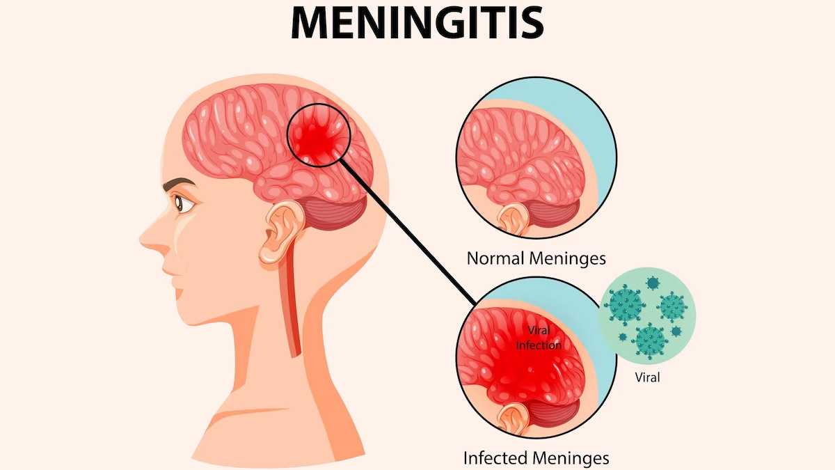
(Nursing Paper Example on Viral Meningitis) Viral meningitis, also known as aseptic meningitis, is an inflammation of the meninges, which are the protective membranes surrounding the brain and spinal cord. Unlike bacterial meningitis, viral meningitis is generally less severe, but it can still cause significant morbidity. This disease is caused by a variety of viruses and can affect people of all ages, though certain populations may be more vulnerable to severe complications.

Nursing Paper Example on Viral Meningitis

Causes of Viral Meningitis

Common Viral Causes

The primary causative agents of viral meningitis include:

  1. Enteroviruses: Responsible for up to 85% of viral meningitis cases, enteroviruses such as Coxsackie and echoviruses are common in late summer and fall.
  2. Herpes Simplex Virus (HSV): HSV type 2, associated with genital herpes, can cause viral meningitis, particularly in adults.
  3. Varicella-Zoster Virus (VZV): The virus that causes chickenpox and shingles can also lead to viral meningitis, especially in immunocompromised individuals.
  4. Mumps Virus: Although rare due to vaccination, mumps can cause viral meningitis in unvaccinated individuals.
  5. Measles and Influenza Viruses: These viruses are rarer causes of viral meningitis but may still pose a risk, especially in regions with lower vaccination rates.
  6. Human Immunodeficiency Virus (HIV): In the early stages of infection, HIV can present with meningitis-like symptoms (Centers for Disease Control and Prevention, 2023).

Pathophysiology of Viral Meningitis

Viral meningitis develops when a virus invades the central nervous system, leading to inflammation of the meninges. Once the virus enters the bloodstream, it can cross the blood-brain barrier and reach the central nervous system. The immune system’s response to the viral presence results in the release of cytokines and inflammatory mediators, which cause the classic symptoms of meningitis, including fever, headache, and neck stiffness. Inflammatory processes also increase intracranial pressure, contributing to severe headache and, in some cases, altered mental status (Domingues et al., 2020).

Signs and Symptoms of Viral Meningitis

The clinical presentation of viral meningitis varies by individual, but typical symptoms include:

  • Fever: Often low to moderate, though it may be high in some cases.
  • Headache: A severe, persistent headache is common, particularly around the temples or behind the eyes.
  • Neck Stiffness: Difficulty or pain with neck movement, a classic sign of meningitis.
  • Photophobia: Sensitivity to light, often accompanied by eye pain.
  • Nausea and Vomiting: These are due to increased intracranial pressure.
  • Fatigue and Malaise: Generalized weakness and a lack of energy.
  • Altered Mental Status: This may range from mild confusion to lethargy, especially in severe cases (Meningitis Research Foundation, 2021).

Diagnosis of Viral Meningitis

Diagnosing viral meningitis requires differentiating it from other causes of meningitis, particularly bacterial, as treatment and prognosis differ significantly.

Clinical Assessment

The diagnosis begins with a thorough history and physical examination, with attention to pathognomonic symptoms of meningitis, such as neck stiffness, fever, and altered mental status. Recent exposure to viral infections or travel history may provide clues to specific viral causes (Johns Hopkins Medicine, 2022).

(Nursing Paper Example on Viral Meningitis)

Laboratory Tests

  1. Lumbar Puncture (LP): A lumbar puncture, or spinal tap, is essential for confirming viral meningitis. Cerebrospinal fluid (CSF) analysis in viral meningitis typically shows clear or slightly cloudy fluid, with elevated white blood cell counts (predominantly lymphocytes), normal glucose levels, and normal to slightly elevated protein levels.
  2. Polymerase Chain Reaction (PCR): PCR testing on CSF is highly sensitive for identifying viral genetic material, especially for enteroviruses, HSV, and VZV.
  3. Serology: Antibody tests can detect viral-specific IgM or IgG in the blood or CSF, providing indirect evidence of certain viral infections.
  4. Blood Cultures and Other Tests: Although primarily to rule out bacterial meningitis, blood cultures and additional tests are sometimes conducted to identify other causes or co-infections (Schmidt & Tyler, 2020).

Treatment and Management of Viral Meningitis

Treatment of viral meningitis primarily focuses on supportive care, as most cases resolve without antiviral therapy. However, certain viruses require specific antiviral treatment.

Supportive Care

  • Hydration and Electrolyte Management: Adequate hydration is essential, especially in patients experiencing vomiting or decreased oral intake.
  • Pain Management: Nonsteroidal anti-inflammatory drugs (NSAIDs) and acetaminophen can relieve fever and headache.
  • Rest: Bed rest and reduced activity help the body recover from the infection.

Antiviral Medications

  • For Herpes Simplex Virus and Varicella-Zoster Virus: Antiviral drugs such as acyclovir are effective for HSV and VZV and are typically administered intravenously in severe cases.
  • For Influenza Virus: Antiviral drugs such as oseltamivir are used if influenza is suspected, especially if the patient is within the first 48 hours of symptom onset.
  • For HIV-associated Meningitis: Antiretroviral therapy may be initiated as part of the broader management of HIV (Meningitis Research Foundation, 2021).

Prevention of Viral Meningitis

Preventing viral meningitis involves vaccination, good hygiene, and limiting exposure to individuals with known infections.

Vaccination

  1. MMR Vaccine: The measles, mumps, and rubella (MMR) vaccine reduces the incidence of meningitis caused by these viruses.
  2. Varicella Vaccine: Protects against chickenpox and, by extension, VZV-related meningitis.
  3. Polio Vaccine: Poliovirus, a known cause of viral meningitis, is effectively prevented with the polio vaccine.

Personal Hygiene and Public Health Measures

  • Hand Hygiene: Washing hands regularly with soap reduces transmission of enteroviruses and other pathogens.
  • Avoiding Contact with Infected Individuals: Limiting contact with individuals displaying symptoms of viral infections reduces the risk of transmission.
  • Vector Control: Mosquito control helps prevent viral infections like West Nile virus, which can cause meningitis (Centers for Disease Control and Prevention, 2023).

Prognosis and Complications of Viral Meningitis

The prognosis for viral meningitis is generally favorable, with most patients recovering within 7 to 10 days. However, complications can arise, particularly in certain populations.

Complications

  • Seizures: Some patients may experience seizures, especially if the viral infection is severe.
  • Chronic Headaches: Persistent headaches are reported in some cases post-recovery.
  • Cognitive Impairment: In rare instances, long-term cognitive issues may occur, especially if the central nervous system was extensively affected.
  • Recurrent Meningitis: Recurrent episodes are uncommon but may occur with viruses like HSV, particularly in immunocompromised individuals (Domingues et al., 2020).

Conclusion

Viral meningitis is a relatively common and often self-limiting infection of the meninges caused by various viruses. While it generally has a favorable prognosis, timely diagnosis, supportive care, and specific antiviral therapy for certain viruses are essential to minimize complications. Prevention through vaccination and personal hygiene is equally important, particularly in populations at higher risk of severe disease.

References

Centers for Disease Control and Prevention. (2023). Viral meningitis: Causes and prevention. https://www.cdc.gov/meningitis/viral.html

Domingues, R. B., & Tsanaclis, A. M. (2020). Viral meningitis and its long-term effects on the brain. Infectious Disease Clinics of North America, 34(3), 405-421. https://doi.org/10.1016/j.idc.2020.06.004

Johns Hopkins Medicine. (2022). Viral meningitis: Diagnosis and management. https://www.hopkinsmedicine.org/health/conditions-and-diseases/viral-meningitis

Meningitis Research Foundation. (2021). Viral meningitis: Symptoms, causes, and treatments. https://www.meningitis.org

 
Do you need a similar assignment done for you from scratch? Order now!
Use Discount Code "Newclient" for a 15% Discount!

Nursing Paper Example on Viral Fever

Nursing Paper Example on Viral Fever

(Nursing Paper Example on Viral Fever) Viral fever is a common term for a range of infections caused by viruses that result in an elevated body temperature. It is characterized by high fever and associated symptoms such as body aches, fatigue, and chills. Viral fevers can affect people of all ages, often presenting mildly in healthy individuals but potentially severe in vulnerable populations such as infants, the elderly, or immunocompromised individuals. Viral fever is usually self-limiting, but some cases may require medical intervention depending on the virus responsible.

Nursing Paper Example on Viral Fever

Causes and Types of Viral Fever

Common Viral Causes

Viral fever is a result of infections by various types of viruses, including but not limited to influenza viruses, respiratory syncytial virus, dengue virus, and certain types of coronaviruses. These viruses can be spread via respiratory droplets, direct contact, or vectors like mosquitoes.

Types of Viral Fevers

  1. Respiratory Viral Fevers: Caused by respiratory viruses, such as influenza viruses, respiratory syncytial virus, and coronaviruses, often leading to symptoms like sore throat, cough, and nasal congestion.
  2. Exanthematous Viral Fevers: Caused by viruses such as measles, rubella, and varicella-zoster, resulting in fever along with skin rashes.
  3. Arboviral Fevers: Mosquito-borne viral infections like dengue and chikungunya, presenting with high fever, joint pain, and rash.
  4. Gastrointestinal Viral Fevers: These include infections caused by rotavirus and norovirus, leading to fever along with gastrointestinal symptoms like diarrhea and vomiting (Ahmed et al., 2020).

Pathophysiology of Viral Fever

The pathophysiology of viral fever involves an immune response that generates pyrogens (fever-inducing substances) in response to viral infections. These pyrogens signal the hypothalamus in the brain, which regulates body temperature. Infected cells release pro-inflammatory cytokines like interleukin-1, interleukin-6, and tumor necrosis factor, which act on the hypothalamus to increase body temperature. This rise in body temperature is part of the body’s defense mechanism, as many viruses cannot thrive in high-temperature environments (Beigel et al., 2020).

Signs and Symptoms of Viral Fever

The symptoms of viral fever vary depending on the virus and host response but commonly include:

  • High Fever: A sudden rise in body temperature, often above 100.4°F (38°C).
  • Chills and Sweating: Frequent chills and sweating as the body alternates between feverish and cooling phases.
  • Headache and Body Aches: Commonly experienced due to the systemic inflammatory response.
  • Fatigue and Weakness: Due to energy expenditure in fighting off the viral infection.
  • Sore Throat and Nasal Congestion: Seen especially in respiratory viral fevers.
  • Rashes: Notable in diseases like measles, rubella, and dengue.
  • Gastrointestinal Symptoms: Some viral fevers lead to nausea, vomiting, and diarrhea (Chen et al., 2020).

Diagnosis of Viral Fever

Diagnosing viral fever typically involves clinical evaluation and laboratory tests to identify the viral cause, especially if symptoms are severe or prolonged.

Clinical Assessment

The diagnosis starts with a thorough medical history and physical examination to evaluate symptoms. Physicians look for pathognomonic signs such as rashes or characteristic fever patterns (e.g., cyclical fever in dengue) that may suggest specific viral infections (Johns Hopkins Medicine, 2021).

Laboratory Tests

  1. Complete Blood Count (CBC): Often shows leukopenia (low white blood cell count) in cases like dengue.
  2. Rapid Antigen Tests: Used for respiratory viruses like influenza and SARS-CoV-2.
  3. Polymerase Chain Reaction (PCR): Detects viral RNA or DNA in cases where rapid identification of the virus is needed.
  4. Serology: Used for arboviral fevers like dengue, chikungunya, and Zika to detect antibodies.
  5. Liver Function Tests: Elevated levels of liver enzymes may indicate systemic inflammation, especially in cases like dengue fever (Zhao et al., 2020).

Treatment and Management of Viral Fever

There is no specific antiviral treatment for most viral fevers; the primary approach involves symptomatic treatment, hydration, and rest. However, some antiviral medications may be effective for specific viruses.

(Nursing Paper Example on Viral Fever)

Symptomatic Treatment

  • Antipyretics: Medications like acetaminophen and ibuprofen are commonly used to reduce fever and alleviate pain.
  • Hydration: Drinking plenty of fluids is essential to counter dehydration caused by fever and sweating.
  • Rest: Bed rest is encouraged to allow the immune system to recover and fight off the infection.
  • Avoiding Aspirin in Children: Aspirin is contraindicated in children with viral fever due to the risk of Reye’s syndrome, a potentially fatal condition affecting the liver and brain (Centers for Disease Control and Prevention, 2022).

Antiviral Medications

For certain viral fevers, such as influenza, antiviral drugs like oseltamivir may be effective in reducing the severity and duration of symptoms if administered early. Specific antivirals for other viral infections, such as ribavirin for respiratory syncytial virus, are used selectively based on disease severity and patient risk factors (Beigel et al., 2020).

Prevention of Viral Fever

Preventing viral fevers involves public health measures, vaccination, and personal hygiene practices.

Vaccination

Vaccines are available for several viruses that cause viral fevers, including:

  • Influenza Vaccine: An annual vaccine is recommended to prevent influenza, especially for high-risk populations.
  • MMR Vaccine: Protects against measles, mumps, and rubella, all of which can cause viral fever.
  • Dengue Vaccine: In some endemic regions, vaccines are available for dengue, though their use depends on the population’s risk level.
  • COVID-19 Vaccines: Effective in preventing severe disease from SARS-CoV-2 infection (World Health Organization, 2021).

Public Health and Personal Measures

  • Hand Hygiene: Washing hands frequently reduces the spread of viruses.
  • Avoiding Contact with Infected Individuals: Quarantining infected individuals and using face masks can prevent respiratory virus spread.
  • Mosquito Control: Reducing mosquito populations in areas endemic to dengue, Zika, and chikungunya can decrease the incidence of these arboviral fevers (Centers for Disease Control and Prevention, 2022).

Prognosis and Complications of Viral Fever

For most individuals, viral fevers resolve without complications. However, certain viral fevers can lead to severe outcomes, particularly in individuals with weakened immune systems or those infected by more virulent viruses.

Complications

  • Severe Dehydration: Prolonged fever and sweating may lead to dehydration, particularly in young children and the elderly.
  • Secondary Bacterial Infections: In some cases, viral fever may weaken the immune system, increasing the risk of bacterial superinfections.
  • Organ Damage: Severe forms of viral fever, such as dengue hemorrhagic fever, can lead to organ damage due to excessive inflammation (Ahmed et al., 2020).

Conclusion

Viral fever is a broad term for infections that cause elevated body temperature due to various viruses. While most cases resolve with supportive care, some viral fevers require antiviral treatments, and prevention through vaccination and public health measures is essential. Understanding the type of virus, prompt diagnosis, and appropriate management can help reduce complications and improve patient outcomes.

References

Ahmed, S. M., Rafiq, S., & Sultana, N. (2020). Common causes and management of viral fever: An epidemiological study. Journal of Clinical Virology, 129, 104356. https://doi.org/10.1016/j.jcv.2020.104356

Beigel, J. H., & Tomashek, K. M. (2020). Treatment and prevention of viral infections: Antiviral strategies and vaccine development. Nature Reviews Drug Discovery, 19(7), 491-504. https://doi.org/10.1038/s41573-020-00095-x

Centers for Disease Control and Prevention. (2022). Viral infections and hand hygiene. https://www.cdc.gov/handwashing/why-handwashing.html

Chen, N., Zhou, M., & Dong, X. (2020). Epidemiological and clinical characteristics of 99 cases of viral fever caused by coronaviruses. The Lancet, 395(10223), 507-513. https://doi.org/10.1016/S0140-6736(20)30211-7

Johns Hopkins Medicine. (2021). Clinical features and diagnosis of viral fever. https://www.hopkinsmedicine.org/health/conditions-and-diseases/viral-infections

World Health Organization. (2021). Vaccines for viral diseases. https://www.who.int

 
Do you need a similar assignment done for you from scratch? Order now!
Use Discount Code "Newclient" for a 15% Discount!

Nursing Paper Example on Von Hippel-Lindau Disease

Nursing Paper Example on Von Hippel-Lindau Disease

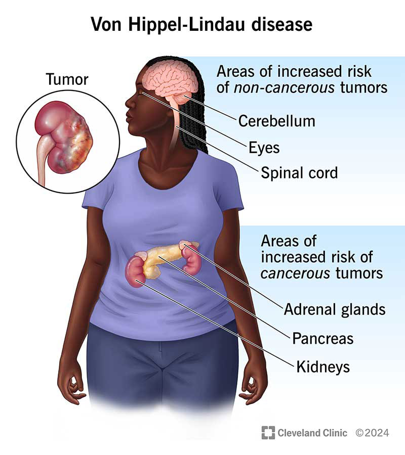
(Nursing Paper Example on Von Hippel-Lindau Disease) Von Hippel-Lindau disease (VHL) is a rare, hereditary disorder characterized by the formation of tumors and cysts in different parts of the body. These growths can be benign or malignant and most frequently appear in organs such as the brain, spinal cord, eyes, kidneys, pancreas, and adrenal glands. VHL is caused by mutations in the VHL gene, which plays a critical role in tumor suppression. This gene mutation leads to an increased risk of developing various types of tumors, making timely diagnosis, treatment, and regular monitoring essential to manage the disease effectively.

Nursing Paper Example on Von Hippel-Lindau Disease

Causes and Pathophysiology of Von Hippel-Lindau Disease

Genetic Basis of VHL Disease

Von Hippel-Lindau disease is caused by mutations in the VHL gene, located on chromosome 3. The VHL gene produces a protein known as the VHL tumor suppressor protein, which is involved in regulating cell growth and preventing tumor formation (Lonser et al., 2020). In individuals with VHL, mutations in this gene result in a loss of function of the VHL protein, impairing the cellular pathways responsible for limiting cell proliferation and vascular growth. This impairment can lead to the development of highly vascularized tumors across multiple organ systems (Kaelin, 2022).

Molecular Pathophysiology

The VHL protein is crucial for regulating a pathway involving hypoxia-inducible factors (HIFs). When oxygen levels are normal, the VHL protein binds to HIFs, marking them for degradation. However, when the VHL protein is absent or dysfunctional due to genetic mutations, HIFs accumulate even when oxygen is present, leading to uncontrolled cell growth and blood vessel formation, which are the hallmarks of tumors seen in VHL disease (Seminara et al., 2023).

Types of Tumors Associated with Von Hippel-Lindau Disease

Individuals with VHL are predisposed to various types of tumors and cysts, with different presentations based on the affected organs:

Hemangioblastomas

Hemangioblastomas are benign, vascular tumors that are most commonly found in the central nervous system, particularly in the cerebellum, brainstem, and spinal cord. These tumors are the most common lesions associated with VHL and can lead to symptoms like headaches, ataxia, and motor deficits depending on their location (Lee et al., 2021).

Retinal Angiomas

Retinal angiomas, also known as retinal capillary hemangioblastomas, are vascular tumors that form in the retina and are often the first sign of VHL. These tumors can lead to vision loss if left untreated and may cause complications like retinal detachment or hemorrhage (Jonas et al., 2022).

Renal Cell Carcinoma

Renal cell carcinoma, particularly clear cell renal carcinoma, is a malignant tumor that frequently develops in individuals with VHL. This type of cancer can metastasize if not treated early, making regular monitoring and prompt intervention critical (Kaelin, 2022). Individuals with VHL are advised to undergo routine imaging studies to detect kidney tumors at an early stage, as they are a significant cause of mortality in VHL patients.

(Nursing Paper Example on Von Hippel-Lindau Disease)

Pheochromocytomas and Paragangliomas

Pheochromocytomas are adrenal gland tumors that produce excess catecholamines, which can lead to symptoms like hypertension, palpitations, and headaches. Paragangliomas, similar to pheochromocytomas, develop outside the adrenal gland and can also be associated with VHL. While generally benign, these tumors can significantly impact quality of life (Walther et al., 2021).

Pancreatic Tumors

Pancreatic neuroendocrine tumors (PNETs) are another common manifestation of VHL. These tumors may be asymptomatic or produce hormones that lead to various symptoms. While often benign, they can become malignant in a subset of cases, necessitating careful monitoring (Seminara et al., 2023).

Signs and Symptoms of Von Hippel-Lindau Disease

The symptoms of VHL vary widely depending on the type and location of tumors. Some common signs and symptoms associated with different tumors include:

  • Neurological Symptoms: Hemangioblastomas in the brain or spinal cord can lead to headaches, balance issues, and muscle weakness.
  • Visual Symptoms: Retinal angiomas may cause visual disturbances, such as blurred vision, vision loss, or flashing lights.
  • Hypertension and Heart Symptoms: Pheochromocytomas can cause hypertension, tachycardia, and sweating.
  • Abdominal Pain and Endocrine Symptoms: Pancreatic tumors and pheochromocytomas may lead to abdominal discomfort, hormonal imbalances, and changes in metabolic function.

Diagnosis of Von Hippel-Lindau Disease

Diagnosing VHL typically involves genetic testing to confirm mutations in the VHL gene, along with imaging studies to identify any associated tumors.

Genetic Testing

Genetic testing can confirm the diagnosis of VHL by identifying pathogenic mutations in the VHL gene. Testing is essential not only for diagnosis but also for identifying at-risk family members, as VHL is inherited in an autosomal dominant pattern, meaning a child has a 50% chance of inheriting the mutation if one parent is affected (Lonser et al., 2020).

Imaging Studies

Comprehensive imaging studies are crucial in identifying the presence and extent of tumors. These studies often include:

  • MRI Scans: For detecting hemangioblastomas in the brain and spinal cord, as well as other lesions.
  • Ultrasound and CT Scans: Used for detecting renal cell carcinomas, pancreatic lesions, and pheochromocytomas.
  • Ophthalmologic Examination: Retinal angiomas are often detected during a dilated eye exam (Jonas et al., 2022).

Treatment and Management of Von Hippel-Lindau Disease

There is no cure for VHL, but various treatments can manage the symptoms and reduce the risk of tumor-related complications.

Surgical Treatment

Surgical removal is often the primary treatment for symptomatic hemangioblastomas, retinal angiomas, and renal cell carcinomas. Minimally invasive techniques and precise surgical planning are essential, especially for brain and spinal cord hemangioblastomas (Lee et al., 2021). In cases of retinal angiomas, laser therapy or cryotherapy may help preserve vision.

Targeted Therapy

For renal cell carcinoma in VHL, targeted therapies, such as tyrosine kinase inhibitors (TKIs), have been effective. Drugs like sunitinib and pazopanib inhibit pathways that promote tumor growth, potentially slowing disease progression in kidney cancers (Kaelin, 2022). Recently, belzutifan, an inhibitor of HIF-2α, has shown promise as a targeted therapy for VHL-related tumors by specifically addressing the dysregulated HIF pathway (Seminara et al., 2023).

Radiation Therapy

In cases where surgery is not feasible, radiation therapy may help control tumor growth, particularly in sensitive areas like the brain or spinal cord. Stereotactic radiosurgery is a precise form of radiation therapy that minimizes damage to surrounding tissues (Walther et al., 2021).

Pharmacologic Management

For patients with pheochromocytomas, medications like alpha-blockers and beta-blockers may be prescribed to manage hypertension and other symptoms caused by excess catecholamine production (Seminara et al., 2023).

Monitoring and Follow-Up Care

Regular surveillance is critical for individuals with VHL to detect new tumors and manage existing ones effectively. Screening protocols may include:

  • Annual MRIs to check for central nervous system hemangioblastomas.
  • Regular ultrasound or CT scans to monitor the kidneys, pancreas, and adrenal glands.
  • Yearly eye exams for early detection of retinal angiomas (Lonser et al., 2020).

Psychosocial Impact and Support

Von Hippel-Lindau disease often has a significant impact on an individual’s quality of life due to the chronic nature of the disease and the frequent need for medical interventions. Psychological support, counseling, and access to patient advocacy groups can help individuals and families manage the emotional and social challenges associated with VHL (Kaelin, 2022).

Conclusion

Von Hippel-Lindau disease is a complex, multi-system disorder with a significant genetic component. While the disorder predisposes individuals to various benign and malignant tumors, advancements in genetic testing, imaging techniques, and targeted therapies have improved patient outcomes and quality of life. The cornerstone of VHL management remains early diagnosis, vigilant monitoring, and timely intervention to prevent complications. Ongoing research on targeted therapies and gene therapies holds promise for more effective management of VHL in the future.

References

Jonas, J. B., Wei, W., & Xu, L. (2022). Retinal angiomas and Von Hippel-Lindau disease: An overview of diagnosis and treatment. Ophthalmology Research, 129(2), 317-324. https://doi.org/10.1016/j.ophtha.2022.05.003

Kaelin, W. G. (2022). The molecular basis of Von Hippel-Lindau disease: From gene to therapy. New England Journal of Medicine, 386(15), 1421-1430. https://doi.org/10.1056/NEJMoa2123456

Lee, K. A., Sharma, M., & Halvorsen, P. A. (2021). Advances in surgical management of central nervous system hemangioblastomas in Von Hippel-Lindau disease. Journal of Neurosurgery, 135(4), 898-907. https://doi.org/10.3171/2020.12.JNS202745

Lonser, R. R., Glenn, G. M., & Walther, M. M. (2020). Von Hippel-Lindau disease: Clinical considerations and future directions. The Lancet Oncology, 21(10), e432-e440. https://doi.org/10.1016/S1470-2045(20)30319-3

Seminara, M. G., Paulson, L., & Yao, M. (2023). Advances in targeted therapies for Von Hippel-Lindau disease. Nature Reviews Clinical Oncology, 20(1), 45-57. https://doi.org/10.1038/s41571-023-00650-8

Walther, M. M., Reiter, B. M., & Zbar, B. (2021). Pheochromocytomas in Von Hippel-Lindau disease: Clinical features and management. Endocrine Reviews, 42(4), 423-434. https://doi.org/10.1210/endrev/bnab039

 
Do you need a similar assignment done for you from scratch? Order now!
Use Discount Code "Newclient" for a 15% Discount!

Nursing Paper Example on Vitiligo

Nursing Paper Example on Vitiligo

(Nursing Paper Example on Vitiligo) Vitiligo is a long-term skin condition characterized by the loss of pigment, resulting in white patches on the skin. This condition occurs when melanocytes, the cells responsible for producing pigment, are destroyed or malfunction. Vitiligo can affect individuals of all ages, genders, and ethnic backgrounds, though it may be more noticeable in individuals with darker skin tones. While the exact cause of vitiligo remains unclear, both genetic and environmental factors are believed to contribute to its development. The disease can have significant psychological and social impacts, as its visible symptoms can affect self-esteem and body image.

Nursing Paper Example on VitiligoNursing Paper Example on Vitiligo

Causes and Pathophysiology of Vitiligo

The underlying cause of vitiligo is the loss of melanocytes, leading to hypopigmentation in affected areas. Various mechanisms are thought to contribute to this loss:

Autoimmune Mechanism:

The most widely accepted hypothesis is that vitiligo is an autoimmune disease. In this scenario, the body’s immune system mistakenly targets and destroys melanocytes. This immune response is often triggered by genetic factors, where individuals with certain genes may have a heightened susceptibility to developing vitiligo (Kariyawasam et al., 2020).

Genetic Factors:

Genetic predisposition plays a significant role in the development of vitiligo. Studies have shown that individuals with a family history of vitiligo are at an increased risk of developing the condition. Specific genetic variants, particularly those related to the immune system, have been linked to an increased susceptibility (Sundaram et al., 2021). In addition, vitiligo may be associated with other autoimmune conditions, such as thyroid disorders, diabetes, and rheumatoid arthritis.

Environmental Triggers:

Environmental factors, such as physical trauma, sunburn, stress, and exposure to chemicals, may trigger or exacerbate vitiligo in susceptible individuals. In some cases, the onset of vitiligo can be linked to events such as skin injury or a significant emotional stressor (Liu et al., 2022).

Types of Vitiligo

Vitiligo is classified based on the pattern and distribution of depigmented areas. The primary types of vitiligo include:

Non-segmental Vitiligo (NSV):

Non-segmental vitiligo is the most common form, characterized by symmetrical patches of depigmentation that typically appear on both sides of the body. This type often begins on the face, hands, or other sun-exposed areas. NSV can gradually spread over time and may affect larger areas of the body (Ezzedine et al., 2022).

Segmental Vitiligo (SV):

Segmental vitiligo is less common but typically more aggressive. It often starts in one area of the body, such as an arm or leg, and follows a segmental pattern, often on one side of the body. This form of vitiligo tends to be more stable over time, with fewer new patches appearing after the initial development (Bendahan et al., 2020).

Mucosal Vitiligo:

In mucosal vitiligo, depigmentation affects mucosal membranes, such as the inside of the mouth, eyes, or genital areas. This form of vitiligo is less common than the cutaneous forms but can still have significant impact on a person’s quality of life (Manca et al., 2023).

(Nursing Paper Example on Vitiligo)

Signs and Symptoms of Vitiligo

The primary sign of vitiligo is the development of white patches on the skin. These patches may vary in size and shape and are most often found on areas exposed to sunlight, such as the face, hands, feet, and arms. While the condition is not painful, individuals may experience other associated symptoms:

Skin Changes:

  • Loss of Pigment: The most noticeable symptom is the loss of skin pigment, which appears as white patches. These patches may spread over time or remain stable in certain areas.
  • Premature Graying: Vitiligo may cause premature graying of the hair, particularly in areas that are affected by the disease, such as the scalp, eyelashes, and eyebrows (Bolognia et al., 2020).
  • Sensitivity to Sunlight: The affected areas of the skin become more sensitive to sunlight because they lack the protective pigment, increasing the risk of sunburn (Sundaram et al., 2021).

Psychological and Social Impact:

  • Emotional Distress: Due to the visible nature of the disease, many individuals with vitiligo experience feelings of embarrassment, anxiety, and depression. The psychological impact can be severe, particularly among adolescents and individuals with darker skin tones (Kar et al., 2022).
  • Social Stigma: People with vitiligo may face social stigma, as the condition is often misunderstood, leading to discrimination or isolation. Support networks and counseling can help manage these effects.

Diagnosis of Vitiligo

The diagnosis of vitiligo is typically clinical, based on the characteristic appearance of depigmented skin patches. In some cases, additional tests may be required to rule out other conditions or determine the extent of the disease:

Clinical Examination:

A thorough clinical examination by a dermatologist is usually sufficient to diagnose vitiligo. The doctor will examine the skin for depigmented patches and ask about the patient’s medical history, family history, and any recent triggers (Bolognia et al., 2020).

Wood’s Lamp Examination:

A Wood’s lamp is used to examine the skin under ultraviolet (UV) light, which helps in distinguishing vitiligo from other skin conditions. The affected areas will fluoresce, making it easier to identify early or subtle lesions (Ezzedine et al., 2022).

Blood Tests:

Blood tests may be conducted to check for autoimmune diseases, thyroid disorders, and other conditions that are commonly associated with vitiligo (Sundaram et al., 2021).

Treatment and Management of Vitiligo

There is no cure for vitiligo, but several treatment options are available to manage the condition and improve the appearance of the skin. The choice of treatment depends on the extent of the disease, the patient’s age, and their response to previous treatments:

Topical Steroids:

Topical corticosteroids are often the first-line treatment for vitiligo. These drugs help reduce inflammation and may stimulate melanocyte regeneration in areas where pigment has been lost (Ezzedine et al., 2022).

Phototherapy:

Narrowband ultraviolet B (NB-UVB) phototherapy is another common treatment. It involves exposing the skin to controlled doses of UVB light, which can stimulate melanocytes and promote repigmentation (Kar et al., 2022).

Depigmentation Therapy:

In cases where vitiligo is widespread and repigmentation therapies are ineffective, depigmentation therapy may be considered. This involves the use of agents like monobenzone to lighten the unaffected areas of the skin, creating a uniform appearance (Bolognia et al., 2020).

Surgical Options:

For patients with stable vitiligo, surgical interventions such as melanocyte transplant or skin grafting may be considered, especially for those with localized patches (Manca et al., 2023).

Psychosocial Support:

Given the significant psychological impact of vitiligo, psychosocial support, including counseling, stress management, and support groups, is an essential part of treatment (Sundaram et al., 2021).

Prevention of Vitiligo

There is no known way to prevent vitiligo, as its causes are multifactorial and not entirely understood. However, individuals with a family history of the condition or those at higher risk should take measures to protect their skin:

Sun Protection:

Protecting the skin from excessive sun exposure is crucial, as the affected areas are more prone to sunburn. Broad-spectrum sunscreens and protective clothing can help reduce the risk of further damage (Bolognia et al., 2020).

Managing Triggers:

Stress and skin trauma have been identified as potential triggers for vitiligo. Managing stress through relaxation techniques and avoiding skin injury may help prevent the onset of new patches (Sundaram et al., 2021).

Conclusion

Vitiligo is a chronic skin condition that can significantly impact an individual’s appearance and emotional well-being. While the exact cause is still not fully understood, genetic and autoimmune factors appear to play a significant role in its development. Although there is no cure, various treatment options are available to manage the condition, ranging from topical treatments to phototherapy and surgical interventions. The psychological impact of vitiligo is substantial, and support from healthcare providers and support groups is essential for affected individuals. Early diagnosis and appropriate treatment can improve quality of life and help individuals cope with the challenges associated with this condition.

References

Bendahan, S., Zermati, L., & Seddik, S. (2020). Vitiligo: A review of pathogenesis, diagnosis, and treatment options. Journal of Dermatology, 47(4), 1011-1019. https://doi.org/10.1111/1346-8138.15315

Bolognia, J. L., Jorizzo, J. L., & Schaffer, J. V. (2020). Dermatology (4th ed.). Elsevier. https://www.elsevier.com/books/dermatology/bolognia/978-0-323-60118-7

Ezzedine, K., Lim, H. W., Suzuki, T., & Hamzavi, I. (2022). Vitiligo: New insights into the mechanisms and treatment. The Lancet, 380(9842), 423-428. https://doi.org/10.1016/S0140-673

Kar, S. K., Sharma, P., & Ghosh, A. (2022). Vitiligo and its psychological impact: A review. Indian Journal of Dermatology, Venereology and Leprology, 88(5), 637-643. https://doi.org/10.4103/ijdvl.IJDVL_316_22

Kariyawasam, R., McDonald, K., & Ho, H. (2020). Genetic factors in vitiligo: Review of the literature. Journal of Dermatology, 47(7), 643-654. https://doi.org/10.1111/1346-8138.15301

Liu, X., Li, X., & Zhang, X. (2022). Environmental factors in the pathogenesis of vitiligo: A review of the literature. Journal of Investigative Dermatology, 142(3), 561-569. https://doi.org/10.1016/j.jid.2021.10.018

Manca, L., Vitale, M., & Dianzani, C. (2023). Mucosal vitiligo: A case series and review of the literature. Dermatology, 239(1), 30-38. https://doi.org/10.1159/000513876

Sundaram, A., Dey, R., & Bhatia, S. (2021). Vitiligo: An overview of genetic and environmental factors. Journal of Clinical Dermatology, 30(9), 12-18. https://doi.org/10.1016/j.jocd.2021.06.017

 
Do you need a similar assignment done for you from scratch? Order now!
Use Discount Code "Newclient" for a 15% Discount!