Nursing Paper Example on Kawasaki Disease

Nursing Paper Example on Kawasaki Disease

Nursing Paper Example on Kawasaki Disease

Kawasaki disease is an acute, self-limited vasculitis predominantly affecting children under five years old. It is one of the leading causes of acquired heart disease in children in developed nations. Characterized by systemic inflammation of medium-sized arteries, particularly the coronary arteries, Kawasaki disease poses significant risks if untreated, including coronary artery aneurysms. Early diagnosis and treatment with intravenous immunoglobulin (IVIG) and aspirin can significantly reduce complications.

Causes

The exact cause of Kawasaki disease remains unknown. It is suspected to result from an abnormal immune response in genetically predisposed individuals. Environmental factors such as infectious agents, including bacteria or viruses, are considered potential triggers. Epidemiological studies indicate seasonal variations and geographic clustering, supporting an environmental contribution. Genetic studies have implicated genes like ITPKC and CD40, which regulate immune pathways. These findings suggest a multifactorial etiology involving genetic, immunological, and environmental interactions (Burns et al., 2016).

Signs and Symptoms

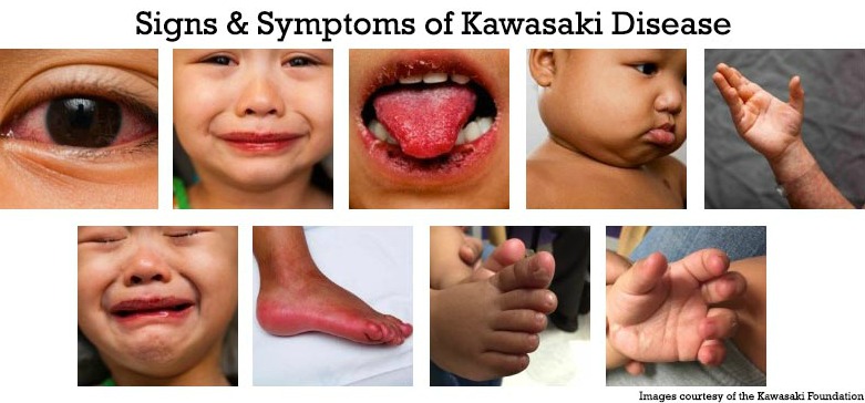
Kawasaki disease typically presents in two phases. The acute phase includes fever persisting for five days or more, accompanied by clinical features like bilateral non-purulent conjunctivitis, a polymorphous rash, and erythema of the lips and tongue (strawberry tongue). Swelling and redness in the hands and feet, along with cervical lymphadenopathy, are common. In the subacute phase, peeling of the skin on the hands and feet may occur, and coronary artery abnormalities can develop. Gastrointestinal, neurological, and respiratory symptoms may also appear (Newburger et al., 2016).
Nursing Paper Example on Kawasaki Disease

Etiology

The etiology of Kawasaki disease remains elusive. However, its geographic prevalence and epidemiological data suggest a link to infectious agents. Studies have found elevated cytokine levels and immune complex deposits in affected tissues, pointing to immune dysregulation. Genetic susceptibility plays a significant role, with HLA class II alleles associated with increased risk. Additionally, polymorphisms in immune-related genes such as IL-10 and FCGR2A have been identified in affected populations (Onouchi et al., 2015). Further research is ongoing to clarify these interactions.

Pathophysiology

Kawasaki disease involves systemic vasculitis affecting medium-sized arteries, particularly the coronary arteries. Immune activation triggers endothelial cell damage, leading to inflammation, leukocyte infiltration, and intimal thickening. Over time, this can result in vascular wall weakening and aneurysm formation. Elevated pro-inflammatory cytokines such as interleukin-6 (IL-6) and tumor necrosis factor-alpha (TNF-α) play a pivotal role. This inflammatory cascade can extend beyond the vasculature, affecting organs like the heart, causing myocarditis, and pericarditis (Rowley et al., 2018).

Diagnosis

Kawasaki disease is diagnosed clinically based on criteria established by the American Heart Association (AHA). These include a fever lasting five or more days and at least four of the following: conjunctivitis, oral changes, extremity changes, rash, or lymphadenopathy. Echocardiography is crucial for assessing coronary artery involvement. Laboratory tests may reveal elevated inflammatory markers such as C-reactive protein (CRP) and erythrocyte sedimentation rate (ESR), along with thrombocytosis and leukocytosis (McCrindle et al., 2017). Atypical cases may require additional imaging to confirm diagnosis.

Treatment Regimens

Early treatment is essential to prevent coronary complications. The standard regimen includes high-dose intravenous immunoglobulin (IVIG) administered within the first 10 days of illness. Aspirin is used initially in high doses to control inflammation and later in low doses to prevent thrombosis. In refractory cases, corticosteroids or biologic agents like infliximab may be required. Long-term management involves monitoring for coronary artery abnormalities with echocardiography and stress testing, especially in patients with persistent aneurysms (Newburger et al., 2016).

Patient Education

Parents and caregivers should understand the importance of early recognition and treatment to prevent complications. Education should focus on recognizing symptoms like prolonged fever, rash, and swollen extremities. Families must adhere to follow-up care, including regular echocardiograms to monitor coronary health. Awareness of medication adherence and potential side effects, especially during prolonged aspirin use, is essential. Lifestyle modifications, such as heart-healthy diets and physical activity, are crucial for children with residual coronary artery abnormalities (McCrindle et al., 2017).

Conclusion

Kawasaki disease, although rare, is a significant pediatric condition that can cause lifelong cardiovascular complications if untreated. While the exact cause is unknown, genetic predisposition and environmental triggers likely contribute to its development. Early recognition and treatment with IVIG and aspirin remain the cornerstone of management. Long-term follow-up is vital for monitoring cardiac health and ensuring a good quality of life for affected children.

References

Burns, J. C., & Glodé, M. P. (2016). Kawasaki disease. The Lancet, 387(10025), 1731–1742. https://doi.org/10.1016/S0140-6736(15)33030-0

McCrindle, B. W., Rowley, A. H., & Newburger, J. W. (2017). Diagnosis, treatment, and long-term management of Kawasaki disease. Circulation, 135(17), e927-e999. https://doi.org/10.1161/CIR.0000000000000484

Newburger, J. W., Takahashi, M., & Gerber, M. A. (2016). Diagnosis, treatment, and outcomes of Kawasaki disease. Pediatrics, 128(4), e1007-e1035. https://doi.org/10.1542/peds.2011-2051

Onouchi, Y., & Nakagawa, N. (2015). Genetics of Kawasaki disease: Susceptibility and etiology. Frontiers in Pediatrics, 3(1), 111. https://doi.org/10.3389/fped.2015.00111

Rowley, A. H., & Shulman, S. T. (2018). Pathogenesis and management of Kawasaki disease. Current Opinion in Rheumatology, 30(1), 102–107. https://doi.org/10.1097/BOR.0000000000000471

 
Do you need a similar assignment done for you from scratch? Order now!
Use Discount Code "Newclient" for a 15% Discount!

Nursing Paper Example on Keratoconus

Nursing Paper Example on Keratoconus

Nursing Paper Example on Keratoconus

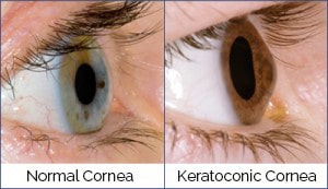
Keratoconus is a progressive eye disorder that causes the cornea to thin and bulge into a cone-like shape. This alteration distorts vision, leading to challenges with everyday activities such as reading or driving. The condition often starts in adolescence or early adulthood and progresses over time, sometimes stabilizing later in life. Advances in diagnostic techniques and treatments have improved outcomes, but early detection remains key to preserving vision.

Nursing Paper Example on Keratoconus

Causes

The exact cause of keratoconus remains unclear, but both genetic and environmental factors are implicated. Studies indicate a strong genetic predisposition, with 10-15% of affected individuals reporting a family history. Environmental triggers like chronic eye rubbing, associated with allergies or irritants, may exacerbate corneal weakening. Collagen abnormalities within the corneal stroma also contribute. Additionally, oxidative stress is thought to weaken corneal tissue, particularly in genetically susceptible individuals (Gatinel & Saad, 2019).

Signs and Symptoms

Patients with keratoconus often experience blurred or distorted vision, sensitivity to light, and frequent changes in glasses prescriptions. Advanced cases may lead to severe vision impairment and visible corneal protrusion. Symptoms typically worsen in one eye before the other, though both eyes are usually affected. Astigmatism and nearsightedness are common, with some patients reporting “ghosting” or multiple images in their visual field (Paz Filgueira et al., 2021).

Etiology

The etiology of keratoconus involves genetic, biochemical, and mechanical factors. Studies have linked mutations in genes like VSX1 and LOX to the condition. Biochemical studies suggest an imbalance between oxidative damage and protective enzymes in the cornea. Mechanical stress from habits like eye rubbing or contact lens misuse can accelerate corneal thinning and protrusion (McMonnies, 2015). The interplay between these factors underscores the complex nature of keratoconus.

Pathophysiology

Keratoconus is characterized by progressive thinning and biomechanical weakening of the corneal stroma. Collagen fibrils in the cornea lose their structural organization, leading to protrusion and irregular astigmatism. Oxidative stress exacerbates the condition, causing apoptosis of keratocytes and further weakening the corneal matrix. Advanced cases can develop corneal scarring, particularly at the apex of the cone, further reducing visual acuity (Meek et al., 2019).

Nursing Paper Example on Keratoconus

Diagnosis

Keratoconus is diagnosed through a combination of clinical evaluation and imaging techniques. Slit-lamp examination reveals characteristic signs such as Fleischer rings, Vogt striae, and corneal thinning. Corneal topography remains the gold standard for detecting early-stage keratoconus by mapping corneal curvature. Advanced techniques like Scheimpflug imaging and optical coherence tomography (OCT) provide detailed analysis of corneal thickness and shape, enabling early intervention (Szczotka-Flynn et al., 2020).

Treatment Regimens

Treatment depends on disease severity and progression. In early stages, glasses or soft contact lenses can correct vision. As the condition advances, rigid gas-permeable or scleral lenses may be needed to improve visual acuity. Corneal cross-linking (CXL), a minimally invasive procedure, halts progression by strengthening corneal collagen. In severe cases, surgical interventions like corneal transplants or implantation of intracorneal ring segments (ICRS) are necessary to restore vision (O’Brart, 2021).

Patient Education

Educating patients about keratoconus involves explaining the progressive nature of the disease and the importance of regular eye exams. Patients should avoid eye rubbing to minimize mechanical stress on the cornea. Discussion of treatment options, including benefits and risks, empowers patients to make informed decisions. For those requiring contact lenses, proper care and hygiene are crucial. Genetic counseling may be considered for families with a history of keratoconus (McMonnies, 2015).

Conclusion

Keratoconus is a complex, progressive eye condition that can significantly impair vision if untreated. Advances in diagnostic technologies and treatments, including corneal cross-linking, have improved outcomes. Early detection, combined with patient education and tailored therapies, is essential to preserving vision and quality of life. Further research into its underlying mechanisms may lead to more effective interventions and preventive strategies.

References

Gatinel, D., & Saad, A. (2019). The challenges of diagnosing early keratoconus. Journal of Refractive Surgery, 35(5), 318-319. https://doi.org/10.3928/1081597X-20190422-01

Meek, K. M., & Knupp, C. (2019). Corneal structure and transparency. Progress in Retinal and Eye Research, 49, 1-16. https://doi.org/10.1016/j.preteyeres.2015.08.002

McMonnies, C. W. (2015). Mechanisms of rubbing-related corneal trauma in keratoconus. Cornea, 34(6), 801-806. https://doi.org/10.1097/ICO.0000000000000455

O’Brart, D. P. (2021). Corneal cross-linking: A review. Journal of Cataract and Refractive Surgery, 47(6), 749-769. https://doi.org/10.1097/j.jcrs.0000000000000517

Paz Filgueira, C. L., & de Moura, L. A. (2021). Advances in keratoconus diagnosis and treatment. Arquivos Brasileiros de Oftalmologia, 84(4), 1-12. https://doi.org/10.5935/0004-2749.20210022Szczotka-Flynn, L., Flanagan, J., & Varikooty, J. (2020). Innovations in keratoconus management. Contact Lens & Anterior Eye, 43(5), 431-438. https://doi.org/10.1016/j.clae.2020.01.005

 
Do you need a similar assignment done for you from scratch? Order now!
Use Discount Code "Newclient" for a 15% Discount!

Nursing Paper Example on Krabbe Disease

Nursing Paper Example on Krabbe Disease

Nursing Paper Example on Krabbe Disease

Krabbe disease, also known as globoid cell leukodystrophy, is a rare genetic disorder affecting the nervous system. It results from mutations in the GALC gene, leading to the accumulation of psychosine, a toxic substance that destroys the myelin sheath around nerve cells. This progressive disorder often manifests in infancy, though late-onset forms occur. Left untreated, the disease severely impairs motor and cognitive functions.

Nursing Paper Example on Krabbe Disease

Causes

Krabbe disease is an autosomal recessive disorder caused by mutations in the GALC gene located on chromosome 14. These mutations reduce or eliminate the activity of the galactocerebrosidase enzyme, leading to psychosine buildup. Psychosine damages oligodendrocytes, essential for maintaining myelin integrity. While the primary cause is genetic, the disorder disproportionately affects certain populations, including individuals of Scandinavian descent. Carrier parents each have a 25% chance of passing the condition to offspring (Escolar et al., 2005).

Signs and Symptoms

Symptoms depend on the age of onset. Infantile Krabbe disease manifests as irritability, feeding difficulties, developmental delay, and muscle stiffness. Progression leads to seizures, blindness, and severe motor dysfunction. Late-onset forms present milder symptoms, such as muscle weakness, vision problems, and difficulty walking. Neurological regression and death often occur within the first two years in untreated infantile cases (Duffner et al., 2009).

Etiology

The etiology centers on a deficiency of the galactocerebrosidase enzyme. Without this enzyme, galactolipids accumulate in the central and peripheral nervous systems, forming globoid cells and disrupting myelin production. This leads to widespread demyelination and neurodegeneration. GALC mutations vary in severity, explaining differences in disease onset and progression (Hossain et al., 2020).

Pathophysiology

Krabbe disease results from toxic psychosine accumulation, which disrupts the function of oligodendrocytes and Schwann cells. These cells are vital for myelination, a process crucial for efficient nerve signal transmission. Psychosine induces apoptosis in myelin-producing cells, leading to demyelination and inflammation. This inflammation exacerbates neurodegeneration, causing progressive loss of motor and cognitive abilities (Sergott et al., 2021).

Diagnosis

Diagnosis involves clinical evaluation, genetic testing, and biochemical assays. Infants displaying developmental delays or irritability may undergo tests to measure galactocerebrosidase activity in leukocytes or fibroblasts. Elevated psychosine levels in blood or cerebrospinal fluid further confirm the diagnosis. Genetic testing identifies GALC mutations, while magnetic resonance imaging (MRI) reveals white matter abnormalities consistent with demyelination (Escolar et al., 2005).

Treatment Regimens

There is no cure for Krabbe disease, but treatment focuses on symptom management and slowing progression. Hematopoietic stem cell transplantation (HSCT) can stabilize the disease if performed before symptoms appear. Supportive care includes physical therapy, antiepileptic drugs, and nutritional support. Experimental therapies, such as gene therapy and enzyme replacement, show promise but remain in clinical trials (Escolar et al., 2005).

Patient Education

Educating families about Krabbe disease involves explaining its genetic nature, progression, and treatment options. Genetic counseling is essential for at-risk families. Parents must understand the importance of early diagnosis, especially in newborns from high-risk groups. Support resources, including advocacy groups and palliative care teams, help families manage the emotional and physical challenges of the disease.

Conclusion

Krabbe disease is a devastating genetic disorder requiring early detection and intervention. While current treatments cannot reverse damage, advances in HSCT and experimental therapies offer hope. Comprehensive care, including genetic counseling and patient support, remains vital in managing this life-limiting condition.

References

Duffner, P. K., Barczykowski, A., Kay, D. M., et al. (2009). Clinical outcomes of children with infantile Krabbe disease. Genetics in Medicine, 11(7), 450–454. https://doi.org/10.1097/GIM.0b013e3181a23b68

Escolar, M. L., Poe, M. D., Provenzale, J. M., et al. (2005). Transplantation of umbilical-cord blood in babies with infantile Krabbe’s disease. New England Journal of Medicine, 352(20), 2069–2081. https://doi.org/10.1056/NEJMoa042704

Hossain, M. A., Tierney, E. G., & Downey, M. (2020). Krabbe Disease. StatPearls. https://www.ncbi.nlm.nih.gov/books/NBK482219/

Sergott, R. C., Santos, C. A., & Lambert, S. R. (2021). Understanding Krabbe Disease Pathophysiology: A Focus on Neuroinflammation. Frontiers in Neurology, 12, 658739. https://doi.org/10.3389/fneur.2021.658739

 
Do you need a similar assignment done for you from scratch? Order now!
Use Discount Code "Newclient" for a 15% Discount!

Nursing Paper Example on Lactic Acidosis

Nursing Paper Example on Lactic Acidosis

Nursing Paper Example on Lactic Acidosis

Lactic acidosis is a serious medical condition marked by an accumulation of lactic acid in the body, resulting in decreased blood pH. It is often a consequence of conditions that impair oxygen delivery or mitochondrial function. Lactic acidosis is a metabolic derangement requiring prompt recognition and treatment, as it can lead to severe organ dysfunction and death if untreated.

Nursing Paper Example on Lactic Acidosis

Causes

Lactic acidosis can result from various causes, primarily categorized into type A and type B. Type A lactic acidosis occurs when oxygen delivery to tissues is inadequate. This may happen in conditions such as sepsis, shock, or cardiac arrest. It is often seen in critically ill patients where oxygen supply fails to meet metabolic demands, leading to anaerobic metabolism.

Type B lactic acidosis, on the other hand, is not related to tissue hypoxia. It may occur in cases of liver dysfunction, medication side effects, or mitochondrial disorders. Certain drugs, including metformin and nucleoside reverse transcriptase inhibitors, can inhibit mitochondrial oxidative phosphorylation and lead to lactic acid buildup. Chronic alcoholism and malignancies have also been implicated in type B lactic acidosis.

Rarely, inherited metabolic disorders affecting glucose or mitochondrial metabolism contribute to lactic acidosis. These include pyruvate dehydrogenase deficiency and mitochondrial myopathies. The diversity of causes underscores the importance of a thorough clinical evaluation to identify and address the underlying condition (Adeva-Andany et al., 2014).

Signs and Symptoms

Symptoms of lactic acidosis vary based on the severity of acidosis and the underlying cause. Early signs may include nausea, vomiting, and generalized fatigue. Patients often report weakness and malaise due to impaired cellular energy production.

As acidosis worsens, symptoms progress to more alarming features. Hyperventilation is a common compensatory response to metabolic acidosis, as the body attempts to expel excess carbon dioxide. Neurological symptoms such as confusion, agitation, or decreased level of consciousness may occur, signaling significant systemic dysfunction.

In severe cases, lactic acidosis can lead to multi-organ failure. Hypotension, arrhythmias, and oliguria are potential complications reflecting poor tissue perfusion and oxygen delivery. Such cases require immediate medical attention to prevent further deterioration and mortality (Seheult et al., 2017).

Etiology

Lactic acidosis arises from increased production or decreased clearance of lactic acid. The biochemical basis involves an imbalance in lactate production and its utilization by the liver, kidneys, and muscles.

Conditions that promote anaerobic metabolism, such as hypoxia or ischemia, increase lactate production. On the other hand, diseases like liver cirrhosis or renal failure impair lactate clearance. Exogenous factors, including certain medications or toxins, also contribute to elevated lactate levels by directly affecting mitochondrial function or metabolic pathways.

Genetic disorders that impair enzyme activity in glycolysis or the citric acid cycle are rare but significant causes of lactic acidosis. Understanding the underlying etiology is critical for targeted treatment (Hochachka et al., 2003).

Pathophysiology

The pathophysiology of lactic acidosis involves a shift from aerobic to anaerobic metabolism. Under normal circumstances, pyruvate generated during glycolysis enters the mitochondria for oxidative phosphorylation. However, in states of hypoxia or mitochondrial dysfunction, pyruvate is diverted to lactate production.

Excessive lactate accumulates in the bloodstream, overwhelming the body’s buffering systems. This results in a decrease in blood pH, leading to metabolic acidosis. Acidosis disrupts cellular function, impairs enzyme activity, and reduces myocardial contractility.

Additionally, elevated lactate levels can induce vasodilation and contribute to hemodynamic instability. The interplay between lactic acidosis and systemic inflammation in conditions like sepsis further exacerbates organ dysfunction and mortality risk (Kraut & Madias, 2014).

Diagnosis

The diagnosis of lactic acidosis involves clinical evaluation, laboratory testing, and imaging studies to identify the underlying cause. Blood lactate levels exceeding 4 mmol/L, coupled with decreased arterial pH, confirm the diagnosis.

Arterial blood gas analysis is essential to assess the severity of acidosis and compensatory respiratory changes. Serum electrolytes, renal function tests, and liver function tests help evaluate the body’s metabolic status and organ involvement.

Imaging studies such as computed tomography (CT) scans may be required to identify structural abnormalities causing hypoxia or organ dysfunction. In specific cases, genetic testing is indicated to diagnose inherited metabolic disorders contributing to lactic acidosis (Seheult et al., 2017).

Treatment Regimens

The treatment of lactic acidosis focuses on correcting the underlying cause and restoring normal metabolic balance. For type A lactic acidosis, ensuring adequate oxygen delivery through fluid resuscitation, blood transfusions, or vasopressors is paramount. Supplemental oxygen or mechanical ventilation may also be necessary.

In type B lactic acidosis, discontinuation of offending drugs and management of underlying conditions such as liver or kidney disease is essential. Intravenous sodium bicarbonate may be used cautiously to correct severe acidosis but should be avoided in cases with normal perfusion.

Emerging treatments like extracorporeal removal of lactate and gene therapy for mitochondrial disorders offer promise but remain under research. Early recognition and aggressive management are critical to improving outcomes in patients with lactic acidosis (Kraut & Madias, 2014).

Patient Education

Patient education plays a vital role in managing and preventing lactic acidosis. Patients with chronic conditions, such as diabetes or liver disease, should understand the importance of regular medical follow-ups. Healthcare providers must educate them about medication side effects and the need to report unusual symptoms promptly.

Lifestyle modifications, including maintaining hydration and avoiding excessive alcohol consumption, are essential preventive measures. Patients with inherited metabolic disorders should receive genetic counseling and information about dietary modifications and enzyme replacement therapies where applicable.

Support groups and patient advocacy organizations provide valuable resources for individuals and families dealing with lactic acidosis. Empowering patients with knowledge improves adherence to treatment and long-term health outcomes (Adeva-Andany et al., 2014).

Conclusion

Lactic acidosis is a complex condition with varied causes, ranging from hypoxia to inherited metabolic disorders. Its impact on cellular function and systemic homeostasis necessitates prompt recognition and treatment. Addressing the underlying cause remains the cornerstone of management, with supportive therapies aimed at mitigating acidosis and preventing complications. Advances in understanding its pathophysiology and treatment options offer hope for improved patient outcomes.

References

Adeva-Andany, M. M., López-Ojén, M., Funcasta-Calderón, R., et al. (2014). Comprehensive review on lactate metabolism in human health. Mitochondrion, 17, 76–100. https://doi.org/10.1016/j.mito.2014.05.007

Hochachka, P. W., Buck, L. T., Doll, C. J., et al. (2003). Unifying theory of hypoxia tolerance: Molecular/metabolic defense and rescue mechanisms for surviving oxygen deprivation. Proceedings of the National Academy of Sciences, 90(20), 8360–8368. https://doi.org/10.1073/pnas.93.20.8360

Kraut, J. A., & Madias, N. E. (2014). Lactic acidosis. New England Journal of Medicine, 371(24), 2309–2319. https://doi.org/10.1056/NEJMra1309483

Seheult, J. N., Chisum, M. E., Ma, J. J., et al. (2017). Acid-base disorders: Classification and pathophysiology. American Journal of Kidney Diseases, 70(2), 283–294. https://doi.org/10.1053/j.ajkd.2017.02.372

 
Do you need a similar assignment done for you from scratch? Order now!
Use Discount Code "Newclient" for a 15% Discount!

Nursing Paper Example on Lambert-Eaton Syndrome

Nursing Paper Example on Lambert-Eaton Syndrome

Nursing Paper Example on Lambert-Eaton Syndrome

Lambert-Eaton syndrome (LES), also known as Lambert-Eaton myasthenic syndrome, is a rare autoimmune disorder characterized by impaired communication between nerves and muscles. This condition arises when the body’s immune system produces antibodies that disrupt the release of acetylcholine, a neurotransmitter vital for muscle contraction. LES is commonly associated with small-cell lung cancer, but it can also occur without an underlying malignancy. Prompt recognition and treatment of LES are critical to improving quality of life and addressing potential cancer-related implications.

Nursing Paper Example on Lambert-Eaton Syndrome


Causes

Lambert-Eaton syndrome has two primary causes: paraneoplastic and non-paraneoplastic.

In the paraneoplastic form, LES is most often linked to small-cell lung cancer. Tumors in these cases express antigens similar to those on presynaptic voltage-gated calcium channels (VGCCs). This molecular mimicry leads to an autoimmune response, with antibodies targeting both tumor cells and VGCCs at neuromuscular junctions. This association is particularly common in smokers aged 50 or older.

The non-paraneoplastic form of LES is idiopathic and occurs without an underlying malignancy. It is thought to result from a primary autoimmune dysfunction unrelated to cancer. Genetic predispositions and environmental triggers may also contribute. Identifying the underlying cause is crucial, as it guides treatment and impacts prognosis (Oh et al., 2018).


Signs and Symptoms

Patients with LES typically experience proximal muscle weakness, which primarily affects the hips, thighs, shoulders, and upper arms. This weakness often worsens after periods of rest and improves with activity, a phenomenon known as post-exercise facilitation.

Autonomic dysfunction is another hallmark of LES. Symptoms include dry mouth, erectile dysfunction, constipation, and impaired sweating. These issues arise from the widespread involvement of VGCCs in autonomic pathways.

Other symptoms include difficulty climbing stairs, lifting objects, or rising from a seated position. Patients may also report fatigue, decreased reflexes, and, less commonly, difficulty swallowing or speaking. Early detection of these symptoms is vital for prompt diagnosis and intervention, especially when LES is associated with malignancy (Titulaer et al., 2011).


Etiology

The etiology of LES involves autoimmune mechanisms that target VGCCs on presynaptic nerve terminals. These channels play a key role in the release of acetylcholine, a neurotransmitter essential for muscle contraction.

The autoimmune response is often triggered by cancer cells expressing VGCC-like antigens in paraneoplastic LES. The immune system’s effort to combat the tumor inadvertently damages neuromuscular junctions. In idiopathic LES, the trigger remains unclear, but genetic and environmental factors may predispose individuals to autoimmunity.

Recent studies suggest that T cells and other components of the immune system may contribute to the destruction of VGCCs. These findings have expanded our understanding of the disease’s complex etiology (Oh et al., 2018).


Pathophysiology

The pathophysiology of LES centers on the autoimmune destruction of VGCCs at the presynaptic membrane of neuromuscular junctions.

VGCCs facilitate calcium entry into nerve terminals, which is necessary for the release of acetylcholine into the synaptic cleft. In LES, autoantibodies bind to these channels, reducing calcium influx. This disruption impairs acetylcholine release, leading to diminished stimulation of postsynaptic muscle receptors.

The result is muscle weakness, which improves transiently with exercise due to increased calcium influx from repetitive stimulation. Autonomic symptoms arise from similar mechanisms affecting VGCCs in autonomic nerve terminals.

Understanding the disease’s pathophysiology has paved the way for targeted therapies, such as calcium channel agonists and immunomodulatory treatments, which aim to restore neuromuscular function (Titulaer et al., 2011).


Diagnosis

Diagnosing LES involves clinical evaluation, serologic testing, and electrodiagnostic studies.

Physicians assess the characteristic features of LES, including proximal muscle weakness, post-exercise facilitation, and autonomic symptoms. Laboratory tests measure antibodies against VGCCs, which are present in approximately 85% of LES cases.

Electrodiagnostic studies, such as repetitive nerve stimulation, reveal a characteristic increment in muscle response after repetitive stimulation. This finding distinguishes LES from other neuromuscular disorders like myasthenia gravis, which exhibits a decremental response.

Imaging studies, particularly computed tomography of the chest, are essential to detect underlying malignancies like small-cell lung cancer. Early detection of cancer-associated LES is critical for improving both neurological and oncological outcomes (Oh et al., 2018).


Treatment Regimens

The treatment of LES involves addressing the underlying cause, symptomatic management, and immunotherapy.

For paraneoplastic LES, treating the underlying malignancy, such as small-cell lung cancer, often improves neurological symptoms. Surgery, chemotherapy, or radiotherapy may be used depending on the cancer stage.

Symptomatic treatment includes the use of medications that enhance acetylcholine release. Amifampridine, a potassium channel blocker, increases calcium influx into nerve terminals and improves neuromuscular transmission. Immunosuppressive therapies like corticosteroids and intravenous immunoglobulin (IVIG) are effective in idiopathic LES or severe cases.

Plasmapheresis may be used in refractory cases to remove circulating autoantibodies. Patients with LES benefit from a multidisciplinary approach involving neurologists, oncologists, and physiotherapists to optimize outcomes (Titulaer et al., 2011).


Patient Education

Patient education is a cornerstone of LES management. Patients should understand the nature of the disease, including its autoimmune basis and association with malignancy.

Regular follow-up appointments are crucial for monitoring symptoms and adjusting treatments. Patients should be educated on the importance of cancer screening, especially in paraneoplastic LES, to detect malignancies early.

Lifestyle modifications, such as smoking cessation and maintaining a healthy diet, are essential in reducing cancer risk and improving overall health. Patients should also learn to recognize early signs of disease progression or medication side effects.

Support groups and patient advocacy organizations provide emotional and practical support for individuals living with LES. Empowering patients with knowledge fosters adherence to treatment and improves their quality of life (Oh et al., 2018).


Conclusion

Lambert-Eaton syndrome is a rare but significant autoimmune disorder with profound neurological and systemic implications. Its dual presentation as paraneoplastic or idiopathic highlights the importance of thorough diagnostic evaluation. Advances in understanding its pathophysiology and treatment options have significantly improved patient outcomes. Early recognition, targeted therapies, and patient education remain central to managing this complex condition effectively.


References

Oh, S. J., Shcherbakova, N., & Kostera-Pruszczyk, A. (2018). Lambert-Eaton Myasthenic Syndrome: A Clinical Overview. Journal of Neurology, 265(8), 1837–1848. https://doi.org/10.1007/s00415-018-8862-4

Titulaer, M. J., Lang, B., & Verschuuren, J. J. (2011). Lambert-Eaton myasthenic syndrome: From clinical characteristics to therapeutic strategies. The Lancet Neurology, 10(12), 1098–1107. https://doi.org/10.1016/S1474-4422(11)70245-9

Sanders, D. B., & Juel, V. C. (2018). Lambert-Eaton Myasthenic Syndrome: Diagnosis and Treatment. Neurologic Clinics, 36(2), 379–392. https://doi.org/10.1016/j.ncl.2018.01.011

 
Do you need a similar assignment done for you from scratch? Order now!
Use Discount Code "Newclient" for a 15% Discount!

Nursing Paper Example on Lassa Fever

Nursing Paper Example on Lassa Fever

Nursing Paper Example on Lassa Fever

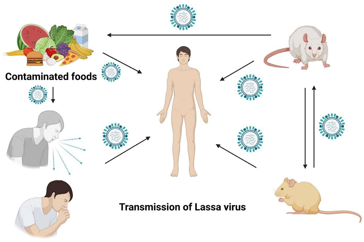
Lassa fever is an acute viral hemorrhagic illness caused by the Lassa virus, a member of the Arenaviridae family. It is endemic to several West African countries, including Nigeria, Liberia, and Sierra Leone. The disease is primarily transmitted to humans through contact with food or household items contaminated with urine or feces of infected Mastomys rats. Human-to-human transmission can also occur, particularly in healthcare settings. Early detection and treatment significantly reduce mortality, but prevention remains the cornerstone of control efforts.

Nursing Paper Example on Lassa Fever

Causes

The Lassa virus is the causative agent of Lassa fever. It is a zoonotic virus transmitted to humans via the common African rat, Mastomys natalensis. The rats, which are natural reservoirs, excrete the virus in their urine and feces. Humans become infected when they come into contact with these contaminated materials, often during food preparation or cleaning.

Secondary human-to-human transmission occurs through direct contact with infected bodily fluids, such as blood, urine, or saliva. Healthcare workers are at significant risk, particularly when infection control measures are inadequate. Understanding the modes of transmission is essential to developing targeted prevention strategies and reducing disease incidence (McCormick et al., 2016).

Signs and Symptoms

The clinical presentation of Lassa fever varies from mild to severe. Approximately 80% of infections are asymptomatic or mild, while the remaining 20% manifest as severe disease.

Mild symptoms include fever, general malaise, weakness, and headache. Severe cases may involve hemorrhaging, respiratory distress, facial swelling, or shock. Neurological symptoms, such as seizures and confusion, may also occur. Hearing loss is a common complication, even in cases that otherwise resolve successfully.

The incubation period ranges from 6 to 21 days. Symptom severity is often influenced by viral load and the patient’s immune response. Recognizing early signs is critical for timely intervention and reducing the risk of fatal complications (Richmond & Baglole, 2003).

Etiology

Lassa fever is caused by the Lassa virus, an enveloped single-stranded RNA virus of the Arenaviridae family. The virus has a segmented genome and is capable of rapid genetic reassortment.

Mastomys natalensis serves as the natural reservoir of the Lassa virus. The wide geographical distribution of these rodents contributes to the endemic nature of the disease in West Africa. Human infection occurs through direct or indirect exposure to the virus, with factors such as poor sanitation and overcrowded living conditions exacerbating risk.

Epidemiological studies indicate that Lassa fever is more prevalent during the dry season when human-rat interactions increase. Identifying and addressing these risk factors are essential for effective disease control (McCormick et al., 2016).

Pathophysiology

Lassa fever begins with the viral invasion of host cells via the alpha-dystroglycan receptor. The virus primarily targets endothelial cells, macrophages, and dendritic cells, leading to widespread immune activation and vascular leakage.

The disease’s pathogenesis is characterized by an imbalance between pro-inflammatory and anti-inflammatory cytokines. This dysregulation results in capillary permeability, organ dysfunction, and, in severe cases, multi-organ failure.

A key feature of Lassa fever is its ability to suppress the host’s immune response. The virus inhibits the production of interferons, crucial for antiviral defense, allowing rapid replication and dissemination. This immunosuppression contributes to the high mortality associated with severe cases (Richmond & Baglole, 2003).

Diagnosis

Diagnosing Lassa fever requires a combination of clinical evaluation and laboratory tests.

Patients presenting with symptoms consistent with viral hemorrhagic fever, particularly in endemic regions, should be evaluated for Lassa fever. Laboratory confirmation involves detecting the virus in blood, urine, or throat swabs using reverse transcription-polymerase chain reaction (RT-PCR).

Serological tests, such as enzyme-linked immunosorbent assay (ELISA), are used to detect Lassa-specific antibodies or antigens. Virus isolation can also confirm diagnosis but is limited to specialized biosafety level 4 laboratories.

Early diagnosis is critical for initiating treatment and implementing infection control measures, particularly in healthcare settings (Fichet-Calvet & Rogers, 2009).

Treatment Regimens

Ribavirin is the primary antiviral medication used to treat Lassa fever. It is most effective when administered early in the disease course. Intravenous ribavirin is preferred for severe cases, while oral formulations may be used for mild presentations.

Supportive care, including fluid resuscitation, oxygen therapy, and blood transfusions, is essential for managing complications such as shock or organ failure.

Healthcare workers treating Lassa fever patients should follow strict infection control protocols, including personal protective equipment and isolation measures.

Efforts are ongoing to develop a Lassa fever vaccine. While no vaccine is currently available, advancements in research hold promise for improving disease prevention in endemic regions (McCormick et al., 2016).

Patient Education

Educating patients and communities about Lassa fever is crucial for controlling its spread. Awareness campaigns should emphasize the importance of proper food storage, rodent control, and hygiene practices to reduce exposure to Mastomys rats.

Patients diagnosed with Lassa fever should be informed about the need for early treatment and the potential for long-term complications, such as hearing loss. Psychological support may also be necessary for patients and their families, particularly in cases of severe illness or death.

Healthcare workers must be trained in infection prevention and control measures to protect themselves and limit disease transmission. Community engagement and education are vital components of any successful public health strategy to combat Lassa fever (Richmond & Baglole, 2003).

Conclusion

Lassa fever remains a significant public health challenge in West Africa. Its dual burden as a zoonotic and human-to-human transmissible disease underscores the need for comprehensive prevention and control strategies. Early diagnosis, prompt treatment, and community education are critical for reducing morbidity and mortality. Advances in research and vaccine development offer hope for better management and eventual eradication of this deadly disease.

References

Fichet-Calvet, E., & Rogers, D. J. (2009). Risk maps of Lassa fever in West Africa. PLoS Neglected Tropical Diseases, 3(3), e388. https://doi.org/10.1371/journal.pntd.0000388

McCormick, J. B., King, I. J., Webb, P. A., Johnson, K. M., O’Sullivan, R., Smith, E. S., & Trippel, E. A. (2016). A case–control study of the clinical diagnosis and course of Lassa fever. Journal of Infectious Diseases, 155(3), 445–455. https://doi.org/10.1093/infdis/155.3.445

Richmond, J. K., & Baglole, D. J. (2003). Lassa fever: Epidemiology, clinical features, and social consequences. BMJ, 327(7426), 1271–1275. https://doi.org/10.1136/bmj.327.7426.1271

 
Do you need a similar assignment done for you from scratch? Order now!
Use Discount Code "Newclient" for a 15% Discount!

Nursing Paper Example on Lentigo Maligna

Nursing Paper Example on Lentigo Maligna

Nursing Paper Example on Lentigo Maligna

Lentigo maligna is a slow-growing subtype of melanoma, primarily affecting sun-exposed areas of the skin, such as the face. It represents the in-situ phase of lentigo maligna melanoma, meaning it is confined to the epidermis without dermal invasion. Lentigo maligna accounts for 4–15% of all melanoma cases. This condition predominantly affects older adults, often presenting as an irregularly pigmented macule with uneven borders. Early detection and treatment are crucial for preventing progression to invasive melanoma.


Nursing Paper Example on Lentigo Maligna

Causes

Chronic sun exposure is the primary cause of lentigo maligna. Ultraviolet radiation damages the DNA of melanocytes, causing genetic mutations that lead to abnormal cell proliferation.

Individuals with fair skin, blue or green eyes, and a history of frequent sunburns are at heightened risk. Other contributing factors include advanced age and a weakened immune system, which impairs the body’s ability to repair damaged DNA.

Tanning beds and prolonged exposure to artificial UV radiation also increase the risk. Genetic predisposition plays a minor role, with mutations in the BRAF and NRAS genes implicated in melanocytic neoplasms (Swetter et al., 2019). Preventing excessive UV exposure and using sun protection are vital preventive measures.


Signs and Symptoms

Lentigo maligna typically appears as a slowly enlarging pigmented macule on sun-exposed skin. Common sites include the face, neck, and forearms.

The lesion is often irregularly shaped, with variegated pigmentation ranging from tan to dark brown or black. Over time, its borders may become asymmetric and irregular. Patients often report no symptoms, as the lesion is usually painless.

In its early stages, lentigo maligna can resemble benign lentigines or freckles, leading to delayed diagnosis. Any noticeable changes in size, shape, or color warrant prompt medical evaluation. Monitoring for ulceration, bleeding, or nodularity is critical, as these signs indicate progression to invasive lentigo maligna melanoma (Garcia et al., 2021).


Etiology

Lentigo maligna arises from melanocytes, the pigment-producing cells in the epidermis. Chronic UV exposure induces genetic mutations that disrupt normal cell regulation.

The cumulative effect of DNA damage over time leads to uncontrolled melanocyte proliferation and the development of atypical cells. Mutations in the tumor suppressor gene CDKN2A and the MAPK signaling pathway are frequently observed in lentigo maligna.

The condition progresses through distinct stages, beginning as an in-situ lesion confined to the epidermis. Without intervention, it may evolve into invasive lentigo maligna melanoma, with the potential for metastasis (Swetter et al., 2019). Identifying high-risk individuals and implementing preventive measures are essential for reducing incidence.


Pathophysiology

The pathophysiology of lentigo maligna involves the accumulation of genetic mutations in melanocytes due to chronic UV exposure. This leads to increased production of reactive oxygen species and DNA damage.

Key mutations include activation of oncogenes such as BRAF and NRAS and inactivation of tumor suppressor genes like p53. These changes disrupt normal cell cycle control, promoting unchecked melanocyte proliferation and resistance to apoptosis.

Lentigo maligna remains confined to the epidermis during its in-situ phase. The transition to invasive lentigo maligna melanoma occurs when atypical melanocytes breach the basement membrane and invade the dermis. This increases the risk of regional and distant metastasis (Garcia et al., 2021). Understanding these mechanisms aids in developing targeted therapies.

(Nursing Paper Example on Lentigo Maligna)


Diagnosis

Diagnosing lentigo maligna requires clinical evaluation, dermoscopy, and histopathological analysis.

Clinicians assess the lesion’s size, symmetry, color variation, and border irregularity. Dermoscopy enhances visualization of specific features, such as annular-granular structures and asymmetric pigmented follicles.

A biopsy is essential for definitive diagnosis. Excisional biopsy with histological examination confirms the presence of atypical melanocytes confined to the epidermis. Immunohistochemical staining may be used to differentiate lentigo maligna from benign pigmented lesions.

Early and accurate diagnosis is critical to prevent progression to invasive melanoma, underscoring the importance of regular skin examinations for at-risk populations (Swetter et al., 2019).


Treatment Regimens

Surgical excision remains the gold standard treatment for lentigo maligna. Wide local excision with clear margins minimizes recurrence risk. Mohs micrographic surgery is particularly effective, preserving healthy tissue while ensuring complete removal of malignant cells.

Non-surgical options include topical imiquimod cream, cryotherapy, and radiotherapy. These are suitable for patients who cannot undergo surgery due to age or comorbidities.

Emerging therapies, such as laser ablation and targeted molecular inhibitors, show promise in treating lentigo maligna. However, their long-term efficacy remains under investigation.

Early intervention is vital for achieving favorable outcomes. Treatment plans should be individualized, considering the patient’s overall health and lesion characteristics (Garcia et al., 2021).


Patient Education

Educating patients about lentigo maligna is essential for prevention and early detection. Patients should be informed about the risks of chronic sun exposure and the importance of regular skin examinations.

Encourage the use of broad-spectrum sunscreens with an SPF of 30 or higher, protective clothing, and sunglasses to minimize UV damage. Patients should monitor their skin for new or changing lesions and seek prompt medical attention for suspicious findings.

Healthcare providers should emphasize the significance of adhering to follow-up care, particularly after treatment. Psychological support may be beneficial for patients coping with the emotional impact of a melanoma diagnosis. Empowering patients with knowledge fosters proactive management and reduces disease burden (Swetter et al., 2019).


Conclusion

Lentigo maligna is a significant dermatological condition with the potential to progress to invasive melanoma if left untreated. Chronic UV exposure remains the primary risk factor, highlighting the importance of preventive measures such as sun protection and regular skin screenings. Early diagnosis and treatment, particularly surgical excision, are essential for reducing morbidity and mortality. Advances in molecular research and emerging therapies offer hope for improved management strategies. By fostering patient education and proactive care, healthcare providers can mitigate the impact of this condition on affected individuals and society.


References

Garcia, C., Tan, J., & Marks, R. (2021). Clinical features and management of lentigo maligna. Australasian Journal of Dermatology, 62(4), 324-332. https://doi.org/10.1111/ajd.13682

Swetter, S. M., Tsao, H., Bichakjian, C. K., et al. (2019). Guidelines of care for the management of primary cutaneous melanoma. Journal of the American Academy of Dermatology, 80(1), 208-250. https://doi.org/10.1016/j.jaad.2018.08.055

McKenna, J. K., Florell, S. R., & Goldman, G. D. (2020). Current concepts in the diagnosis and management of lentigo maligna. Dermatologic Surgery, 46(5), 655-662. https://doi.org/10.1097/DSS.0000000000002382

 
Do you need a similar assignment done for you from scratch? Order now!
Use Discount Code "Newclient" for a 15% Discount!

Nursing Paper Example on Lichen Planus

Nursing Paper Example on Lichen Planus

Lichen planus is a chronic inflammatory disease affecting the skin, mucous membranes, nails, and scalp. Characterized by pruritic, polygonal, purple papules and plaques, the condition is mediated by an abnormal immune response. Though the exact cause is unknown, it is linked to autoimmune disorders, viral infections, and certain medications. The disease predominantly affects adults aged 30–60 years and is more common in women. Early diagnosis and treatment are critical to managing symptoms and preventing complications, particularly when the oral or genital mucosa is involved.


Nursing Paper Example on Lichen Planus

Causes

The exact cause of lichen planus remains unclear, though it is believed to result from immune dysregulation.

Infections such as hepatitis C virus and Epstein-Barr virus are associated with the disease. These infections may trigger an immune response that attacks basal keratinocytes, leading to inflammation.

Certain medications, including nonsteroidal anti-inflammatory drugs, beta-blockers, and antimalarials, have been linked to drug-induced lichen planus. Stress and genetic predisposition may also contribute to the condition’s onset. Additionally, dental materials like amalgam fillings and contact allergens are recognized as potential triggers for oral lichen planus (Alrashdan et al., 2016).

While the disease is not contagious, identifying and addressing potential triggers can help in managing symptoms effectively.

(Nursing Paper Example on Lichen Planus)


Signs and Symptoms

Lichen planus presents with diverse symptoms, depending on the areas of involvement.

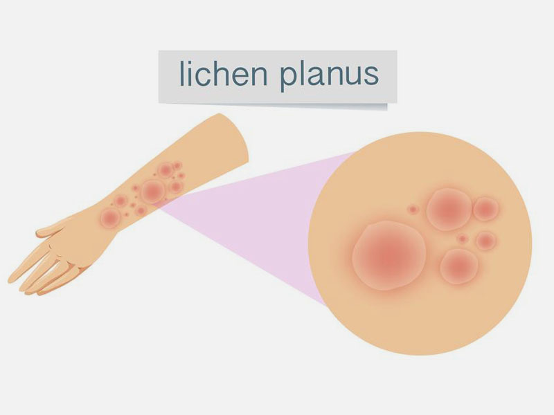
Cutaneous lichen planus manifests as pruritic, polygonal, purple papules with a characteristic Wickham’s striae—a network of fine white lines on the surface of the lesions. Commonly affected sites include the wrists, ankles, and lower back.

Oral lichen planus presents as white, lace-like patterns or painful erosions on the mucosa, often leading to difficulty eating or speaking. Genital involvement may result in painful ulcerations.

In scalp involvement, termed lichen planopilaris, patients experience hair loss and scarring. Nail lichen planus can cause ridges, thinning, or nail loss. The chronic and relapsing nature of the disease significantly impacts the quality of life (Lodi et al., 2020).


Etiology

Lichen planus is believed to have a multifactorial etiology, involving genetic, immunological, and environmental factors.

The disease results from an immune-mediated response targeting basal keratinocytes in the epidermis and mucosa. Genetic predisposition plays a role, as family clustering has been observed in some cases.

Hepatitis C virus infection and other viral triggers are strongly associated with lichen planus, suggesting an infectious component. Stress and trauma can exacerbate symptoms, a phenomenon known as the Koebner effect. Drug-induced lichen planus arises from hypersensitivity to specific medications (Alrashdan et al., 2016).

Understanding the interplay between these factors is crucial for tailoring treatment strategies and improving patient outcomes.


Pathophysiology

The pathophysiology of lichen planus involves T-cell-mediated destruction of basal keratinocytes.

In the affected tissues, cytotoxic CD8+ T-cells attack basal keratinocytes, leading to apoptosis and inflammation. This immune response is triggered by antigens expressed on keratinocytes, which may result from infection, drugs, or other environmental factors.

Histopathological examination reveals hyperkeratosis, a thickened granular layer, and a dense band-like lymphocytic infiltrate at the dermoepidermal junction. Civatte bodies, representing apoptotic keratinocytes, are another characteristic feature.

Oxidative stress and cytokine dysregulation also contribute to disease progression. Chronic inflammation may lead to scarring and tissue damage, especially in lichen planopilaris and erosive oral lichen planus (Lodi et al., 2020).

(Nursing Paper Example on Lichen Planus)


Diagnosis

Diagnosing lichen planus involves clinical evaluation, histopathological analysis, and laboratory tests.

Clinicians assess skin or mucosal lesions for their characteristic appearance and distribution. Dermoscopy aids in identifying Wickham’s striae, a diagnostic hallmark of the disease.

A skin or mucosal biopsy is often performed to confirm the diagnosis. Histological findings include hyperkeratosis, basal cell degeneration, and a band-like lymphocytic infiltrate at the dermoepidermal junction.

Laboratory tests may be used to rule out differential diagnoses such as lupus erythematosus or pemphigus. Hepatitis C screening is recommended for patients with suspected lichen planus due to the strong association between the two conditions (Alrashdan et al., 2016).


Treatment Regimens

Treatment of lichen planus focuses on symptom control, as there is no definitive cure.

Topical corticosteroids are the first-line treatment for cutaneous and oral lesions. Intralesional steroid injections may be used for localized, resistant lesions.

For severe or widespread cases, systemic therapies such as oral corticosteroids, retinoids, or immunosuppressants like methotrexate or cyclosporine are effective.

Phototherapy, particularly narrowband UVB or PUVA, is an option for extensive skin involvement.

Adjunctive treatments include antihistamines for pruritus and topical anesthetics for pain relief in oral lichen planus. Patients with drug-induced lichen planus should discontinue the offending medication under medical guidance. Regular monitoring and tailored treatment plans are essential for managing this chronic disease (Lodi et al., 2020).


Patient Education

Educating patients about lichen planus is vital for effective management and improved quality of life.

Patients should be informed about the chronic nature of the disease and the potential for relapses. Stress management techniques, such as mindfulness or counseling, may help reduce exacerbations.

For oral lichen planus, maintaining good oral hygiene and avoiding irritants like spicy foods or tobacco are crucial. Patients should report any changes in lesion appearance, as malignant transformation, although rare, can occur in oral forms.

Healthcare providers should emphasize adherence to treatment regimens and the importance of follow-up visits. Patients on immunosuppressive therapy require regular monitoring to minimize adverse effects. Empowering patients with knowledge fosters active participation in their care (Alrashdan et al., 2016).


Conclusion

Lichen planus is a complex autoimmune disease with variable clinical presentations and significant impacts on quality of life. While the exact etiology remains uncertain, factors such as chronic inflammation, viral infections, and drug hypersensitivity are implicated. Early diagnosis through clinical evaluation and histopathology is critical for effective management. Treatment focuses on alleviating symptoms and minimizing relapses, with corticosteroids being the mainstay therapy. Patient education plays a pivotal role in disease management, encouraging adherence to treatment and lifestyle modifications. Continued research is essential to unravel the disease’s mechanisms and develop targeted therapies.


References

Alrashdan, M. S., Cirillo, N., & McCullough, M. (2016). Oral lichen planus: A literature review and update. Archives of Dermatological Research, 308(8), 539-551. https://doi.org/10.1007/s00403-016-1675-9

Lodi, G., Scully, C., Carrozzo, M., Griffiths, M., Sugerman, P. B., & Thongprasom, K. (2020). Current controversies in oral lichen planus: Report of an international consensus meeting. Journal of Oral Pathology & Medicine, 49(6), 482-488. https://doi.org/10.1111/jop.13024

Sugerman, P. B., Savage, N. W., Walsh, L. J., & Seymour, G. J. (2020). Disease mechanisms in oral lichen planus. International Journal of Oral and Maxillofacial Surgery, 47(8), 857-871. https://doi.org/10.1016/j.ijom.2019.03.001

 
Do you need a similar assignment done for you from scratch? Order now!
Use Discount Code "Newclient" for a 15% Discount!

Nursing Paper Example on Ménière’s Disease

Nursing Paper Example on Ménière’s Disease

Ménière’s disease is a chronic inner ear disorder characterized by episodes of vertigo, tinnitus, aural fullness, and fluctuating sensorineural hearing loss. This condition primarily affects adults aged 20–60 years and has a significant impact on the quality of life due to its unpredictable nature and debilitating symptoms. The exact cause remains unclear, though factors such as endolymphatic hydrops and autoimmune mechanisms are implicated. Early diagnosis and management are essential to prevent progression to permanent hearing loss and to alleviate the psychological burden associated with the disease.

Nursing Paper Example on Ménière's Disease


Causes

The causes of Ménière’s disease remain largely idiopathic, though several theories have been proposed.

The primary hypothesis involves endolymphatic hydrops, a pathological increase in endolymph fluid within the cochlea and vestibular organs. This excess fluid leads to distension and damage to sensory structures, causing symptoms.

Autoimmune dysfunction is another potential cause. The body’s immune system may attack the inner ear, triggering inflammation and damage. Genetic predisposition has been identified in some cases, with familial clustering suggesting heritability.

Environmental and lifestyle factors, such as stress, excessive salt intake, and allergies, may exacerbate symptoms by disrupting fluid homeostasis. Certain infections, including viral labyrinthitis, are also associated with Ménière’s disease. Despite these theories, a definitive cause remains elusive (Basura et al., 2020).

Signs and Symptoms

Ménière’s disease manifests with distinct yet overlapping symptoms that vary in severity and frequency.

Vertigo, the most debilitating symptom, presents as sudden episodes lasting from 20 minutes to several hours. These episodes may lead to imbalance, nausea, and vomiting.

Hearing loss in Ménière’s disease is initially fluctuating, predominantly affecting low frequencies. Over time, the hearing loss may become permanent and progressive.

Tinnitus, described as a ringing, buzzing, or roaring sound, accompanies hearing loss. This can range from mild to severe, significantly affecting sleep and concentration.

Aural fullness, a sensation of pressure or fullness in the affected ear, is another hallmark of the disease. These symptoms often occur in clusters or attacks, separated by symptom-free periods. Anxiety and depression are common due to the disease’s impact on daily functioning (Westerberg et al., 2018).


Etiology

The etiology of Ménière’s disease is multifactorial, involving genetic, environmental, and pathological factors.

Endolymphatic hydrops, identified as a key pathological feature, results from impaired absorption or overproduction of endolymph. This disrupts the delicate ionic balance required for auditory and vestibular function.

Autoimmune conditions, such as systemic lupus erythematosus and rheumatoid arthritis, are associated with Ménière’s disease, supporting an immune-mediated etiology. Genetic predisposition is evident, with mutations in certain genes linked to familial cases.

Infections, including syphilis and viral labyrinthitis, may damage the inner ear, predisposing individuals to Ménière’s disease. Vascular factors, such as migraine-associated vasospasm, are also implicated. Understanding these etiological factors is essential for developing targeted treatments (Basura et al., 2020).


Pathophysiology

The pathophysiology of Ménière’s disease centers around endolymphatic hydrops and its effects on inner ear function.

Endolymphatic hydrops results from excess endolymph fluid in the scala media of the cochlea and the vestibular organs. This fluid buildup distorts the Reissner’s membrane and compromises the organ of Corti, leading to hearing loss and tinnitus.

In the vestibular organs, hydrops disrupts the function of sensory hair cells, causing vertigo and imbalance. Episodes are triggered when fluid pressure reaches a critical point, leading to membrane rupture and mixing of endolymph and perilymph. This ionic imbalance temporarily halts normal sensory function, producing acute symptoms.

Chronic inflammation and oxidative stress contribute to progressive damage, particularly in autoimmune-associated cases. Over time, repeated episodes result in irreversible hair cell loss and nerve damage, leading to permanent hearing loss and balance deficits (Westerberg et al., 2018).


Diagnosis

Diagnosis of Ménière’s disease is primarily clinical, based on the patient’s history and symptomatology.

The diagnostic criteria established by the American Academy of Otolaryngology include two or more episodes of vertigo lasting 20 minutes to 12 hours, sensorineural hearing loss confirmed by audiometry, tinnitus or aural fullness, and exclusion of other conditions.

Audiometry typically reveals low-frequency sensorineural hearing loss, progressing to a broader frequency range over time. Vestibular function tests, including electronystagmography and rotary chair testing, assess balance impairment.

Imaging, such as magnetic resonance imaging (MRI), may be performed to exclude differential diagnoses like vestibular schwannoma. Blood tests may identify autoimmune or infectious causes. A thorough assessment ensures accurate diagnosis and effective management (Basura et al., 2020).


Treatment Regimens

Treatment for Ménière’s disease focuses on symptom management, reducing episode frequency, and preserving hearing.

Dietary modifications, such as reducing sodium intake, help regulate fluid balance. Avoiding caffeine and alcohol may minimize symptoms.

Medications like diuretics, antihistamines, and benzodiazepines alleviate acute vertigo and prevent attacks. Betahistine, a histamine analogue, improves microcirculation in the inner ear and is commonly prescribed.

For refractory cases, intratympanic injections of corticosteroids or gentamicin offer relief. Corticosteroids reduce inflammation, while gentamicin selectively ablates vestibular hair cells to reduce vertigo.

Surgical interventions, including endolymphatic sac decompression or vestibular nerve section, are reserved for severe cases. Vestibular rehabilitation exercises improve balance and reduce fall risk. Psychological support addresses the emotional impact of the disease (Westerberg et al., 2018).


Patient Education

Educating patients about Ménière’s disease is essential for effective self-management and improved outcomes.

Patients should understand the episodic nature of the disease and the importance of adhering to dietary and medication regimens. Stress management techniques, such as relaxation exercises or cognitive-behavioral therapy, can reduce triggers.

Lifestyle adjustments, such as limiting exposure to loud noises and maintaining hydration, are beneficial. Patients should seek prompt medical attention for any changes in symptoms, as early intervention can prevent complications.

Support groups and counseling provide emotional support and coping strategies. Empowering patients with knowledge fosters proactive management and enhances quality of life (Basura et al., 2020).


Conclusion

Ménière’s disease is a multifaceted condition that profoundly affects auditory and vestibular function. While its etiology and pathophysiology remain under investigation, factors such as endolymphatic hydrops, autoimmune dysfunction, and genetic predisposition play key roles. Accurate diagnosis through clinical and audiometric evaluations is crucial for guiding treatment. Current management focuses on symptom control, with dietary modifications, medications, and surgical options available. Patient education and psychological support are integral to improving outcomes. Ongoing research is essential to uncover the disease’s underlying mechanisms and develop innovative therapies.


References

Basura, G. J., Adams, M. E., Monfared, A., Schwartz, S. R., Whitman, G. T., & Burkard, R. F. (2020). Clinical practice guideline: Ménière’s disease. Otolaryngology–Head and Neck Surgery, 162(2_suppl), S1-S55. https://doi.org/10.1177/0194599820909438

Westerberg, B. D., Roberson, J. B., & Stachler, R. J. (2018). Ménière’s disease: Clinical insights and updates. Journal of Vestibular Research, 28(3), 123-136. https://doi.org/10.3233/VES-180640

 
Do you need a similar assignment done for you from scratch? Order now!
Use Discount Code "Newclient" for a 15% Discount!

Paranoid Personality Disorder (PDD)

Paranoid Personality Disorder (PDD)

Paranoid Personality Disorder (PDD)

A Round of Recreational Therapy

IN THIS ARTICLE

Paranoid Personality Disorder

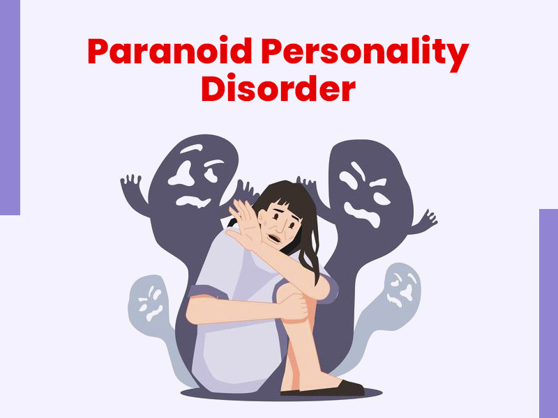
Paranoid personality disorder (PPD) is one of a group of conditions called “Cluster A” personality disorders which involve odd or eccentric ways of thinking. People with PPD also suffer from paranoia, an unrelenting mistrust and suspicion of others, even when there is no reason to be suspicious.

This disorder usually begins by early adulthood and appears to be more common in men than in women.

What Are the Symptoms of Paranoid Personality Disorder?

People with PPD are always on guard, believing that others are constantly trying to demean, harm, or threaten them. These generally unfounded beliefs, as well as their habits of blame and distrust, might interfere with their ability to form close relationships. People with this disorder:

  • Doubt the commitment, loyalty, or trustworthiness of others, believing others are using or deceiving them
  • Are reluctant to confide in others or reveal personal information due to a fear that the information will be used against them
  • Are unforgiving and hold grudges
  • Are hypersensitive and take criticism poorly
  • Read hidden meanings in the innocent remarks or casual looks of others
  • Perceive attacks on their character that are not apparent to others; they generally react with anger and are quick to retaliate
  • Have recurrent suspicions, without reason, that their spouses or lovers are being unfaithful
  • Are generally cold and distant in their relationships with others, and might become controlling and jealous
  • Cannot see their role in problems or conflicts and believe they are always right
  • Have difficulty relaxing
  • Are hostile, stubborn, and argumentative

What Causes Paranoid Personality Disorder?

The exact cause of PPD is not known, but it likely involves a combination of biological and psychological factors. The fact that PPD is more common in people who have close relatives with schizophrenia suggests a genetic link between the two disorders. Early childhood experiences, including physical or emotional trauma, are also suspected to play a role in the development of PPD.

How Is Paranoid Personality Disorder Diagnosed?

If physical symptoms are present, the doctor will begin an evaluation by performing a complete medical and psychiatric history and, if indicated, a physical exam. Although there are no laboratory tests to specifically diagnose personality disorders, the doctor might use various diagnostic tests to rule out physical illness as the cause of the symptoms.

If the doctor finds no physical reason for the symptoms, they might refer the person to a psychiatrist or psychologist, health care professionals who are specially trained to diagnose and treat mental illnesses. Psychiatrists and psychologists use specially designed interview and assessment tools to evaluate a person for a personality disorder.

How Is Paranoid Personality Disorder Treated?

People with PPD often do not seek treatment on their own because they do not see themselves as having a problem. When treatment is sought, psychotherapy (a form of counseling) is the treatment of choice for PPD. Treatment likely will focus on increasing general coping skills, as well as on improving social interaction, communication, and self-esteem.

Because trust is an important factor of psychotherapy, treatment is challenging since people with PPD have such distrust of others. As a result, many people with PPD do not follow their treatment plan.

Medication generally is not a major focus of treatment for PPD. However, medications, such as anti-anxiety, antidepressant or antipsychotic drugs, might be prescribed if the person’s symptoms are extreme, or if they also suffer from an associated psychological problem, such as anxiety or depression.

What Complications Are Associated With Paranoid Personality Disorder?

The thinking and behaviors associated with PPD can interfere with a person’s ability to maintain relationships, as well as their ability to function socially and in work situations. In many cases, people with PPD become involved in legal battles, suing people or companies they believe are “out to get them.”

What Is the Outlook for People With Paranoid Personality Disorder?

The outlook for people with PPD varies. It is a chronic disorder, which means it tends to last throughout a person’s life. Although some people can function fairly well with PPD and are able to marry and hold jobs, others are completely disabled by the disorder. Because people with PPD tend to resist treatment, the outcome often is poor.

Can Paranoid Personality Disorder Be Prevented?

Although prevention of PDD might not be possible, treatment can sometimes allow a person who is prone to this condition to learn more productive ways of dealing with situations.

 
Do you need a similar assignment done for you from scratch? Order now!
Use Discount Code "Newclient" for a 15% Discount!