Nursing Paper Example on Cushing’s Syndrome

Nursing Paper Example on Cushing’s Syndrome

Cushing’s syndrome is a rare endocrine disorder caused by prolonged exposure to high levels of cortisol. It can result from endogenous overproduction or exogenous corticosteroid use. The condition leads to a wide array of systemic manifestations, significantly impacting a patient’s physical and metabolic health. Early diagnosis and appropriate treatment are crucial to prevent severe complications.


Nursing Paper Example on Cushing's Syndrome

Causes of Cushing’s Syndrome

Cushing’s syndrome can result from endogenous or exogenous factors.

Exogenous Causes

Chronic corticosteroid therapy: Often prescribed for conditions like asthma, rheumatoid arthritis, or organ transplant.

Topical or inhaled corticosteroids: When used excessively over long periods.

Endogenous Causes

  • Adrenocorticotropic hormone (ACTH)-dependent:

Pituitary adenomas (Cushing’s disease): Most common endogenous cause.

Ectopic ACTH production: Seen in small cell lung cancer and other tumors.

  • ACTH-independent:

Adrenal adenomas or carcinomas: Lead to excessive cortisol production.

Macronodular adrenal hyperplasia: Rare cause of cortisol overproduction.


Signs and Symptoms

Cushing’s syndrome presents with a spectrum of clinical features, many of which are due to hypercortisolism’s catabolic effects.

Physical Features

  • Central obesity with thin extremities.
  • Moon facies (round, puffy face).
  • Dorsocervical fat pad (buffalo hump).
  • Purple striae on the abdomen, thighs, and breasts.
  • Easy bruising and delayed wound healing.

Systemic Symptoms

Musculoskeletal: Proximal muscle weakness, osteoporosis, and fractures.

Metabolic: Hyperglycemia, insulin resistance, and dyslipidemia.

Cardiovascular: Hypertension, increased risk of thromboembolism.

Neuropsychiatric: Mood swings, depression, anxiety, or psychosis.

Reproductive: Irregular menstruation, infertility, or decreased libido.


Etiology

The etiology of Cushing’s syndrome varies depending on its endogenous or exogenous origin.

Pituitary Tumors (Cushing’s Disease)

ACTH-secreting pituitary adenomas are the most common endogenous cause.

Ectopic ACTH Production

Neuroendocrine tumors (e.g., small cell lung cancer, thymic tumors) produce ACTH aberrantly.

Adrenal Causes

Adenomas, carcinomas, or hyperplasia can independently produce excessive cortisol.

Iatrogenic Causes

Prolonged corticosteroid use for chronic inflammatory or autoimmune conditions is the leading cause of exogenous Cushing’s syndrome.


Pathophysiology

Cushing’s syndrome results from sustained hypercortisolism, disrupting various physiological processes.

Mechanism of Disease

Excess cortisol dysregulates carbohydrate, protein, and fat metabolism.

Persistent hyperglycemia contributes to insulin resistance.

Protein catabolism leads to muscle wasting and thinning of the skin.

Fat redistribution occurs, leading to central obesity and characteristic facial features.

Cardiovascular Effects

Cortisol elevates blood pressure by enhancing vascular sensitivity to catecholamines and suppressing nitric oxide production.

Immune Effects

Suppressed inflammatory responses increase susceptibility to infections.


Diagnosis

Diagnosing Cushing’s syndrome involves clinical assessment and laboratory confirmation of hypercortisolism.

Screening Tests

24-hour urinary free cortisol (UFC): Elevated levels confirm hypercortisolism.

Low-dose dexamethasone suppression test: Failure to suppress cortisol indicates Cushing’s syndrome.

Late-night salivary cortisol: Elevated levels are highly specific for hypercortisolism.

Differential Diagnosis

Measurement of ACTH helps differentiate ACTH-dependent from ACTH-independent causes.

Imaging studies (e.g., MRI of the pituitary, CT of the adrenal glands) localize the source.


Treatment Regimens

Treatment for Cushing’s syndrome depends on the underlying cause and aims to normalize cortisol levels.

Surgical Management

Transsphenoidal surgery: Preferred for ACTH-secreting pituitary adenomas.

Adrenalectomy: Indicated for adrenal adenomas or carcinomas.

Resection of ectopic ACTH-producing tumors: Essential for source control.

Medical Management

Steroidogenesis Inhibitors: Metyrapone, ketoconazole, or osilodrostat reduce cortisol production.

Pituitary-Directed Therapy: Pasireotide (a somatostatin analog) inhibits ACTH secretion.

Glucocorticoid Receptor Antagonists: Mifepristone is used in severe hyperglycemia cases.

Radiation Therapy

Used in cases of recurrent or persistent pituitary tumors after surgery.


Patient Education

Understanding the Disease

Educate patients about the cause and symptoms of Cushing’s syndrome.

Stress the importance of follow-up and adherence to prescribed treatments.

Managing Medication

Gradual tapering of corticosteroids prevents withdrawal and adrenal insufficiency.

Lifestyle Modifications

Encourage weight loss and regular physical activity to mitigate metabolic complications.

Stress reduction techniques may benefit patients with neuropsychiatric symptoms.

Support Systems

Provide resources for counseling and support groups to help cope with the emotional burden.


Additional Considerations

Complications

Cardiovascular disease, infections, and osteoporosis are common in untreated cases.

Long-term use of medications like ketoconazole requires monitoring for hepatotoxicity.

Prognosis

Early intervention improves outcomes, but untreated Cushing’s syndrome has a poor prognosis.


Conclusion

Cushing’s syndrome is a complex endocrine disorder requiring a multidisciplinary approach for diagnosis and treatment. Early recognition and management are critical to reduce morbidity and mortality associated with the condition. Continued research into targeted therapies offers hope for improving patient outcomes.


References

Bertagna, X., Guignat, L., Groussin, L., & Bertherat, J. (2009). Cushing’s disease. Best Practice & Research Clinical Endocrinology & Metabolism, 23(5), 607-623. https://www.sciencedirect.com/science/article/pii/S1521690X09000789

Lacroix, A., Feelders, R. A., Stratakis, C. A., & Nieman, L. K. (2015). Cushing’s syndrome. The Lancet, 386(9996), 913-927. https://www.thelancet.com/journals/lancet/article/PIIS0140-6736(14)61375-1/fulltext

Nieman, L. K., Biller, B. M., Findling, J. W., Newell-Price, J., Savage, M. O., & Stewart, P. M. (2008). The diagnosis of Cushing’s syndrome: an Endocrine Society Clinical Practice Guideline. Journal of Clinical Endocrinology & Metabolism, 93(5), 1526-1540. https://academic.oup.com/jcem/article/93/5/1526/2597367

National Institutes of Health. (2023). Cushing’s Syndrome. https://www.niddk.nih.gov/health-information/endocrine-diseases/cushings-syndrome

Mayo Clinic. (2023). Cushing syndrome. https://www.mayoclinic.org/diseases-conditions/cushing-syndrome/symptoms-causes/syc-20351310

 
Do you need a similar assignment done for you from scratch? Order now!
Use Discount Code "Newclient" for a 15% Discount!

Nursing Paper Example on Creutzfeldt-Jakob Disease

Nursing Paper Example on Creutzfeldt-Jakob Disease

(Nursing Paper Example on Creutzfeldt-Jakob Disease) Creutzfeldt-Jakob disease (CJD) is a rare, fatal neurodegenerative disorder caused by the accumulation of abnormal prion proteins in the brain. It belongs to the group of transmissible spongiform encephalopathies (TSEs) and is characterized by rapid progression and severe neurological decline. Although CJD is relatively rare, its devastating effects and lack of curative treatments make it a critical focus in neurology and infectious disease research.


Nursing Paper Example on Creutzfeldt-Jakob Disease

Causes of Creutzfeldt-Jakob Disease

CJD arises from the misfolding of normal prion proteins into an abnormal conformation.

Types of CJD

  • Sporadic CJD: Accounts for 85-90% of cases; the cause is unknown but may involve spontaneous prion misfolding.
  • Hereditary CJD: Linked to mutations in the PRNP gene, which encodes the prion protein.
  • Acquired CJD: Transmitted through exposure to infectious prions via contaminated surgical instruments, corneal transplants, or tainted beef products (variant CJD).

Signs and Symptoms

Initial Symptoms

  • Subtle memory lapses and difficulty concentrating.
  • Behavioral changes, including irritability and depression.

Neurological Symptoms

  • Cognitive Decline: Rapid progression to severe dementia.
  • Movement Disorders: Myoclonus (sudden muscle jerks), ataxia, and tremors.
  • Visual Disturbances: Blurred vision or cortical blindness.

End-Stage Symptoms

  • Mutism, coma, and loss of voluntary muscle control.
  • Death typically occurs within 6-12 months of symptom onset.

Etiology

Prion Proteins

The misfolded prion protein (PrP^Sc) is resistant to protease digestion and accumulates in the brain.

Genetics

Mutations in the PRNP gene predispose individuals to hereditary CJD. Common mutations include E200K and D178N.

Transmission

  • Direct contact with infected tissue or contaminated medical equipment can transmit prions.
  • Variant CJD is linked to the consumption of prion-infected beef.

Pathophysiology

The pathophysiology of CJD involves the transformation of normal cellular prion proteins (PrP^C) into an abnormal, disease-causing form (PrP^Sc).

Mechanisms of Disease

  • PrP^Sc induces a conformational change in PrP^C, perpetuating its own production.
  • These abnormal prions aggregate, forming amyloid plaques and causing neurotoxicity.

Brain Changes

  • Spongiform changes: Microscopic vacuoles in the brain’s gray matter.
  • Neuronal loss: Widespread degeneration of brain tissue.
  • Gliosis: Reactive astrocytes and microglia surround the damaged areas.

Diagnosis

Clinical Criteria

  • Rapidly progressive dementia and two or more neurological features (e.g., myoclonus, ataxia).
  • Exclusion of other causes of dementia through history and physical examination.

Diagnostic Tools

  • Electroencephalogram (EEG): Periodic sharp wave complexes are highly suggestive.
  • Cerebrospinal Fluid (CSF): Detection of 14-3-3 protein and other prion-specific biomarkers.
  • Magnetic Resonance Imaging (MRI): Hyperintensities in the caudate nucleus and putamen on diffusion-weighted imaging (DWI).
  • Brain Biopsy/Autopsy: Definitive diagnosis is achieved through histopathological examination.

(Nursing Paper Example on Creutzfeldt-Jakob Disease)


Treatment Regimens

There is no cure for CJD, and treatment is primarily palliative.

Symptom Management

  • Antiepileptics: Used to control myoclonus and seizures.
  • Pain Relief: Analgesics for physical discomfort.
  • Psychological Support: Counseling for patients and families.

Experimental Therapies

  • Antibodies targeting prion proteins are being explored in clinical trials.
  • Small molecules that stabilize the normal conformation of PrP^C show potential.

Patient Education

Understanding the Disease

  • Educate patients and families about the nature and progression of the disease.
  • Emphasize the absence of curative treatments but highlight ongoing research.

Preventive Measures

  • Proper sterilization protocols for surgical equipment to prevent iatrogenic transmission.
  • Avoid consumption of potentially contaminated beef in endemic regions.

Supportive Resources

  • Refer families to support groups and organizations specializing in neurodegenerative disorders.

Additional Considerations

Public Health Implications

  • Surveillance programs monitor prion diseases to identify outbreaks of variant CJD.
  • Blood donation policies exclude individuals with potential exposure to infectious prions.

Differential Diagnosis

  • Conditions such as Alzheimer’s disease, Lewy body dementia, and paraneoplastic syndromes may mimic CJD, necessitating thorough evaluation.

Conclusion

Creutzfeldt-Jakob disease is a rare but devastating neurodegenerative disorder with no known cure. The condition is characterized by rapid progression, severe cognitive and motor decline, and ultimately, death. Early recognition, exclusion of alternative diagnoses, and palliative care are essential for managing patients. Continued research into prion biology holds promise for developing targeted therapies and improving outcomes for this fatal disease.


References

Centers for Disease Control and Prevention. (2022). Creutzfeldt-Jakob Disease, Classic (CJD). https://www.cdc.gov/prions/cjd/classic-cjd.html

Collinge, J. (2016). Mammalian prions and their wider relevance in neurodegenerative diseases. Nature Reviews Neuroscience, 17(5), 322-333. https://www.nature.com/articles/nrn.2016.48

Mead, S., & Reilly, M. M. (2015). Aetiology and genetic basis of human prion diseases. Nature Reviews Neurology, 11(7), 416-428. https://www.nature.com/articles/nrneurol.2015.103

National Institutes of Health. (2023). Prion Diseases Information Page. https://www.ninds.nih.gov/disorders/all-disorders/prion-diseases-information-page

World Health Organization. (2020). Variant Creutzfeldt-Jakob Disease. https://www.who.int/news-room/fact-sheets/detail/variant-creutzfeldt-jakob-disease

 
Do you need a similar assignment done for you from scratch? Order now!
Use Discount Code "Newclient" for a 15% Discount!

Nursing Paper Example on Costochondritis

Nursing Paper Example on Costochondritis

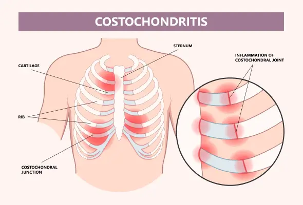
Costochondritis is a common musculoskeletal condition that involves inflammation of the costochondral, costosternal, or costovertebral joints. These are the points where the ribs attach to the sternum or spine. While it is not life-threatening, it can mimic more serious conditions, such as cardiac-related chest pain, leading to significant patient distress. Understanding its causes, clinical presentation, and management strategies is essential to ensure effective treatment and reassurance for affected individuals.


Nursing Paper Example on Costochondritis

Causes of Costochondritis

Mechanical Stress

Repetitive physical activity, such as heavy lifting, coughing, or trauma, can strain the chest wall, leading to inflammation.

Infections

Though rare, viral or bacterial infections can affect the costochondral joints, especially after surgical procedures or thoracic trauma.

Arthritic Conditions

Underlying systemic inflammatory diseases such as rheumatoid arthritis, ankylosing spondylitis, or psoriatic arthritis may contribute to costochondral inflammation.

Idiopathic

In many cases, the exact cause remains unknown. These idiopathic cases are generally self-limiting and resolve without significant intervention.


Signs and Symptoms

Chest Pain

Sharp, stabbing, or aching pain localized to the costochondral or costosternal joints.

Pain often worsens with movement, deep breathing, or palpation of the affected joints.

Tenderness

Physical examination reveals tenderness at one or more costochondral junctions, typically in the second to fifth ribs.

Non-Radiating Pain

Unlike cardiac pain, costochondritis pain does not radiate to other areas such as the arms or jaw.

Absence of Systemic Symptoms

Fever, fatigue, and other systemic symptoms are generally absent, distinguishing costochondritis from infections or inflammatory diseases.


Etiology

Costochondritis is considered a benign condition, with several potential etiologies:

Trauma or Overuse: Often seen in athletes or individuals engaging in repetitive chest wall movements.

Infections: Rarely, organisms such as Staphylococcus aureus or Mycobacterium tuberculosis can affect the costochondral joints.

Post-Surgical Inflammation: Procedures involving the sternum, such as coronary artery bypass grafting, can predispose individuals to localized inflammation.


Pathophysiology

The pathophysiology of costochondritis involves localized inflammation of the cartilage that connects the ribs to the sternum.

Inflammatory Response

Mechanical stress or microtrauma triggers an inflammatory cascade, leading to swelling and increased sensitivity of nerve endings in the region.

Chronic Irritation

In recurrent cases, prolonged inflammation may result in fibrosis or cartilage degradation, contributing to persistent discomfort.


Diagnosis

Clinical Examination

Diagnosis is primarily clinical, based on history and physical findings such as tenderness over the costochondral junctions.

Pain reproduction upon palpation is a key diagnostic feature.

Exclusion of Cardiac Causes

Electrocardiogram and cardiac biomarkers may be necessary to rule out myocardial infarction or angina.

Imaging studies, such as chest X-rays, are often unremarkable but help exclude other causes of chest pain.

Advanced Imaging

MRI or ultrasound may be considered in atypical or refractory cases to assess soft tissue inflammation.

Bone scans can help differentiate costochondritis from conditions like Tietze syndrome, which involves visible swelling of the affected joints.


Treatment Regimens

Non-Pharmacological Treatments

Rest and Activity Modification: Avoid activities that exacerbate chest pain.

Heat or Cold Therapy: Alternating applications can reduce inflammation and pain.

Physical Therapy: Stretching and strengthening exercises improve posture and reduce chest wall strain.

Pharmacological Treatments

Nonsteroidal Anti-Inflammatory Drugs (NSAIDs): Ibuprofen or naproxen is commonly prescribed to reduce pain and inflammation.

Topical Analgesics: Lidocaine patches or diclofenac gel may provide localized relief.

Corticosteroid Injections: In refractory cases, local steroid injections can provide rapid symptom relief.

Surgical Interventions

Rarely required, surgery may be considered in chronic cases with persistent symptoms unresponsive to conservative treatments.


Patient Education

Understanding the Condition

Reassure patients that costochondritis is benign and self-limiting in most cases.

Emphasize that the condition is unrelated to cardiac or life-threatening causes of chest pain.

Self-Care Strategies

Encourage the use of proper ergonomics and body mechanics during activities to minimize strain.

Teach patients to recognize and avoid activities that trigger symptoms.

When to Seek Help

Advise patients to seek medical evaluation if chest pain is associated with symptoms such as shortness of breath, dizziness, or radiating pain, as these may indicate a cardiac origin.


Additional Considerations

Prognosis

The prognosis for costochondritis is excellent, with most cases resolving within weeks to months.

Chronic cases may require ongoing management to prevent recurrence.

Complications

While rare, untreated costochondritis may result in chronic pain or overlapping symptoms with other musculoskeletal disorders.

Differentiating from Tietze Syndrome

Unlike costochondritis, Tietze syndrome is characterized by visible swelling at the costochondral junctions. Proper differentiation is important for accurate management.


Conclusion

Costochondritis is a common, non-life-threatening condition that can cause significant discomfort and anxiety due to its similarity to cardiac chest pain. Early diagnosis, effective symptom management, and reassurance are key to improving patient outcomes. With appropriate care, most patients experience full resolution of symptoms and return to normal activities.


References

Clark, S., & Kruse, R. J. (2018). Costochondritis: Diagnosis and treatment. American Family Physician, 97(8), 485-490. https://www.aafp.org/pubs/afp/issues/2018/0415/p485.html

Gaitini, D., & Be’ery, I. (2019). Imaging of chest wall pain: Ultrasound as a first-line diagnostic tool. Clinical Radiology, 74(1), 11-20. https://www.clinicalradiologyonline.net/article/S0009-9260(18)30164-2/fulltext

Jansen, C. H., et al. (2020). Differentiation between costochondritis and Tietze syndrome: A clinical perspective. Pain Medicine, 21(5), 1010-1016. https://academic.oup.com/painmedicine/article/21/5/1010/5824253

Mayo Clinic. (2023). Costochondritis. https://www.mayoclinic.org/diseases-conditions/costochondritis/symptoms-causes/syc-20371175

Medscape. (2023). Costochondritis: Overview and treatment. https://emedicine.medscape.com/article/93326-overview

 
Do you need a similar assignment done for you from scratch? Order now!
Use Discount Code "Newclient" for a 15% Discount!

Nursing Paper Example on Cold Agglutinin Disease

Nursing Paper Example on Cold Agglutinin Disease

Cold agglutinin disease is a rare autoimmune hemolytic anemia characterized by the premature destruction of red blood cells due to the activity of cold agglutinins. These are autoantibodies that become activated at low temperatures, typically below 37°C (98.6°F). The condition is often associated with underlying infections, lymphoproliferative disorders, or may occur idiopathically. Patients with cold agglutinin disease experience a range of symptoms, including fatigue, hemolysis-related anemia, and cold-induced symptoms such as acrocyanosis. Early diagnosis and tailored management strategies are essential to improve patient outcomes.


Nursing Paper Example on Cold Agglutinin Disease

Causes of Cold Agglutinin Disease

Primary Cold Agglutinin Disease

This idiopathic form has no identifiable underlying cause and accounts for most cases in older adults.

Secondary Cold Agglutinin Disease

This form is associated with other conditions, including:

Infections: Mycoplasma pneumoniae, Epstein-Barr virus, and cytomegalovirus infections are common triggers.

Lymphoproliferative Disorders: Conditions such as Waldenström macroglobulinemia or non-Hodgkin lymphoma often result in cold agglutinin production.

Autoimmune Diseases: Rarely linked to systemic lupus erythematosus or other autoimmune conditions.

Genetic and Environmental Factors

While not directly inherited, certain genetic predispositions may influence immune system dysregulation, increasing susceptibility.


Signs and Symptoms

Hemolysis-Related Symptoms

Fatigue and pallor due to anemia.

Jaundice from elevated bilirubin levels caused by red blood cell breakdown.

Dark urine from hemoglobinuria.

Cold-Induced Symptoms

Acrocyanosis: Bluish discoloration of extremities, especially fingers, toes, and ears, due to red blood cell agglutination.

Raynaud’s phenomenon: Cold-induced discoloration and pain in the fingers and toes.

General Symptoms

Weakness and dizziness.

Splenomegaly, though less common than in other hemolytic anemias.


Etiology

Cold agglutinin disease results from the production of cold-reactive immunoglobulins, particularly IgM autoantibodies, that target red blood cell antigens. These antibodies activate the complement system, leading to intravascular and extravascular hemolysis.

Secondary forms are frequently associated with:

Infections: Mycoplasma pneumoniae induces transient cold agglutinin production during infection.

Lymphoproliferative Disorders: Abnormal B-cell activity contributes to chronic antibody production.


Pathophysiology

The disease mechanism involves:

Cold Agglutinin Activation

Autoantibodies bind to red blood cells in cooler parts of the body, such as the extremities. This agglutination clogs microvasculature and causes hemolysis.

Complement Activation

Cold agglutinins activate the complement cascade, particularly C3b deposition, leading to:

Intravascular Hemolysis: Direct red blood cell destruction in circulation.

Extravascular Hemolysis: Phagocytosis of complement-coated red blood cells in the liver.

Temperature Sensitivity

The antibodies become inactive at higher core body temperatures, confining hemolysis to areas exposed to cold.


Diagnosis

Clinical History and Physical Examination

Documenting cold sensitivity and hemolysis-related symptoms.

Identifying triggers such as infections or cold exposure.

Laboratory Tests

Complete Blood Count: Anemia with elevated reticulocyte count.

Peripheral Blood Smear: Evidence of agglutinated red blood cells.

Cold Agglutinin Titer: Confirmatory test showing high titers of cold-reactive IgM antibodies.

Complement Testing: Reduced C3 and C4 levels indicate complement activation.

Direct Antiglobulin Test: Positive for complement components but not for IgG.

Imaging and Further Testing

Chest X-ray: Rule out underlying Mycoplasma pneumoniae infection.

Bone Marrow Biopsy: Assess for lymphoproliferative disorders in unexplained cases.


Treatment Regimens

General Management

Avoiding Cold Exposure: Critical for reducing symptoms and preventing hemolysis.

Transfusions: Used for severe anemia but must be warmed to body temperature.

Pharmacological Treatments

Rituximab: First-line treatment targeting B-cells producing cold agglutinins.

Corticosteroids: Less effective but occasionally used to reduce inflammation.

Immunosuppressive Therapy: Agents like cyclophosphamide or bortezomib for refractory cases.

Treatment for Underlying Conditions

Infections: Antibiotic or antiviral therapy for secondary cold agglutinin disease caused by Mycoplasma pneumoniae or Epstein-Barr virus.

Lymphoproliferative Disorders: Chemotherapy or targeted therapies for conditions such as Waldenström macroglobulinemia.


Patient Education

Self-Management

Dress warmly, especially in cold climates, to reduce cold exposure.

Use gloves and other protective clothing to avoid exacerbation of symptoms.

Symptom Monitoring

Patients should be vigilant for worsening anemia symptoms, such as increased fatigue or jaundice.

Vaccinations

Routine influenza and pneumococcal vaccinations to prevent infections that may exacerbate symptoms.

Regular Follow-Up

Patients need ongoing evaluation to monitor disease progression and treatment response.


Additional Considerations

Prognosis

Primary cold agglutinin disease has a generally favorable prognosis with appropriate management.

Secondary forms improve with treatment of the underlying cause.

Complications

Severe anemia requiring repeated transfusions.

Thrombosis due to agglutinated red blood cells impairing blood flow.

Future Research

Advancements in targeted therapies, such as complement inhibitors, show promise in improving outcomes for patients with refractory disease.


Conclusion

Cold agglutinin disease is a rare but manageable autoimmune condition characterized by cold-induced hemolysis. Early diagnosis, avoidance of cold exposure, and targeted therapies, such as rituximab, significantly improve patient outcomes. Ongoing research and tailored patient education remain essential to further enhance quality of life and reduce disease burden.


References

Berentsen, S., & Sundic, T. (2015). Red blood cell destruction in autoimmune hemolytic anemia: Role of complement and potential new targets for therapy. Frontiers in Immunology, 6(395), 1-10. https://www.frontiersin.org/articles/10.3389/fimmu.2015.00395/full

Brodsky, R. A. (2021). Warm and cold autoimmune hemolytic anemias. New England Journal of Medicine, 384(15), 1401-1414. https://www.nejm.org/doi/full/10.1056/NEJMra2033980

Swiecicki, P. L., Hegerova, L. T., & Gertz, M. A. (2013). Cold agglutinin disease. Blood, 122(7), 1114-1121. https://doi.org/10.1182/blood-2013-02-453217

Harvard Health Publishing. (2023). Cold agglutinin disease. https://www.health.harvard.edu/a_to_z/cold-agglutinin-disease-a-to-z

American Society of Hematology. (2023). Cold agglutinin disease overview. https://www.hematology.org/education/patients/anemia/cold-agglutinin-disease

 
Do you need a similar assignment done for you from scratch? Order now!
Use Discount Code "Newclient" for a 15% Discount!

Nursing Paper Example on Chronic Obstructive Pulmonary Disease (COPD)

Nursing Paper Example on Chronic Obstructive Pulmonary Disease (COPD)

(Nursing Paper Example on Chronic Obstructive Pulmonary Disease (COPD)) Chronic obstructive pulmonary disease (COPD) is a progressive respiratory condition characterized by persistent airflow limitation and chronic inflammatory responses in the lungs. It primarily results from long-term exposure to noxious substances such as cigarette smoke and environmental pollutants. COPD encompasses chronic bronchitis and emphysema, with varying degrees of overlap between the two conditions. The disease is a leading cause of morbidity and mortality worldwide, significantly affecting patients’ quality of life.


Nursing Paper Example on Chronic Obstructive Pulmonary Disease (COPD)

Causes of COPD

Cigarette Smoking

The most common cause of COPD is long-term cigarette smoking, accounting for approximately 85% of cases.

Environmental Pollutants

  • Prolonged exposure to air pollutants, including dust, chemicals, and industrial emissions, increases COPD risk.
  • Biomass fuel smoke from indoor cooking in poorly ventilated spaces is a significant cause in developing countries.

Genetic Factors

  • Deficiency in alpha-1 antitrypsin, a protease inhibitor, predisposes individuals to early-onset COPD.

Respiratory Infections

Recurrent respiratory infections in childhood can impair lung development and increase vulnerability to COPD.

Other Factors

  • Aging and gender influence the prevalence and progression of the disease due to lung capacity and hormonal differences.

Signs and Symptoms

Primary Symptoms

  • Chronic cough: Often productive with sputum, especially in chronic bronchitis.
  • Dyspnea: Shortness of breath that progressively worsens, limiting physical activity.
  • Wheezing: Indicative of airway obstruction.

Other Symptoms

  • Fatigue due to increased respiratory effort.
  • Frequent respiratory infections.
  • Cyanosis: Bluish discoloration of skin due to low oxygen levels in advanced stages.

Complications

  • Acute exacerbations triggered by infections or environmental factors.
  • Pulmonary hypertension and right-sided heart failure (cor pulmonale).

Etiology

Pathogens and Exposures

The primary etiological factors in COPD are chronic inhalation of harmful substances, including tobacco smoke, occupational hazards, and environmental pollutants.

Genetic Susceptibility

  • Alpha-1 antitrypsin deficiency: This rare genetic disorder reduces the ability to neutralize proteolytic enzymes, leading to lung tissue damage.
  • Other genetic polymorphisms may contribute to inflammatory responses and tissue remodeling.

Pathophysiology

The hallmark of COPD is irreversible airflow obstruction caused by structural and inflammatory changes in the respiratory system.

Chronic Bronchitis

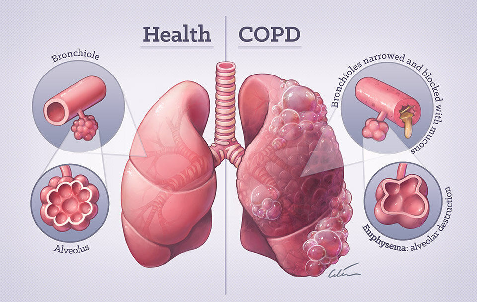
  • Inflammation of the bronchial walls leads to mucosal thickening and excessive mucus production.
  • Chronic obstruction occurs due to mucus plugging and airway narrowing.

Emphysema

  • Destruction of alveolar walls reduces surface area for gas exchange.
  • Loss of elastic recoil results in airway collapse during expiration.

Airflow Limitation

  • Persistent airway obstruction increases the work of breathing.
  • Gas trapping and hyperinflation occur, further reducing lung capacity.

Chronic hypoxemia and hypercapnia develop in advanced stages, resulting in systemic complications.

(Nursing Paper Example on Chronic Obstructive Pulmonary Disease (COPD))


DSM-5 Diagnosis

COPD does not fall under the purview of psychiatric disorders classified by the DSM-5. However, its impact on mental health is significant, with conditions like depression and anxiety commonly observed in COPD patients. Psychological assessments may be conducted alongside medical evaluation to address these comorbidities.


Diagnosis

Clinical Assessment

  • Evaluation of symptom history, including chronic cough, sputum production, and dyspnea.
  • Consideration of risk factors such as smoking history or occupational exposure.

Pulmonary Function Tests

  • Spirometry: A critical tool for diagnosis, demonstrating reduced forced expiratory volume in one second (FEV1) and a lowered FEV1/FVC ratio (<70%).

Imaging

  • Chest X-ray: Identifies hyperinflation, flattened diaphragms, or bullae typical of emphysema.
  • CT scan: Provides detailed imaging to assess emphysematous changes and rule out other conditions.

Laboratory Tests

  • Arterial blood gases: Evaluate oxygen and carbon dioxide levels in advanced disease.
  • Alpha-1 antitrypsin testing in younger patients or those without smoking history.

Treatment Regimens

Effective management of COPD requires a combination of pharmacological and non-pharmacological approaches.

Pharmacological Treatment

  • Bronchodilators:
    • Short-acting beta-agonists and anticholinergics provide immediate relief.
    • Long-acting formulations maintain airflow and reduce exacerbations.
  • Inhaled Corticosteroids: Reduce airway inflammation and decrease exacerbation frequency.
  • Combination Therapies: Inhaled corticosteroids combined with long-acting beta-agonists enhance efficacy.
  • Phosphodiesterase-4 Inhibitors: Used in severe cases to control inflammation.
  • Antibiotics and Antivirals: Treat or prevent exacerbations caused by infections.

Non-Pharmacological Interventions

  • Smoking Cessation: The most critical step to halt disease progression.
  • Pulmonary Rehabilitation: Combines exercise training, nutrition advice, and psychological support.
  • Oxygen Therapy: Indicated in patients with chronic hypoxemia.
  • Surgical Options: Lung volume reduction surgery or transplantation for select severe cases.

Patient Education

Empowering patients with knowledge about their condition is essential for effective self-management.

Lifestyle Modifications

Quit smoking and avoid exposure to environmental pollutants.

Engage in regular physical activity to maintain lung capacity and overall health.

Medication Adherence

Proper use of inhalers and regular medication compliance are crucial for symptom control.

Symptom Monitoring

Recognize early signs of exacerbation and seek prompt medical attention.

Vaccinations

Annual influenza and pneumococcal vaccinations to reduce infection risk.

Psychological Support

Manage stress and anxiety through counseling or support groups.


Additional Considerations

Global Burden

COPD is a leading cause of death worldwide, with significant economic and healthcare implications.

Comorbidities

  • Cardiovascular diseases, osteoporosis, and diabetes are common in COPD patients.
  • Mental health disorders require integrated care approaches.

Future Directions

  • Novel therapies, including biologics targeting inflammatory pathways, are under investigation.
  • Public health measures to reduce smoking and air pollution can significantly decrease COPD prevalence.

Conclusion

Chronic obstructive pulmonary disease is a multifaceted condition with profound effects on respiratory function and overall health. Early diagnosis, lifestyle modifications, and personalized management plans can improve outcomes and enhance the quality of life for individuals living with COPD. Continued research into innovative therapies and preventive strategies remains essential to mitigate the global burden of this debilitating disease.


References

Global Initiative for Chronic Obstructive Lung Disease. (2023). Global strategy for the diagnosis, management, and prevention of chronic obstructive pulmonary disease. https://goldcopd.org

Centers for Disease Control and Prevention. (2023). What is COPD? https://www.cdc.gov/copd/

World Health Organization. (2023). Chronic obstructive pulmonary disease (COPD). https://www.who.int/news-room/fact-sheets/detail/chronic-obstructive-pulmonary-disease-(copd)

Agusti, A., & Hogg, J. C. (2019). Update on the pathogenesis of chronic obstructive pulmonary disease. New England Journal of Medicine, 381(13), 1248-1256. https://www.nejm.org/doi/full/10.1056/NEJMra1900475

Rabe, K. F., & Watz, H. (2017). Chronic obstructive pulmonary disease. The Lancet, 389(10082), 1931-1940. https://doi.org/10.1016/S0140-6736(17)31222-9

 
Do you need a similar assignment done for you from scratch? Order now!
Use Discount Code "Newclient" for a 15% Discount!

Nursing Paper Example on Chikungunya

Nursing Paper Example on Chikungunya

Chikungunya is a viral disease transmitted to humans through the bites of infected Aedes mosquitoes, primarily Aedes aegypti and Aedes albopictus. First identified in Tanzania in 1952, chikungunya outbreaks have since occurred in various parts of the world, including Asia, Africa, Europe, and the Americas. The disease’s name is derived from a Makonde word meaning “that which bends up,” describing the stooped posture caused by severe joint pain, a characteristic feature of the illness.


Nursing Paper Example on Chikungunya

Causes of Chikungunya

Chikungunya is caused by the chikungunya virus, a single-stranded RNA virus belonging to the Togaviridae family and the Alphavirus genus.

Vectors:

  • Aedes aegypti: A primary vector found predominantly in tropical and subtropical climates.
  • Aedes albopictus: A secondary vector adapted to cooler climates, expanding the virus’s geographical range.

Transmission cycle:

  • Humans become infected through the bite of a mosquito that has fed on an infected person.
  • The virus can also infect nonhuman primates and other mammals, which may act as reservoirs.

Environmental conditions such as warm temperatures and stagnant water favor mosquito breeding, increasing transmission risk.


Signs and Symptoms

The clinical presentation of chikungunya includes an acute febrile phase followed by chronic symptoms in some individuals.

Acute symptoms:

  • Fever: High-grade fever lasting 3–5 days.
  • Polyarthralgia: Severe joint pain, particularly in the wrists, knees, and ankles.
  • Rash: Maculopapular rash affecting the trunk, limbs, and occasionally the face.
  • Headache, myalgia, and fatigue.

Chronic symptoms:

  • Persistent joint pain: Some patients experience arthritis-like symptoms lasting months to years.
  • Relapsing joint pain is more common in older adults and those with preexisting joint disorders.

Complications are rare but may include neurological, cardiac, and ocular manifestations, particularly in severe cases or immunocompromised individuals.


Etiology

The chikungunya virus’s ability to cause disease stems from its structural proteins and interactions with host immune responses.

Viral properties:

  • Envelope glycoproteins E1 and E2 mediate cell entry and fusion.
  • Nonstructural proteins facilitate replication in host cells.

Host factors:

  • Individuals with weakened immune systems or comorbidities are more susceptible to severe forms of the disease.
  • Genetic predispositions may influence the intensity of immune responses and symptom severity.

The virus’s high replication rate contributes to rapid symptom onset and systemic inflammation.


Pathophysiology

The chikungunya virus primarily affects epithelial and endothelial cells, fibroblasts, and macrophages.

Virus entry: Following a mosquito bite, the virus enters dermal fibroblasts and begins replication.

Immune activation: Infected cells release proinflammatory cytokines, leading to fever, joint inflammation, and systemic symptoms.

End-organ effects: Chronic joint symptoms result from immune-mediated inflammation rather than ongoing viral replication.

The virus’s tropism for joint and muscle tissues explains the persistent pain and swelling seen in chronic cases.


DSM-5 Diagnosis

As chikungunya is an infectious disease, it is not classified under psychiatric or psychological disorders in the DSM-5. However, psychological impacts such as depression or anxiety related to chronic pain or prolonged recovery may require evaluation using DSM-5 criteria.


Diagnosis

Accurate diagnosis of chikungunya relies on clinical assessment and laboratory confirmation.

Clinical evaluation:

Consider travel history to endemic regions.

Assess symptom onset, particularly fever and joint pain, along with physical examination findings such as rash or joint swelling.

Laboratory tests:

  • Serology:

Detection of chikungunya-specific immunoglobulin M and immunoglobulin G antibodies.

  • Polymerase Chain Reaction (PCR):

Identifies viral RNA in the early stages of infection.

  • Viral culture:

Performed in specialized laboratories to confirm the presence of the virus.

Differentiating chikungunya from other mosquito-borne illnesses, such as dengue or Zika virus, is critical for appropriate management.


Treatment Regimens

Currently, no specific antiviral treatment exists for chikungunya. Management focuses on relieving symptoms and preventing complications.

Supportive care:

  • Analgesics and antipyretics:

Paracetamol is recommended to alleviate fever and joint pain.

Nonsteroidal anti-inflammatory drugs are avoided in suspected dengue co-infection due to bleeding risks.

  • Hydration:

Oral or intravenous fluids to prevent dehydration.

  • Rest:

Encouraged during the acute phase to reduce stress on joints.

Chronic symptom management:

  • Physical therapy:

Exercises to improve joint mobility and reduce stiffness.

  • Disease-modifying agents:

Rarely required for persistent arthritis-like symptoms.

Research into chikungunya-specific antivirals and vaccines is ongoing.


Patient Education

Effective education minimizes disease spread and aids recovery.

Preventive measures:

Avoid mosquito bites through protective clothing, insect repellents, and mosquito nets.

Eliminate standing water to reduce mosquito breeding sites.

Symptom recognition:

Early detection of fever and joint pain can prompt timely medical attention.

Recovery strategies:

Follow medical advice regarding pain management and physical therapy.

Maintain hydration and a balanced diet to support immune recovery.

Empowering patients with accurate information reduces complications and transmission risks.


Additional Considerations

Complications:

  • Neonatal chikungunya: Severe outcomes in infants born to infected mothers.
  • Rare cardiac or neurological sequelae in severe cases.

Epidemiology:

  • Reemergence in nonendemic regions due to globalization and climate change.

Research directions:

  • Vaccine development is progressing, with several candidates in clinical trials.
  • Understanding viral evolution and immunity for better prevention and treatment.

Conclusion

Chikungunya is a significant public health concern, particularly in regions with endemic mosquito populations. Its rapid onset of symptoms, particularly debilitating joint pain, requires prompt diagnosis and supportive care. Preventive strategies, including vector control and public education, are critical to reducing disease burden. Advances in research on vaccines and antivirals offer hope for more effective management in the future.


References

Centers for Disease Control and Prevention. (2023). Chikungunya virus: Symptoms and treatment. https://www.cdc.gov/chikungunya/symptoms/index.html

World Health Organization. (2023). Chikungunya. https://www.who.int/news-room/fact-sheets/detail/chikungunya

Weaver, S. C., & Lecuit, M. (2015). Chikungunya virus and the global spread of a mosquito-borne disease. New England Journal of Medicine, 372(13), 1231-1239. https://www.nejm.org/doi/full/10.1056/NEJMra1406035

Pialoux, G., Gaüzère, B. A., Jauréguiberry, S., & Strobel, M. (2007). Chikungunya, an epidemic arbovirosis. The Lancet Infectious Diseases, 7(5), 319-327. https://doi.org/10.1016/S1473-3099(07)70107-X

Pan American Health Organization. (2023). Epidemiological alerts and updates: Chikungunya. https://www.paho.org/en/topics/chikungunya

 
Do you need a similar assignment done for you from scratch? Order now!
Use Discount Code "Newclient" for a 15% Discount!

Nursing Paper Example on Cauda Equina Syndrome

Nursing Paper Example on Cauda Equina Syndrome

Cauda equina syndrome is a rare but severe neurological condition characterized by compression of the cauda equina, a bundle of nerve roots located at the lower end of the spinal cord. This condition requires immediate medical intervention due to its potential to cause significant, often irreversible, damage to motor, sensory, and autonomic functions. Cauda equina syndrome poses a considerable risk to quality of life due to its debilitating symptoms.


Nursing Paper Example on Cauda Equina Syndrome

Causes of Cauda Equina Syndrome

The condition arises from any process that exerts pressure on the cauda equina nerve roots.

Herniated lumbar discs:  Severe or large disc herniations at the lumbar spine are the most common cause.

Trauma: Fractures or dislocations of the spine may compress nerve roots.

Spinal tumors: Primary or metastatic growths in the spine can invade the cauda equina region.

Infections: Epidural abscesses or spinal tuberculosis can create compressive lesions.

Spinal stenosis: Narrowing of the spinal canal, often due to degenerative changes, may lead to nerve root compression.

Iatrogenic causes: Surgical complications or epidural anesthesia errors may result in cauda equina syndrome.


Signs and Symptoms

Cauda equina syndrome presents with a distinct constellation of motor, sensory, and autonomic disturbances.

Sensory symptoms:

Saddle anesthesia: Numbness in areas corresponding to the perineum, buttocks, inner thighs, and genitalia.

Motor symptoms:

Weakness in the lower extremities, particularly affecting the ability to walk or lift the legs.

Autonomic symptoms:

Bladder dysfunction: Retention or incontinence due to impaired detrusor muscle activity.

Bowel dysfunction: Fecal incontinence or constipation.

Sexual dysfunction: Reduced sensation or erectile dysfunction.

These symptoms may develop rapidly or progressively, depending on the underlying cause and severity.


Etiology

Cauda equina syndrome stems from compression or inflammation of the cauda equina nerve roots.

Mechanical compression: Most cases result from physical pressure due to disc herniation, trauma, or tumors.

Vascular compromise: Reduced blood supply due to compression may lead to ischemic damage.

Inflammation: Conditions like arachnoiditis can trigger inflammation of the nerve roots.

Understanding the etiology is vital for determining the appropriate course of treatment.


Pathophysiology

The pathogenesis of cauda equina syndrome involves direct nerve root compression, leading to dysfunction in sensory, motor, and autonomic pathways.

Mechanical compression: Space-occupying lesions increase pressure within the cauda equina region, disrupting normal nerve signaling.

Nerve ischemia: Prolonged compression reduces blood supply, causing ischemic nerve damage.

Inflammatory processes: Secondary inflammation exacerbates nerve root injury.

Loss of function: Impaired nerve transmission results in sensory loss, motor weakness, and autonomic dysfunction.

Timely intervention is critical to halt the progression of these processes and preserve neurological function.


DSM-5 Diagnosis

Cauda equina syndrome is a neurological disorder and is not classified within the DSM-5 as it is not a psychiatric condition.


Diagnosis

Accurate and rapid diagnosis is crucial to prevent permanent neurological damage.

Clinical evaluation:

Detailed history of symptom onset, progression, and triggers.

Physical examination focusing on motor, sensory, and reflex abnormalities.

Diagnostic tests:

Magnetic Resonance Imaging (MRI): The gold standard for visualizing nerve root compression.

Computed Tomography (CT) with myelography: Useful when MRI is contraindicated.

Neurological tests:

Assess lower extremity strength and sensation.

Evaluate bladder and bowel function.

Electrodiagnostic studies: May assess the extent of nerve damage in ambiguous cases.


Treatment Regimens

Management of cauda equina syndrome focuses on relieving nerve root compression and addressing underlying causes.

Emergency surgical intervention:

Decompression surgery:

Laminectomy or discectomy to relieve pressure on the cauda equina.

Optimal outcomes are achieved when surgery occurs within 48 hours of symptom onset.

Medical management:

Corticosteroids: Reduce inflammation and edema in acute cases.

Antibiotics: Treat underlying infections, such as epidural abscesses.

Radiation or chemotherapy: Address metastatic tumors causing compression.

Supportive care: Pain management with nonsteroidal anti-inflammatory drugs or opioids.

Physical therapy to restore motor function and mobility post-surgery.


Patient Education

Educating patients about cauda equina syndrome helps in early detection, management, and recovery.

Symptom recognition: Patients should understand the significance of symptoms like saddle anesthesia and bladder dysfunction.

Post-treatment care: Emphasis on rehabilitation and adherence to follow-up appointments.

Lifestyle modifications: Encouraging exercises to strengthen back muscles and maintain spinal health.

Preventive measures: Avoid activities that increase spinal strain, such as heavy lifting or prolonged sitting.

Patient education fosters better outcomes by encouraging prompt medical attention and compliance with treatment protocols.


Additional Considerations

Complications:

Permanent bladder or bowel incontinence.

Chronic pain and weakness in the lower limbs.

Prognosis:

Outcomes are highly favorable when treated promptly.

Delayed treatment increases the risk of irreversible damage.

Research directions:

Exploring neuroprotective agents to enhance recovery.

Advances in minimally invasive surgical techniques.


Conclusion

Cauda equina syndrome is a critical medical emergency requiring prompt diagnosis and intervention to prevent lasting neurological impairment. Its multifaceted etiology, involving mechanical compression, ischemia, and inflammation, underscores the complexity of its pathogenesis. Timely imaging and surgical intervention are paramount to reversing nerve damage. Patient education and follow-up care play a vital role in improving functional outcomes and quality of life. Continued research offers hope for refining diagnostic and therapeutic strategies for this debilitating condition.


References

Greenberg, M. S. (2020). Handbook of Neurosurgery (9th ed.). Thieme Medical Publishers. https://www.thieme.com/for-reviewers/books/book/1699-handbook-of-neurosurgery

Gleave, J. R., & Macfarlane, R. (2019). Cauda equina syndrome: What is the evidence for early intervention? Nature Reviews Neurology, 15(5), 254-262. https://doi.org/10.1038/s41582-019-0172-5

National Institute of Neurological Disorders and Stroke. (2023). Cauda equina syndrome fact sheet. https://www.ninds.nih.gov/health-information/disorders/cauda-equina-syndrome

AANS (American Association of Neurological Surgeons). (2023). Cauda equina syndrome. https://www.aans.org/en/Patients/Neurosurgical-Conditions-and-Treatments/Cauda-Equina-Syndrome

MedlinePlus. (2023). Cauda equina syndrome. https://medlineplus.gov/caudaequinasyndrome.html

 
Do you need a similar assignment done for you from scratch? Order now!
Use Discount Code "Newclient" for a 15% Discount!

Nursing Paper Example on Carpal Tunnel Syndrome

Nursing Paper Example on Carpal Tunnel Syndrome

Carpal tunnel syndrome is a common peripheral neuropathy caused by the compression of the median nerve within the carpal tunnel of the wrist. This condition leads to sensory and motor impairments in the hand, primarily affecting individuals engaged in repetitive hand and wrist movements. It significantly impacts quality of life and work productivity, emphasizing the need for timely diagnosis and management.


Nursing Paper Example on Carpal Tunnel Syndrome

Causes of Carpal Tunnel Syndrome

Carpal tunnel syndrome develops due to increased pressure on the median nerve within the confined space of the carpal tunnel.

Repetitive motion and overuse: Prolonged repetitive activities, such as typing or using hand tools, can irritate the tendons, leading to swelling.

Trauma or injury: Wrist fractures or sprains may alter the anatomy of the carpal tunnel, increasing pressure on the median nerve.

Medical conditions: Rheumatoid arthritis, diabetes mellitus, and hypothyroidism increase susceptibility through inflammation or fluid retention.

Hormonal changes: Pregnancy and menopause can cause fluid retention, narrowing the carpal tunnel.

Understanding these causes aids in identifying at-risk populations and implementing preventive strategies.


Signs and Symptoms

Carpal tunnel syndrome presents with a characteristic set of sensory and motor impairments, typically affecting the thumb, index, and middle fingers.

Sensory symptoms:

  • Numbness, tingling, and pain in the affected fingers.
  • Symptoms often worsen at night or with repetitive activities.

Motor symptoms:

  • Weakness in the hand, particularly in thumb opposition.
  • Difficulty gripping or holding objects, leading to frequent dropping of items.

Advanced symptoms:

  • Muscle atrophy at the base of the thumb (thenar eminence).
  • Persistent pain and sensory loss if left untreated.

These symptoms may vary in intensity and progression based on the underlying cause and duration of nerve compression.


Etiology

Carpal tunnel syndrome is primarily caused by mechanical compression of the median nerve.

Anatomical factors: A narrow carpal tunnel or variations in the anatomical structure may predispose individuals to the condition.

Inflammatory conditions: Chronic inflammation from conditions like rheumatoid arthritis can narrow the carpal tunnel.

Genetic predisposition: Family history of carpal tunnel syndrome may increase susceptibility.

Understanding the etiology provides insights into the interplay between genetic and environmental risk factors.


Pathophysiology

The pathogenesis of carpal tunnel syndrome involves increased pressure within the carpal tunnel, leading to nerve ischemia and dysfunction.

Increased intra-tunnel pressure: Swelling of tendons or fluid retention reduces space within the carpal tunnel.

Ischemia of the median nerve: Prolonged compression impairs blood flow, causing nerve ischemia.

Nerve dysfunction: Reduced oxygenation and mechanical injury disrupt nerve signal transmission, resulting in sensory and motor symptoms.

Chronic changes: Persistent compression leads to nerve fibrosis and irreversible damage if untreated.

This pathophysiology highlights the importance of early intervention to prevent long-term complications.


DSM-5 Diagnosis

Carpal tunnel syndrome is not classified as a psychiatric disorder and is therefore not included in the DSM-5.


Diagnosis

Diagnosing carpal tunnel syndrome involves a combination of clinical evaluation and diagnostic tests.

Clinical evaluation:

  • Detailed history of symptoms, including their onset, duration, and triggers.
  • Physical examination to assess sensory and motor deficits.

Provocative tests:

  • Tinel’s sign: Tapping over the carpal tunnel elicits tingling or pain in the fingers.
  • Phalen’s test: Flexing the wrist for 60 seconds reproduces symptoms.

Electrodiagnostic tests:

  • Nerve conduction studies: Measure the speed of nerve signals to confirm median nerve compression.
  • Electromyography: Identifies muscle dysfunction associated with nerve injury.

Imaging:

  • Ultrasound or MRI may be used to assess structural abnormalities or exclude other causes.

Treatment Regimens

Carpal tunnel syndrome treatment ranges from conservative management to surgical intervention, depending on symptom severity.

Conservative management:

  • Wrist splints: Immobilize the wrist to reduce pressure on the median nerve, especially during sleep.
  • Nonsteroidal anti-inflammatory drugs: Provide temporary relief from pain and inflammation.
  • Corticosteroid injections: Reduce inflammation and swelling within the carpal tunnel.

Lifestyle modifications:

  • Ergonomic adjustments to reduce repetitive strain.
  • Regular breaks during repetitive activities.

Physical therapy:

  • Stretching and strengthening exercises to improve wrist and hand function.

Surgical intervention:

  • Carpal tunnel release surgery: Relieves pressure on the median nerve by cutting the transverse carpal ligament.
  • Indicated for severe or refractory cases.

Early treatment is critical to prevent permanent nerve damage and restore hand functionality.


Patient Education

Educating patients about carpal tunnel syndrome is vital for effective management and prevention.

  • Symptom monitoring:

Recognizing early symptoms and seeking timely medical attention.

  • Ergonomic practices:

Proper wrist positioning during work to reduce strain.

  • Preventive strategies:

Regular hand and wrist exercises.

Managing underlying conditions like diabetes or hypothyroidism.

  • Post-treatment care:

Adherence to physical therapy and follow-up appointments after surgery.

Patient education empowers individuals to manage their condition proactively and maintain a high quality of life.


Additional Considerations

Complications:

Chronic pain and sensory loss.

Muscle atrophy and permanent hand weakness in untreated cases.

Prognosis:

Early diagnosis and treatment yield excellent outcomes.

Advanced cases may result in residual deficits despite intervention.

Research directions:

Development of minimally invasive surgical techniques.

Investigation of novel therapies targeting nerve regeneration.


Conclusion

Carpal tunnel syndrome is a common yet debilitating condition that requires timely recognition and management. The interplay of repetitive strain, inflammation, and anatomical predisposition underpins its pathogenesis. Diagnosis relies on a combination of clinical assessment and electrodiagnostic tests, while treatment ranges from conservative measures to surgical intervention. Patient education and preventive strategies play a vital role in mitigating risk and improving outcomes. Ongoing research into innovative therapies holds promise for advancing the care of individuals affected by this condition.


References

Atroshi, I., et al. (2019). Epidemiology of carpal tunnel syndrome. Clinical Orthopaedics and Related Research, 476(2), 255-261. https://doi.org/10.1007/s11999-018-03034-2

Bland, J. D. P. (2020). Carpal tunnel syndrome. BMJ, 370, m2023. https://doi.org/10.1136/bmj.m2023

American Academy of Orthopaedic Surgeons. (2023). Carpal tunnel syndrome. https://www.orthoinfo.org/en/diseases–conditions/carpal-tunnel-syndrome/

National Institute of Neurological Disorders and Stroke. (2023). Carpal tunnel syndrome fact sheet. https://www.ninds.nih.gov/health-information/disorders/carpal-tunnel-syndrome

U.S. National Library of Medicine. (2023). Carpal tunnel syndrome overview. https://medlineplus.gov/carpaltunnelsyndrome.html

 
Do you need a similar assignment done for you from scratch? Order now!
Use Discount Code "Newclient" for a 15% Discount!

Nursing Paper Example on Calciphylaxis

Nursing Paper Example on Calciphylaxis

Calciphylaxis, also known as calcific uremic arteriolopathy, is a rare but severe disorder characterized by systemic vascular calcification, ischemic skin lesions, and high mortality rates. It predominantly occurs in individuals with end-stage renal disease (ESRD) but may also affect those without kidney disease. The condition results from calcium-phosphate metabolism disturbances, vascular injury, and pro-inflammatory processes. Timely diagnosis and management are critical to improving patient outcomes.


Nursing Paper Example on Calciphylaxis

Causes of Calciphylaxis

Calciphylaxis arises from multifactorial causes, including disturbances in mineral metabolism, vascular injury, and systemic inflammation.

Disturbed calcium-phosphate metabolism: Hyperphosphatemia and hypercalcemia in ESRD patients contribute to vascular calcification. Secondary hyperparathyroidism exacerbates imbalances in calcium and phosphate levels.

Medication-related factors: Calcium-based phosphate binders and vitamin D analogs increase calcium-phosphate product levels, promoting vascular calcification. Warfarin inhibits matrix Gla-protein, a natural inhibitor of vascular calcification.

Systemic inflammation and vascular injury: Chronic inflammation and oxidative stress damage vascular endothelium, facilitating calcification.

Risk factors: Female sex, obesity, diabetes mellitus, and hypoalbuminemia increase calciphylaxis risk.

Understanding these factors highlights the importance of managing metabolic abnormalities to prevent calciphylaxis.


Signs and Symptoms

Calciphylaxis manifests with a combination of cutaneous, systemic, and metabolic features.

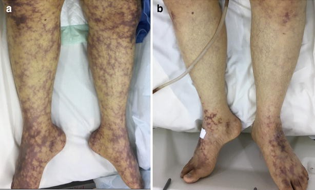
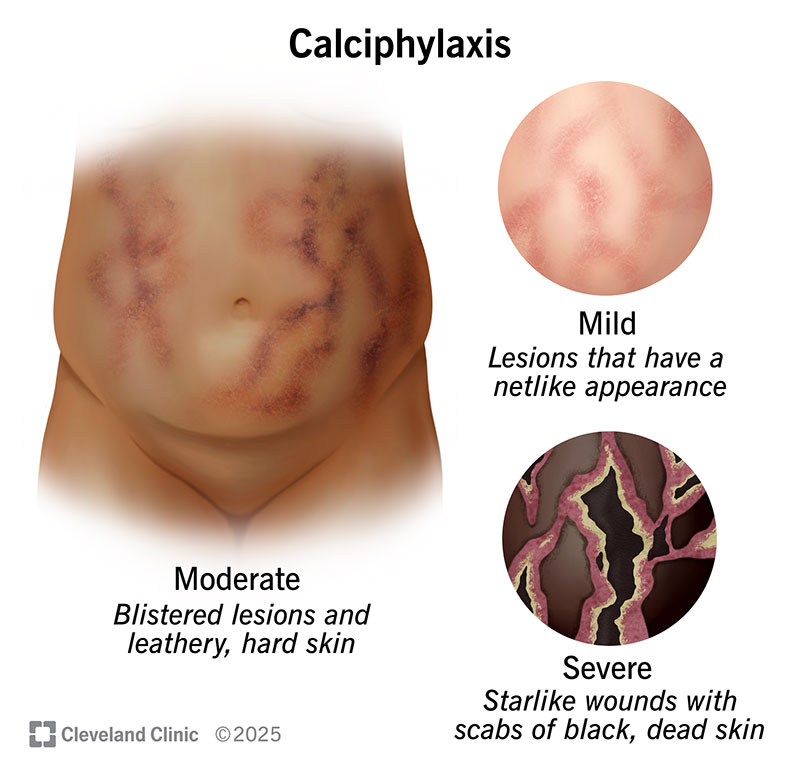
Cutaneous manifestations: Painful skin lesions that progress to necrotic ulcers. Lesions are commonly located on the thighs, abdomen, and buttocks. Surrounding skin may appear violaceous or mottled.

Systemic features: Fever and malaise. Secondary bacterial infections, which may lead to sepsis.

Metabolic abnormalities: Persistent hyperphosphatemia and hypercalcemia. Elevated parathyroid hormone levels due to secondary hyperparathyroidism.

These symptoms often overlap with other conditions, complicating diagnosis.


Etiology

Calciphylaxis primarily results from a complex interplay of vascular injury, pro-calcification factors, and inflammatory processes.

Vascular calcification: Calcium-phosphate deposition occurs in the medial layer of small arteries. Matrix Gla-protein inhibition contributes to unregulated calcification.

Inflammation and endothelial dysfunction: Chronic inflammation promotes endothelial injury, enhancing calcium deposition and thrombotic occlusion.

Pro-coagulant state: Thrombosis of microvasculature worsens ischemia, leading to necrotic skin lesions.

This pathophysiology underscores the systemic nature of calciphylaxis and its high morbidity and mortality.


Pathophysiology

The pathogenesis of calciphylaxis involves several interconnected mechanisms:

Calcium-phosphate imbalance: Hyperphosphatemia and hypercalcemia lead to vascular smooth muscle cell transdifferentiation into osteoblast-like cells, promoting calcification.

Vascular injury and thrombosis: Endothelial injury and hypercoagulability contribute to thrombosis and ischemia in small arterioles.

Inflammatory processes: Cytokine release exacerbates vascular damage and calcification.

Adipose tissue involvement: Calcification predominantly affects arterioles within subcutaneous adipose tissue, leading to skin necrosis.

These processes result in ischemic tissue damage, non-healing ulcers, and systemic complications.


DSM-5 Diagnosis

As calciphylaxis is not a psychiatric condition, it is not classified under the DSM-5.


Diagnosis

Diagnosis of calciphylaxis is primarily clinical but may require supportive investigations.

Clinical evaluation:

Assessment of painful skin lesions and systemic symptoms.

Risk factors such as ESRD, diabetes, and hyperparathyroidism should be evaluated.

Laboratory tests:

Elevated calcium-phosphate product.

High parathyroid hormone levels.

Inflammatory markers such as C-reactive protein.

Imaging:

Plain X-rays or CT scans may reveal vascular calcification.

Bone scintigraphy highlights areas of calcification and inflammation.

Skin biopsy:

Confirms diagnosis by showing calcification of small vessel walls and thrombotic occlusion.

Biopsy carries a risk of poor wound healing.

Prompt recognition and diagnosis are essential for initiating effective treatment.


Treatment Regimens

Calciphylaxis management involves a combination of medical, surgical, and supportive interventions.

Medical therapies:

Sodium thiosulfate:

Chelates calcium, reduces oxidative stress, and improves wound healing.

Vitamin K supplementation:

Restores matrix Gla-protein function, inhibiting vascular calcification.

Parathyroidectomy:

Indicated for refractory secondary hyperparathyroidism.


Dialysis modifications:

Use of low-calcium dialysate to reduce calcium-phosphate product levels.

Intensified dialysis to improve phosphate clearance.

Wound care:

Aggressive management of skin ulcers with debridement and infection control.

Analgesia:

Multimodal pain management, including opioids and non-opioid analgesics.

Timely and multidisciplinary care is crucial for improving survival and quality of life.


Patient Education

Educating patients about calciphylaxis is essential for promoting adherence to treatment and preventing complications.

Risk factor modification: Adherence to dietary phosphate restrictions and prescribed medications. Avoiding calcium-based phosphate binders.

Early recognition: Prompt reporting of new skin lesions or worsening pain to healthcare providers.

Lifestyle modifications: Maintaining a healthy weight and managing diabetes. Engaging in smoking cessation to improve vascular health.

Support groups and counseling services can help patients cope with the psychological burden of this chronic condition.


Additional Considerations

Complications: Non-healing ulcers and secondary infections. Sepsis is a leading cause of mortality in calciphylaxis.

Prognosis: High mortality rates, exceeding 50% in some cohorts. Early diagnosis and aggressive treatment improve outcomes.

Research and future directions: Investigating novel therapies targeting vascular calcification pathways. Evaluating the role of emerging biomarkers in early diagnosis.


Conclusion

Calciphylaxis is a complex and life-threatening condition requiring a multidisciplinary approach to diagnosis and treatment. The interplay of metabolic, inflammatory, and vascular factors underpins its pathogenesis, resulting in severe morbidity and high mortality rates. Comprehensive management strategies, including medical therapy, dialysis modifications, and patient education, can significantly improve outcomes. Continued research into innovative treatment modalities holds promise for reducing disease burden and improving patient survival.


References

Brandenburg, V. M., & Kramann, R. (2020). Calciphylaxis: A still unmet challenge. Clinical Kidney Journal, 13(2), 282-284. https://doi.org/10.1093/ckj/sfz178

Nigwekar, S. U., et al. (2018). Calciphylaxis: Risk factors, diagnosis, and treatment. American Journal of Kidney Diseases, 66(1), 133-146. https://doi.org/10.1053/j.ajkd.2015.12.030

Weenig, R. H. (2017). Calciphylaxis: Current concepts in pathogenesis, diagnosis, and treatment. Journal of the American Academy of Dermatology, 56(4), 569-579. https://doi.org/10.1016/j.jaad.2006.08.064

National Kidney Foundation. (2023). Understanding calciphylaxis. https://www.kidney.org/atoz/content/calciphylaxis

U.S. National Library of Medicine. (2023). Calciphylaxis: Overview and management. https://medlineplus.gov/calciphylaxis.html

 
Do you need a similar assignment done for you from scratch? Order now!
Use Discount Code "Newclient" for a 15% Discount!

Nursing Paper Example on Burkitt Lymphoma

Nursing Paper Example on Burkitt Lymphoma

Burkitt lymphoma is a rare but highly aggressive type of non-Hodgkin lymphoma that originates from B lymphocytes. It is characterized by rapid proliferation, making early diagnosis and treatment critical for survival. Burkitt lymphoma is most commonly associated with the Epstein-Barr virus (EBV) and occurs in three clinical variants: endemic, sporadic, and immunodeficiency-associated forms. Despite its aggressiveness, it is curable with prompt and appropriate treatment.


Nursing Paper Example on Burkitt Lymphoma

Causes of Burkitt Lymphoma

Burkitt lymphoma arises from genetic and environmental factors that drive abnormal cell growth.

Genetic mutations: Translocation of the MYC oncogene, most frequently t(8;14)(q24;q32). This translocation juxtaposes MYC with the immunoglobulin heavy-chain enhancer, leading to uncontrolled cell proliferation.

Viral infections: Epstein-Barr virus (EBV) infection is strongly associated with the endemic form and some sporadic cases. The virus promotes cell proliferation and evasion of apoptosis.

Immunosuppression: Occurs in individuals with HIV/AIDS or those undergoing immunosuppressive therapy.

Understanding these causes helps in identifying at-risk populations and informing targeted interventions.


Signs and Symptoms

The clinical presentation of Burkitt lymphoma varies depending on the subtype and disease stage.

Endemic Burkitt Lymphoma: Common in African children. Frequently presents as a rapidly growing jaw or facial bone tumor. Abdominal swelling may occur due to visceral organ involvement.

Sporadic Burkitt Lymphoma: Predominantly affects the abdomen, presenting with: Abdominal pain and swelling. Bowel obstruction or gastrointestinal bleeding. Enlarged liver or spleen.

Immunodeficiency-Associated Burkitt Lymphoma: Occurs in HIV-positive individuals. May involve lymph nodes, central nervous system (CNS), or bone marrow.

Other general symptoms include fever, night sweats, unexplained weight loss, and fatigue.


Etiology

Burkitt lymphoma originates from B lymphocytes within germinal centers of lymphoid tissues.

MYC translocations: Abnormal activation of the MYC gene is central to Burkitt lymphoma development. It drives unregulated cellular proliferation and metabolism.

Role of EBV: The virus infects B lymphocytes, inducing their proliferation. Latent membrane protein 1 (LMP1) expressed by EBV mimics signaling pathways of the tumor necrosis factor receptor, promoting lymphomagenesis.

HIV and immunosuppression: Chronic immunosuppression impairs the ability to control EBV-infected cells, increasing lymphoma risk.

This multifactorial etiology highlights the complex interplay between genetic mutations, infectious agents, and immune dysregulation.


Pathophysiology

The pathogenesis of Burkitt lymphoma involves several key mechanisms:

MYC gene dysregulation: The translocation of the MYC oncogene leads to overexpression of MYC protein, promoting cellular proliferation and survival.

Metabolic reprogramming: MYC overexpression enhances glycolysis, even in the presence of oxygen (Warburg effect), to meet the energy demands of rapidly dividing cells.

Immune evasion: EBV infection facilitates immune evasion by downregulating immune surveillance mechanisms.

Tumor microenvironment: The release of cytokines and growth factors within lymphoid tissues further supports tumor growth and invasion.

These processes underscore the aggressive nature and rapid progression of Burkitt lymphoma.


DSM-5 Diagnosis

As Burkitt lymphoma is not a psychiatric condition, it is not classified under the DSM-5. Its diagnosis relies on clinical, histological, and molecular criteria.


Diagnosis

Accurate diagnosis of Burkitt lymphoma involves a multi-modal approach:

Clinical evaluation: Assessment of symptoms and risk factors, such as geographic location or immunosuppressive conditions.

Laboratory tests: Complete blood count (CBC) may reveal cytopenias or elevated lactate dehydrogenase (LDH) levels. Peripheral blood smear may show circulating lymphoma cells.

Imaging studies: CT or PET scans identify tumor location and extent.

Tissue biopsy: Histopathology shows a “starry sky” pattern due to macrophages interspersed among rapidly dividing tumor cells. Immunohistochemistry confirms markers such as CD10, CD20, and BCL6.

Cytogenetic and molecular testing: Fluorescence in situ hybridization (FISH) detects MYC translocations.  PCR or EBV serology confirms viral association.

Early and accurate diagnosis is crucial for initiating effective treatment.


Treatment Regimens

Burkitt lymphoma requires aggressive treatment due to its rapid progression.

Chemotherapy: High-intensity, multi-agent regimens such as CODOX-M/IVAC or R-CHOP are standard. CNS prophylaxis with intrathecal chemotherapy is essential to prevent relapse.

Targeted therapy: Rituximab, an anti-CD20 monoclonal antibody, is commonly added to chemotherapy regimens to improve outcomes.

Supportive care: Management of tumor lysis syndrome with hydration and medications like allopurinol or rasburicase. Antiviral therapy for EBV-positive cases in immunosuppressed individuals.

Stem cell transplantation: Considered in refractory or relapsed cases.

Prompt initiation of therapy significantly improves survival rates, even in advanced stages.


Patient Education

Educating patients about Burkitt lymphoma enhances adherence to treatment and aids early recognition of relapse.

Awareness of symptoms: Recognize signs of relapse, such as new lymphadenopathy or systemic symptoms.

Importance of follow-up: Regular follow-up with oncologists to monitor for recurrence or late treatment effects.

Lifestyle modifications: Maintaining a healthy immune system through balanced nutrition and infection prevention strategies.

Support groups and counseling services can help address psychological impacts of the disease and its treatment.


Additional Considerations

Complications: CNS involvement and tumor lysis syndrome are common complications requiring prompt management.

Prognosis: Cure rates exceed 80% with appropriate treatment, especially in localized cases. Advanced-stage disease or delayed treatment initiation worsens prognosis.

Research developments: Ongoing studies on novel targeted therapies and immunotherapies hold promise for improving outcomes further.


Conclusion

Burkitt lymphoma is a highly aggressive but treatable cancer with distinct clinical and molecular features. Early recognition, accurate diagnosis, and prompt initiation of intensive chemotherapy regimens are key to achieving favorable outcomes. Comprehensive patient education and supportive care enhance treatment adherence and quality of life. Continued research into molecular pathways and novel therapies may further improve prognosis and reduce disease burden globally.


References

Ferry, J. A. (2020). Burkitt lymphoma: Clinicopathologic features and differential diagnosis. American Journal of Clinical Pathology, 153(6), 682-694. https://doi.org/10.1093/ajcp/aqaa080

IARC Working Group. (2020). Epstein-Barr virus and lymphoma: Mechanistic insights. IARC Scientific Publications, 254(3), 55-70. https://www.iarc.who.int/news-events/epstein-barr-virus-and-lymphoma/

Leukemia & Lymphoma Society. (2023). Burkitt lymphoma: Overview and treatment. https://www.lls.org/lymphoma/non-hodgkin-lymphoma/burkitt-lymphoma

National Cancer Institute. (2023). Burkitt lymphoma treatment (PDQ®)–Health professional version. https://www.cancer.gov/types/lymphoma/hp/burkitt-treatment-pdq

Wilson, W. H., et al. (2021). Advances in the management of Burkitt lymphoma. Blood, 137(4), 454-461. https://doi.org/10.1182/blood.2020008336

 
Do you need a similar assignment done for you from scratch? Order now!
Use Discount Code "Newclient" for a 15% Discount!