Discussion Post Change Agent

Discussion Post Change Agent

How I see yourself as a change agent guiding others to move from a process focus to outcomes focus

Change is rapid, disruptive, and expected in the nursing environment. Most changes are adopted to improve healthcare processes and their efficiency to optimize outcomes, which is the primary focus of most healthcare organizations today. As care delivery becomes more patient-centered, there is a shift to outcomes rather than processes because patients are more concerned about their health outcomes than the processes adopted to achieve them. Therefore, I see myself as a change agent when I advocate for my patients and adopt positive changes like technology and evidence-based practice that are shaping the efficacy of the healthcare system (Charlotte Davis, 2019). I encourage reception to change because it is imminent and often unpredictable.

Discussion Post Change Agent

I collaborate and communicate with my team to identify barriers and actors affecting the efficiency and effectiveness of healthcare processes and people. I invite collective and meaningful engagement to develop solutions or interventions to improve processes and people to optimize patient and organizational outcomes. I also engage the public, colleagues, and policy-makers regarding healthcare policies that can help address issues such as understaffing, resource acquisition and availability, healthcare affordability, healthcare access, and adverse events that impact patient and organization outcomes (Rafferty, 2018). Driving these changes helps efforts to improve patient outcomes, including safety, satisfaction, and experience.

I am a registered nurse and see myself as an educator in the field, educating my patients, colleagues, and new nurses throughout my daily shifts. I like sharing information that can help improve service delivery and patient outcomes. I engage colleagues and patients on factors affecting care and perform environmental screening to identify these factors and potential solutions. I emphasize patient education because it is the best preventative measure that seeks to empower patients to take control of their health and advocate for themselves. I educate patients regarding healthy habits and lifestyles and attitudes towards treatment adherence and seeking healthcare services proactively.

(Discussion Post Change Agent)

The rationale behind the change from process to outcomes

Many healthcare actors, especially patients, financiers, and health insurance companies, adopt outcome measures rather than process measures to evaluate the performance of healthcare organizations and practitioners. The healthcare reforms have seen a shift to patient-centered, holistic care, value-based systems, and high-reliability organizations, all of which appreciate outcomes, the byproduct of effective and efficient processes and people (Pantaleon, 2019). There is also an emphasis on the relationship between healthcare services and health outcomes. This shift is behind the change from process to outcome.

References

Charlotte Davis, B. S. N. (2019). Change agents wanted. Nursing Made Incredibly Easy.

Pantaleon L. (2019). Why measuring outcomes is important in health care. Journal of veterinary internal medicine33(2), 356–362. https://doi.org/10.1111/jvim.15458

Rafferty A. M. (2018). Nurses as change agents for a better future in health care: the politics of drift and dilution. Health economics, policy, and law13(3-4), 475–491. https://doi.org/10.1017/S1744133117000482

 
Do you need a similar assignment done for you from scratch? Order now!
Use Discount Code "Newclient" for a 15% Discount!

Parse s Human Becoming Theory Case Study Assignment

 Parse’s Human Becoming Theory Case Study Assignment

 Introduction

(Parse s Human Becoming Theory Case Study Assignment)

Nurses’ primary role is to help patients and families through their illnesses and recovery journey. Nursing theory guides nursing practice and provides insights into how nurses should relate to patients and what approach they should take to ensure the utmost patient experience. Hospice nursing is an area demanding compassion and high-level interpersonal skills. Hospice nurses emphasize care, comfort, and quality of life as individuals approach the end of life. Ann is at a point where it is impossible to care for her illnesses, and the benevolent thing would be to ensure she is as comfortable as possible. The theory of human becoming is suitable in such situations as its main emphasis is the quality of life and the consideration of the wholeness of an individual, including their spirituality and perception of life and death. This paper analyzes the application of human Becoming theory to a case study involving hospice care, the theory’s characteristics, strengths, and weaknesses, challenges in healthcare that call for applying this theory, and alternative theories that can be applied in similar situations.

(Parse s Human Becoming Theory Case Study Assignment)

How the Nurse Should Actualize Parse’s Theory of Human Becoming

The human becoming theory emphasizes the wholeness of an individual and the adoption of holistic care that integrates biological, psychological, sociological, cultural, and spiritual aspects. According to the theory, people cannot be separated from the environment because they continuously interact with it (Mitchell, 2019). The nurse has to adopt the themes of meaning, rhythmicity, and transcendence to actualize the theory. The nurse has to be present, listen to Ann and Ben, invite or ask open-ended questions, understand the patient’s and family’s perception of the situation, and develop alternative meanings and paths for the patient and family. It is possible when the nurse begins by establishing a therapeutic and strong nurse-patient relationship, which views the patient as a whole person living experiences via their environment rather than focusing on fixing problems. With a strong nurse-patient relationship, the nurse can help the patient find personal meaning in this situation and visualize the reality per the patient’s experiences with the environment (Barros et al., 2017). In this case study, Ann has lived a long life as a woman, mother, and wife, and the husband describes some of the experiences Ann had with her environment. Through these experiences, the hospice nurse can help the patient find personal meaning in life as a mother and wife and other lived experiences she admires and appreciates.

The nurse must help the patient and family achieve rhythmicity or develop patterns relating to the universe or the environment. Rhythmicity is about people co-creating with the environment in rhythmic patterns (Barros et al., 2017). It is about the nurse acknowledging the values and beliefs of Ann and Ben. For instance, the husband mentions that their daughter died giving birth. Once, they saw two shooting starts and believed they represented their daughter and the baby. The husband believes in life after death, and Ann will join their daughter and grandchild. It implies that the family is spiritual and relates to the universe. The nurse should respect these beliefs, guide the patient and the family through their spirituality, and complement Ben’s belief that the two shooting stars are their daughter and baby, and Ann will join them as the third shooting starts. This way, Ben will feel that their beliefs and spirituality are respected and understood, fostering the care experience. The end of life can be a lonely and frightening journey. The nurse must transcend with the patient or create an environment that allows transcendence, which is going beyond the limits an individual sets (Barros et al., 2017). The nurse should accompany the patient as she travels beyond the limits and be with her throughout the end-of-life journey. It is possible through spiritual care, which involves trust in transcendent. The ultimate end should be the connection with reality and helping the patient and family, Ben, in this case, let go and separate from life because it has been lived. The nurse should guide the process of reflecting and detaching to ensure a successful and comfortable transition from life.

(Parse s Human Becoming Theory Case Study Assignment)

 Parse s Human Becoming Theory Case Study Assignment

Characteristics, Strengths, and Weaknesses of Human Becoming Theory

Characteristics and Assumptions of Human Becoming Theory

The theory of Human becoming guides nurses to focus on the quality of life from each individual’s perspective. It combines biological, psychological, sociological, and spiritual attributes and perceives a person as a unitary actor in pervasive interaction with their environment. The theory is founded around the primary themes of meaning, rhythmicity, and transcendence (Barros et al., 2017). The theory assumes that people coexist while co-creating rhythmic patterns with the universe; people are open, free choosing meaning in situations and are responsible for decisions made; a person is a unitary, persistently co-creating relational patterns; and people are transcending multidimensionally with the possibles. It describes a person as more than the sum of the parts and cannot be separated from the environment. The theory perceives nursing as a human science and an art of caring that adopt an abstract body of knowledge to help individuals (Barros et al., 2017). It encourages nurses to form strong bonds and relationships with their patients because the focus is not on fixing problems, but rather quality of life.

Strengths and Weaknesses of Human Becoming Theory

The theory of Human Becoming separates nursing from other fields, guiding care and valuable administration. The theory adopts the humanistic and interactive approach that perceives people as humans before patients. The theory also encourages individuals to explore their views of their unique experiences, and it does not seek to fix problems but instead maintain the quality of life. However, the research linked to this theory is perceived as a closed circle, and the results are unquantifiable (Marginson, 2019). Comparing results to other research studies is challenging due to the lack of a control group or standardized questions. Furthermore, the Human Becoming theory does not adopt the nursing process and invalidates the idea that each individual participates in a unique lived experience. The theory is inaccessible to new nurses and is challenging to apply in acute, emergent care.

Challenges Existing for Healthcare Institutions to Switch to This Nursing Approach

Healthcare institutions face issues relating to the quality of care, compassion fatigue, patient satisfaction, and scope of nursing and identity (Oleribe et al., 2019). The Human Becoming Theory can be applied to address these challenges. The theory describes nursing as a human science and an art of caring. It emphasizes the quality of life, achieved through proper, holistic, and interactive care. It emphasizes nurses developing therapeutic relationships with their patients, which can aid in understanding patients better, including their needs, preferences, beliefs, and values, and integrating these into healthcare plans to ensure the utmost patient experience and positive patient outcomes. Additionally, therapeutic relationships have proven effective in addressing compassion fatigue, a barrier to quality of care and patient satisfaction and experience with care.

How Parse \’S Understanding of Transcendence Might Guide the Nurse, As Ann \’S death Became a Reality to Ben

Parse’s theory describes transcendence as the human going beyond reality and the desired hopes and dreams via pushing-resisting to create alternatives to viewing reality. Per this theme or principle, the human can create a path moving from the current moment to one that is not yet here. The person can overcome destitution, change views, and start a new life. The understanding of transcending can help the nurse aid Ben in detaching and separating from a life already lived and starting a new one (Pentaris & Tripathi, 2022). The nurse will help Ben reflect on the life already lived and detach from it as Ann’s death becomes more of a reality. The nurse will be present and demonstrate compassion, give hope and affirmation to Ben of life as fruitful, and help him find meaning in existence without Ann.

Alternative Nursing Theory and Plan of Care

Watson’s Theory of Transpersonal Care is also applicable to this case study. The theory focuses on how nurses express care to their clients, emphasizing humanistic nursing aspects as they interact with scientific knowledge and nursing practice. Per the theory, nursing care is about health promotion, prevention of illness, caring for the sick, and restoring health (Aghaei et al., 2020). In hospice care, only the role of caring for the sick applies and should be actualized through a holistic approach that takes into account spirituality. It persists that the human should be valued, respected, nurtured, understood, and care for as a functional integrated self.

A plan of care linked to this case study would focus on grieving and death anxiety as Ann breathes her last. Ben might be experiencing death anxiety and grieving that can be related to the perceived death of Ann, evidenced by worrying about the impact of Ann’s death, powerlessness over Ann’s death, and the fear of the process of dying. The desired outcome would be that Ben identifies and expresses feelings appropriately, continues with normal living and plans for the future, verbalizes understanding of the dying process and associated feelings, and demonstrates personal empowerment in spiritual strengths to find purpose and meaning in life, loss, and grief. The Human Becoming theory’s primary themes of meaning and transcending and the Transpersonal care theory’s assumption of nursing as a holistic approach that considers spirituality and spiritual practice would be the theoretical underpinning of this care plan. The nurse would assess the patient’s view of life and death and monitor signs of hopelessness, expression of anger, guilt, despair, and intrapersonal conflict. The nurse should also determine how Ben will understand and respond to death. Possible interventions include creating an open, nonjudgmental environment, encouraging Ben to verbalize thoughts and concerns like sadness and anger and acknowledge that these feelings are normal, and developing a trusting relationship with Ben to help him appropriately cope and continue with life.

(Parse s Human Becoming Theory Case Study Assignment)

Conclusion

Nursing focuses on care for individuals, families, and communities to help attain, maintain, or recover optimal health and achieve a quality life. According to the Human Becoming theory, nursing is differentiated from other healthcare dimensions because the primary focus is not solving problems but rather the quality of life. Human becoming theory can be actualized by being present and compassionate, offering hope and affirmation, helping patients and families find meaning in life, reflect on their experiences, and transcend. The theory of Transpersonal Care is an alternative to the Human Becoming theory and also adopts the holistic approach to care.

References

Aghaei, M. H., Vanaki, Z., & Mohammadi, E. (2020). Watson’s human caring theory-based palliative care: a discussion paper. International Journal of Cancer Management13(6).

Barros, L. B. F., Silva, L. D. F. D., Guedes, M. V. C., & Pessoa, V. L. M. D. P. (2017). Clinical care of nursing reasoned in Parse: Contribution in the transcendence process of cardiac transplantation. Revista Gaucha De Enfermagem38.

Marginson, S. (2019). Limitations of human capital theory. Studies in Higher Education44(2), 287-301.

Mitchell, G. J. (2019). Chapter 24: Humanbecoming. https://nursekey.com/humanbecoming/

Oleribe, O. O., Momoh, J., Uzochukwu, B. S., Mbofana, F., Adebiyi, A., Barbera, T., Williams, R., & Taylor-Robinson, S. D. (2019). Identifying Key Challenges Facing Healthcare Systems In Africa And Potential Solutions. International journal of general medicine12, 395–403. https://doi.org/10.2147/IJGM.S223882

Pentaris, P., & Tripathi, K. (2022). Palliative Professionals’ Views on the Importance of Religion, Belief, and Spiritual Identities toward the End of Life. International Journal of Environmental Research and Public Health19(10), 6031.

 
Do you need a similar assignment done for you from scratch? Order now!
Use Discount Code "Newclient" for a 15% Discount!

Nursing Paper Example on Juvenile Idiopathic Arthritis

Nursing Paper Example on Juvenile Idiopathic Arthritis

Juvenile idiopathic arthritis (JIA) is the most common type of arthritis in children. It is characterized by chronic inflammation of the joints, leading to pain, stiffness, and swelling. JIA can affect one or multiple joints and may be associated with systemic features. The disease primarily affects children under 16 years of age and can vary in severity. Early diagnosis and effective management are critical to reducing long-term complications and improving the quality of life for affected children.

Nursing Paper Example on Juvenile Idiopathic Arthritis

Causes

The exact cause of juvenile idiopathic arthritis remains unknown, but it is believed to result from a combination of genetic predisposition and environmental factors. Genetic studies have shown associations with specific genes, such as those related to the immune system, including the human leukocyte antigen (HLA) complex. These genes may make certain children more susceptible to autoimmune responses.

Environmental triggers, such as infections or environmental toxins, may activate these underlying genetic factors, leading to the onset of JIA. However, no single environmental factor has been definitively linked to the disease.

Signs and Symptoms

The symptoms of juvenile idiopathic arthritis can vary, but common features include joint pain, swelling, and stiffness. In some cases, the inflammation is limited to one or a few joints (oligoarticular JIA), while in other cases, multiple joints may be affected (polyarticular JIA). Morning stiffness that improves with movement is a hallmark symptom. The affected joints may appear swollen and warm to the touch.

In addition to joint symptoms, children with JIA may also experience systemic signs such as fever, fatigue, and weight loss. In more severe cases, there may be growth disturbances, as well as damage to the joints leading to functional impairment.

(Nursing Paper Example on Juvenile Idiopathic Arthritis)

Etiology

The etiology of juvenile idiopathic arthritis is complex and involves both genetic and environmental factors. It is thought that an abnormal immune response triggers inflammation in the joints. In children with JIA, the immune system mistakenly attacks healthy tissues, particularly the synovium—the lining of the joints. This leads to chronic inflammation and damage.

Genetic factors, such as the presence of certain HLA genes, play a significant role in determining susceptibility to JIA. Environmental factors like infections or trauma may also serve as triggers, but more research is needed to fully understand their role. The exact mechanism by which these factors lead to the development of JIA is still under investigation.

Pathophysiology

The pathophysiology of juvenile idiopathic arthritis involves an autoimmune response in which the body’s immune system attacks its own tissues, particularly the synovial lining of the joints. This results in inflammation, characterized by the infiltration of immune cells such as T-cells and macrophages into the synovium. These immune cells produce cytokines and other inflammatory mediators that contribute to further joint damage.

The chronic inflammation leads to the destruction of cartilage and bone, causing joint deformities and pain. In some cases, the inflammation may also affect other organs, including the eyes, leading to uveitis, or the heart and lungs. The persistence of inflammation can also affect growth and development, especially in children with systemic forms of JIA.

DSM-5 Diagnosis

There is no specific diagnosis of juvenile idiopathic arthritis in the Diagnostic and Statistical Manual of Mental Disorders (DSM-5), as JIA is a physical condition and not a psychiatric disorder. The diagnosis is primarily clinical, based on the patient’s medical history, physical examination, and laboratory tests. The American College of Rheumatology (ACR) criteria are commonly used to classify JIA, which involves the presence of persistent arthritis lasting for at least six weeks in a child under the age of 16.

Blood tests may reveal elevated levels of markers of inflammation, such as C-reactive protein (CRP) and erythrocyte sedimentation rate (ESR). Imaging studies, such as X-rays or MRI, may be used to assess joint damage. A thorough clinical assessment is essential to distinguish JIA from other pediatric inflammatory conditions.

(Nursing Paper Example on Juvenile Idiopathic Arthritis)

Treatment Regimens

Treatment of juvenile idiopathic arthritis focuses on controlling inflammation, relieving pain, and preventing joint damage. The primary approach involves the use of nonsteroidal anti-inflammatory drugs (NSAIDs) to reduce pain and inflammation. If NSAIDs are not sufficient, disease-modifying antirheumatic drugs (DMARDs), such as methotrexate, are often prescribed to slow disease progression.

In cases where inflammation is more severe, biologic therapies like tumor necrosis factor (TNF) inhibitors or interleukin-1 inhibitors may be recommended. Corticosteroids are sometimes used to control flare-ups, but their long-term use is limited due to potential side effects.

Physical therapy is an important component of treatment to maintain joint function and mobility. In some cases, surgery may be needed to correct joint deformities or to replace damaged joints. The treatment regimen is tailored to each child based on the severity of the disease and their response to therapy.

Patient Education

Educating parents and caregivers about juvenile idiopathic arthritis is crucial for effective management of the disease. It is important to emphasize the need for early diagnosis and treatment to prevent joint damage and improve the child’s quality of life. Parents should be instructed on how to administer medications, including NSAIDs and DMARDs, and to watch for any potential side effects. Encouraging regular physical activity and exercise is vital to maintain joint function and flexibility, but activities should be adjusted based on the child’s pain and limitations.

Families should be informed about the potential complications of JIA, such as uveitis or growth disturbances, and the importance of routine screenings for these issues. Regular follow-ups with pediatric rheumatologists are essential to monitor disease progression and adjust treatment as needed. Emotional support is also necessary, as chronic illness can be challenging for both the child and their family.

(Nursing Paper Example on Juvenile Idiopathic Arthritis)

Conclusion

Juvenile idiopathic arthritis is a chronic autoimmune disorder that affects children, leading to joint inflammation and pain. Although the exact cause remains unclear, genetic and environmental factors are thought to play a role in its development. Early diagnosis and treatment are essential to managing symptoms, preventing joint damage, and improving the child’s quality of life. The treatment plan typically includes NSAIDs, DMARDs, biologics, and physical therapy, with the goal of controlling inflammation and maintaining joint function. Patient education plays a key role in managing the disease effectively and ensuring long-term health outcomes.

References

Beukelman, T., & Gertz, R. M. (2017). Juvenile idiopathic arthritis. The Journal of Rheumatology, 44(5), 835-845. https://doi.org/10.3899/jrheum.161060

Giannini, E. H., & Ruperto, N. (2020). Clinical management of juvenile idiopathic arthritis. The Lancet, 395(10217), 2431-2442. https://doi.org/10.1016/S0140-6736(20)31283-9

Société Française de Rhumatologie. (2019). Juvenile idiopathic arthritis: Diagnosis and management. Clinical Rheumatology, 38(5), 1101-1112. https://doi.org/10.1007/s10067-019-04337-3

 
Do you need a similar assignment done for you from scratch? Order now!
Use Discount Code "Newclient" for a 15% Discount!

Nursing Paper Example on Ichthyosis

Nursing Paper Example on Ichthyosis

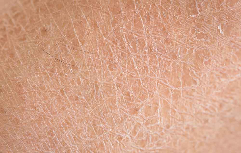
Ichthyosis refers to a group of skin disorders characterized by dry, thickened, and scaly skin. The condition can range from mild to severe, with varying degrees of impact on the patient’s quality of life. There are several forms of ichthyosis, including inherited types, which can manifest from birth, and acquired types, which develop later in life. The severity of symptoms varies widely, and treatment is focused on managing symptoms and improving skin appearance.

Nursing Paper Example on Ichthyosis

Causes

Ichthyosis is primarily caused by genetic mutations that affect the skin’s ability to retain moisture and regulate cell turnover. Inherited forms of ichthyosis are typically caused by mutations in specific genes, such as the keratin 1 and keratin 10 genes in ichthyosis vulgaris. These mutations lead to abnormal keratin production, which causes the skin cells to pile up, forming scales. Environmental triggers can also contribute to the condition, although the primary cause is genetic.

In some acquired forms, factors such as infections, metabolic disorders, and other systemic diseases may play a role. Inherited ichthyosis is often passed down in an autosomal dominant or recessive manner, depending on the specific form.

Signs and Symptoms

The most prominent symptom of ichthyosis is the presence of thickened, scaly skin that may cover large areas of the body. The scales can range from fine, powdery flakes to large, thick plaques that are often dry and cracked. Common sites of involvement include the limbs, torso, and scalp, although the condition can affect any part of the body. The severity of scaling varies and can worsen in cold weather or with dry skin conditions. In some forms of ichthyosis, the skin may be red, inflamed, or painful, especially during flare-ups.

Other symptoms may include itching, increased sensitivity to temperature changes, and skin fissures that can lead to secondary infections.

Etiology

Ichthyosis is caused by mutations in genes responsible for skin cell function. For example, ichthyosis vulgaris, the most common form, results from a mutation in the filaggrin gene. This protein is important for maintaining the skin’s barrier function, and its absence or dysfunction leads to skin dryness and scaling. X-linked recessive ichthyosis is another inherited form caused by mutations in the steroid sulfatase gene, which leads to a buildup of certain substances that impair skin turnover.

Other rarer forms of ichthyosis are caused by defects in proteins responsible for skin development or lipid metabolism. Acquired ichthyosis, while less common, can be associated with systemic conditions such as HIV, cancer, hypothyroidism, and sarcoidosis, all of which may lead to abnormal skin changes resembling ichthyosis.

Pathophysiology

The pathophysiology of ichthyosis involves a disruption in the normal processes of skin cell production, differentiation, and desquamation. In healthy skin, the stratum corneum, the outermost layer, is composed of dead skin cells that are continuously shed and replaced. In individuals with ichthyosis, abnormal keratinization leads to the accumulation of thickened skin cells, resulting in the formation of scales.

The defective genes responsible for ichthyosis disrupt the structure and function of the skin’s outer layer, impairing its ability to retain moisture and protect against environmental stressors. As a result, the skin becomes dry, rough, and prone to cracking. In some forms of ichthyosis, there may be an overproduction of lipids or altered lipid composition in the skin, further contributing to the formation of scales. This process not only affects the appearance of the skin but also its ability to function as a barrier, leaving individuals more vulnerable to infections and other complications.

DSM-5 Diagnosis

There is no specific diagnostic category for ichthyosis in the Diagnostic and Statistical Manual of Mental Disorders (DSM-5) since it is a dermatological condition rather than a psychiatric one. Diagnosis is based on clinical evaluation, including a thorough patient history and physical examination. In most cases, the characteristic appearance of the skin lesions is enough for diagnosis, although genetic testing can confirm the specific form of ichthyosis. In some cases, a skin biopsy may be performed to assess the thickness and composition of the skin layers.

Family history is also an important factor, as many forms of ichthyosis are inherited in an autosomal dominant or recessive pattern. Genetic counseling may be recommended for families affected by hereditary forms of ichthyosis to understand the inheritance pattern and potential risks for future generations.

Treatment Regimens

Management of ichthyosis focuses on alleviating symptoms and preventing complications, as there is currently no cure for the condition. Topical treatments, such as moisturizers, emollients, and keratolytics (e.g., urea, salicylic acid), are commonly used to hydrate the skin and reduce scaling. For more severe cases, prescription-strength creams containing tretinoin or corticosteroids may be used to help normalize skin cell turnover and reduce inflammation.

Oral retinoids such as acitretin may be prescribed for more widespread or severe cases, particularly in conditions like lamellar ichthyosis. In cases where ichthyosis is associated with an underlying systemic condition, treating the primary disease may help improve the skin symptoms.

It is also essential for patients to avoid triggers such as harsh soaps, hot water, and environmental irritants that can worsen dryness and scaling. Regular bathing with lukewarm water and the use of soap substitutes are often recommended to help maintain skin hydration.

Patient Education

Educating patients with ichthyosis about proper skin care is critical to managing the condition effectively. Regular moisturizing is essential, as it helps prevent the skin from becoming dry and cracked. Patients should be advised to apply moisturizers immediately after bathing to lock in moisture. Gentle skin care products, including non-soap cleansers and lukewarm water, should be used to avoid further irritation.

In addition, patients should avoid tight clothing and friction in areas where scaling is prominent, as this can worsen the condition. Sunscreen should be applied regularly to protect the skin from UV damage, as patients with ichthyosis may have increased sensitivity to the sun. Genetic counseling is recommended for patients with hereditary forms of ichthyosis to help them understand the potential risks for family members. Regular follow-up with a dermatologist is important to monitor disease progression and adjust treatment as needed.

Conclusion

Ichthyosis is a group of skin disorders characterized by dry, scaly skin due to defective skin cell production and differentiation. While the condition is often genetic in origin, environmental factors and systemic conditions can also contribute to the disease. Treatment focuses on symptom management, including moisturizing, keratolytics, and oral retinoids for severe cases. Patient education plays a key role in managing the condition, with advice on skin care, avoiding triggers, and protecting the skin from environmental damage. Despite the chronic nature of the disease, early intervention and appropriate treatment can significantly improve the quality of life for individuals affected by ichthyosis.

References

Tobin, D. J., & Gawkrodger, D. J. (2020). Ichthyosis: Clinical presentation and management. American Journal of Clinical Dermatology, 21(3), 347-357. https://link.springer.com/article/10.1007/s40257-020-00507-2

Zouboulis, C. C., & Bechara, F. G. (2021). Ichthyosis: Diagnosis, pathophysiology, and treatment. Dermatologic Clinics, 39(2), 167-183. https://doi.org/10.1016/j.det.2020.11.007

Bialynicki-Birula, R., & Wróblewska, A. (2020). The genetics of ichthyosis: Pathogenesis and clinical presentation. Journal of the European Academy of Dermatology and Venereology, 34(5), 1015-1024. https://doi.org/10.1111/jdv.16456

 
Do you need a similar assignment done for you from scratch? Order now!
Use Discount Code "Newclient" for a 15% Discount!

Nursing Paper Example on Hidradenitis Suppurativa

Nursing Paper Example on Hidradenitis Suppurativa

Hidradenitis suppurativa (HS) is a chronic skin condition characterized by the formation of painful, inflamed lumps and abscesses in areas where skin rubs together, such as the armpits, groin, buttocks, and under the breasts. This condition primarily affects hair follicles and sweat glands, leading to the development of lesions that may drain pus or blood. Over time, repeated flare-ups can result in scar tissue and tunnels under the skin. HS is often misdiagnosed or dismissed as simple acne or boils, making timely treatment critical to reducing complications.

Nursing Paper Example on Hidradenitis Suppurativa

Causes

The exact cause of HS is not fully understood, but genetic and environmental factors are believed to play a role. A major contributing factor is inflammation within the hair follicles and sweat glands. The condition has a strong genetic component, as it is often seen in families, suggesting an inherited predisposition.

Environmental triggers such as obesity, smoking, hormonal changes, and friction from tight clothing can exacerbate symptoms. Lifestyle factors, particularly smoking, are significant risk factors for the disease, worsening the severity and frequency of flare-ups. Additionally, alterations in immune system functioning are thought to play a role in the abnormal inflammatory response associated with HS.

While it is not classified as an autoimmune disorder, the body’s immune system appears to mistakenly attack the skin’s hair follicles, causing the inflammation and abscesses that define the disease. However, not all cases of HS are genetically determined, and environmental triggers can also play a key role in the development and exacerbation of the condition.

Signs and Symptoms

The hallmark symptoms of hidradenitis suppurativa are painful, inflamed lumps or abscesses in the skin’s folds, often leading to abscess formation and pus drainage. These lumps may rupture over time, releasing foul-smelling fluid. These painful lumps can recur in the same areas, causing the formation of tunnel-like structures under the skin called sinus tracts. Over time, repeated flare-ups lead to the thickening of the skin and the formation of scar tissue. The affected areas typically include the armpits, groin, buttocks, and under the breasts, but any skin fold can be involved.

Early-stage symptoms may present as small, pimple-like lumps that progress into larger abscesses. As the disease progresses, these areas may become swollen, red, and tender. Individuals may also experience an increased frequency of flare-ups, leading to chronic discomfort and even disability in severe cases. In addition to visible skin changes, patients with hidradenitis suppurativa may experience systemic symptoms such as fever and fatigue during active flare-ups.

Etiology

The precise etiology of hidradenitis suppurativa remains unclear, though it is thought to involve a combination of genetic, environmental, and immunological factors. A family history of the condition is a strong indicator of a genetic predisposition, suggesting that certain genetic mutations may contribute to its onset. Mutations in the NF-kB pathway, which regulates immune responses and inflammation, have been linked to HS. This pathway’s dysfunction can result in an exaggerated inflammatory response in the skin’s hair follicles and sweat glands.

Environmental factors, particularly smoking, obesity, and excessive friction or sweating, are known to increase the risk of HS flare-ups. Hormonal changes, especially those occurring during puberty or menstruation, are also considered important factors in the development of the disease, with many individuals noticing a worsening of symptoms during these times. The inflammation in HS is not caused by bacterial infection, though infected abscesses can develop secondary to the primary condition.

Pathophysiology

Hidradenitis suppurativa is believed to arise from a combination of hyperkeratinization and inflammation of the hair follicles, particularly in areas of friction. This inflammation leads to the blockage of sweat glands, followed by the formation of painful lumps and abscesses. Over time, these abscesses may rupture, causing the release of pus or blood. Repeated flare-ups lead to the formation of scar tissue, and the development of sinus tracts and fistulas, which are abnormal connections between skin surfaces.

The process is thought to involve an abnormal immune response where the body’s immune system, instead of protecting the skin, triggers inflammation in the affected areas. This chronic inflammation results in the progressive damage to skin structures and the underlying tissue.

Over time, individuals with HS may experience significant scarring, thickened skin, and the development of painful, interconnected tunnels under the skin. In severe cases, extensive scarring can lead to functional and aesthetic impairments, and complications such as secondary infections and mobility limitations may occur.

DSM-5 Diagnosis

Hidradenitis suppurativa is not specifically classified in the DSM-5, as it is a dermatologic condition rather than a psychiatric disorder. Diagnosis is primarily clinical and involves evaluating the patient’s symptoms, medical history, and physical examination findings. No definitive laboratory test or imaging is required for the diagnosis, but it may be confirmed through the identification of characteristic skin lesions in areas of friction.

In some cases, biopsy or culture may be necessary to rule out other conditions or confirm the presence of infection. The diagnosis is typically made based on the presence of recurrent, painful abscesses, sinus tracts, and scarring, often in the armpits, groin, buttocks, and under the breasts. In some cases, associated systemic symptoms such as fever or malaise may also aid in diagnosis.

Treatment Regimens

Management of hidradenitis suppurativa focuses on controlling inflammation, preventing infection, and reducing the frequency and severity of flare-ups. Mild cases may be treated with topical antibiotics or corticosteroids to reduce inflammation and prevent infection. In more severe cases, oral antibiotics such as tetracycline or doxycycline may be prescribed to reduce bacterial colonization and inflammation.

Additionally, biologic agents targeting the immune system, such as adalimumab, have shown promise in reducing flare-ups and managing the condition long-term. Surgical intervention may be necessary in advanced cases to remove abscesses or to drain cysts and sinus tracts. In some instances, patients may require skin grafts to restore the affected areas after significant scarring.

Lifestyle changes, including weight management and smoking cessation, are essential components of treatment to reduce flare-up frequency and severity. Pain management may also be part of the treatment regimen, with nonsteroidal anti-inflammatory drugs (NSAIDs) or opioids being used in severe cases. Early intervention is key to preventing long-term complications and improving quality of life.

Patient Education

Patient education is an integral part of managing hidradenitis suppurativa. Educating patients on the nature of the disease, its triggers, and the importance of early intervention can help reduce the severity of symptoms and improve outcomes.

Smoking cessation is strongly recommended, as tobacco use is a major risk factor for flare-ups. Patients should also be educated on the role of weight management in controlling the condition, as obesity can exacerbate symptoms. In addition, patients should be instructed on proper skin hygiene to reduce irritation and the risk of infection in affected areas. For those with recurrent flare-ups, it is important to avoid tight clothing or friction in areas prone to the condition.

Medications prescribed for HS, including antibiotics or biologics, should be taken as directed, and patients should be aware of potential side effects. In cases of severe disease, patients should be informed about the potential need for surgery, and psychological support may be necessary to address the emotional and social challenges associated with living with a chronic skin condition.

Conclusion

Hidradenitis suppurativa is a chronic and often painful condition that primarily affects areas where skin rubs together, such as the armpits and groin. Although the exact cause remains unclear, it is believed to involve a combination of genetic, environmental, and immunological factors. Early intervention with lifestyle modifications, medications, and, when necessary, surgery, can help manage the disease and improve the quality of life for affected individuals. Educating patients on the importance of avoiding triggers, maintaining proper hygiene, and adhering to treatment plans is essential to managing the condition effectively.

References

Ting, T. Y., & Tey, H. L. (2021). Hidradenitis suppurativa: Epidemiology, pathogenesis, and diagnosis. Journal of Clinical Medicine, 10(11), 2345. https://www.ncbi.nlm.nih.gov/pmc/articles/PMC8002679/

Zouboulis, C. C., & Bechara, F. G. (2020). Hidradenitis suppurativa: Current concepts in pathogenesis and treatment. Journal of Dermatology, 47(7), 712-725. https://doi.org/10.1111/1346-8138.15431

Saunte, D. M. L., & Jemec, G. B. E. (2020). Hidradenitis suppurativa: Advances in diagnosis and treatment. Journal of the American Academy of Dermatology, 82(2), 323-333. https://doi.org/10.1016/j.jaad.2019.06.036

 
Do you need a similar assignment done for you from scratch? Order now!
Use Discount Code "Newclient" for a 15% Discount!

Nursing Paper Example on Henoch-Schönlein purpura

Nursing Paper Example on Henoch-Schönlein purpura

Henoch-Schönlein purpura (HSP) is a form of small-vessel vasculitis that most commonly affects children but can also occur in adults. The disease is characterized by the deposition of immunoglobulin A (IgA) complexes in the blood vessels, which leads to inflammation. HSP typically presents with a purpuric rash, joint pain, abdominal pain, and kidney involvement. The pathophysiology of the disease is complex and not fully understood, though it is often triggered by infections, medications, or other environmental factors.

Nursing Paper Example on Henoch-Schönlein purpura

Causes

Henoch-Schönlein purpura is often triggered by a variety of factors, with infections being the most common cause. Upper respiratory infections, particularly those caused by viral agents such as the adenovirus, parvovirus, and influenza, are frequently linked to the onset of HSP. Streptococcus infections, often in the form of strep throat, are another common trigger.

In addition to infections, certain medications have been implicated in triggering the disease. These include nonsteroidal anti-inflammatory drugs (NSAIDs), antibiotics, and vaccinations, which can induce immune responses that lead to the deposition of immunoglobulin A (IgA) in blood vessel walls.

Genetic factors also play a role in the development of HSP. Individuals with a family history of autoimmune diseases or vasculitis may be more susceptible to the condition. Research indicates that abnormalities in immune system regulation, particularly involving IgA, contribute to the pathogenesis of the disease.

Environmental factors, such as exposure to pollutants or changes in climate, may further contribute to the development of Henoch-Schönlein purpura in genetically predisposed individuals. However, the interplay between genetics, environmental factors, and immune dysregulation remains complex and not fully understood.

Signs and Symptoms

The primary symptom of Henoch-Schönlein purpura is the presence of purpura, which refers to purple spots on the skin caused by bleeding underneath. These spots typically appear on the lower extremities and buttocks, and may also be seen in the arms. The skin lesions can range from small, red dots to larger bruises.

Abdominal pain is another hallmark symptom of HSP, affecting many patients. This pain is often cramp-like and can be intense, sometimes accompanied by nausea and vomiting. It is important to note that abdominal pain may mimic other conditions like appendicitis, making diagnosis challenging.

Joint pain and swelling are also common, particularly affecting the knees and ankles. In some cases, arthritis-like symptoms may occur, leading to discomfort and limited mobility.

Additionally, some individuals with HSP experience kidney involvement, which can manifest as hematuria (blood in the urine) or proteinuria (excess protein in the urine). If left untreated, kidney damage may occur, potentially leading to renal failure.

Fever is present in some patients, and systemic symptoms like fatigue and malaise can contribute to the overall discomfort. These symptoms may occur suddenly and often follow an upper respiratory infection or other triggering event.

Etiology

The exact cause of Henoch-Schönlein purpura (HSP) remains unclear, but it is thought to be triggered by a variety of factors that lead to an immune system response. In many cases, HSP follows a preceding infection, most commonly a respiratory infection caused by viruses like adenovirus or streptococcus. These infections seem to prompt the immune system to produce antibodies, which then form immune complexes that deposit in blood vessels, leading to inflammation and the hallmark symptoms of HSP, such as purpura.

Genetics may also play a role in the development of HSP, as certain family members may have an increased risk of the disease, although no specific gene has been definitively linked to the disorder. HSP is more common in children than adults, with male children being more affected than females.

Other factors that might contribute to HSP include drug reactions, such as to antibiotics or nonsteroidal anti-inflammatory drugs (NSAIDs), and autoimmune conditions. The inflammatory response in HSP is thought to result in vasculitis, which affects small blood vessels and causes leakage of blood and fluid into the surrounding tissue, leading to symptoms like purpura and organ involvement.

Despite these associations, the precise etiology remains complex and likely involves both genetic predisposition and environmental factors.

(Nursing Paper Example on Henoch-Schönlein purpura)

Pathophysiology

The pathophysiology of Henoch-Schönlein purpura (HSP) is primarily characterized by the formation of immune complexes that deposit in small blood vessels, causing vasculitis. The immune complexes, typically consisting of immunoglobulin A (IgA), are triggered by an initial infection or other stimuli. Once these complexes accumulate in the walls of blood vessels, especially in the skin, kidneys, joints, and gastrointestinal tract, they lead to inflammation.

The deposition of these complexes activates the complement system, which amplifies the inflammatory response. This results in the damage and leakage of blood vessels, causing the characteristic symptoms of HSP. The skin manifestations, such as purpura, occur due to hemorrhage from the ruptured blood vessels, while the renal involvement can lead to glomerulonephritis. This can sometimes progress to kidney damage if left untreated.

In the gastrointestinal tract, inflammation may result in abdominal pain, nausea, and vomiting, while joint involvement leads to swelling and pain. The overall inflammation seen in HSP is a result of both the immune complexes and the subsequent activation of inflammatory pathways, including T-cell-mediated responses, further promoting vasculitis. This disorder primarily affects small vessels but can involve medium-sized vessels in severe cases, leading to more widespread tissue damage.

DSM-5 Diagnosis

Henoch-Schönlein purpura (HSP) is not explicitly listed as a separate disorder in the DSM-5, as it is primarily considered a physical, autoimmune condition rather than a psychiatric disorder. Therefore, HSP does not have specific diagnostic criteria outlined in the DSM-5. Instead, its diagnosis is based on clinical features and laboratory findings, including the presence of palpable purpura, abdominal pain, kidney involvement (hematuria or proteinuria), and arthritis or arthralgia.

The clinical diagnosis of HSP is often confirmed by identifying characteristic skin lesions and the presence of IgA deposits in affected tissues. In addition, a thorough history and clinical presentation are essential for differentiating HSP from other vasculitides or systemic diseases that may present with similar symptoms.

While the DSM-5 does not address HSP, clinicians can use it to rule out other psychiatric or psychosomatic disorders if the patient presents with associated anxiety, depression, or other psychological symptoms secondary to the physical condition. It is important to note that managing HSP often requires a multidisciplinary approach, including pediatricians, rheumatologists, and nephrologists, rather than a focus on psychiatric diagnosis alone.

Treatment Regimens

The treatment of Henoch-Schönlein purpura (HSP) primarily focuses on managing symptoms, preventing complications, and addressing underlying causes when possible. Most cases are self-limiting, requiring only supportive care. However, in severe cases, especially those involving kidney dysfunction or gastrointestinal symptoms, more intensive interventions may be necessary.

For mild cases, the use of nonsteroidal anti-inflammatory drugs (NSAIDs) can help alleviate joint pain and inflammation. Corticosteroids, such as prednisone, are commonly used for moderate to severe cases, particularly when there is significant kidney involvement, gastrointestinal bleeding, or severe purpura. These medications help to reduce inflammation and prevent further damage to organs.

In cases where renal involvement progresses to nephrotic syndrome or significant proteinuria, immunosuppressive therapies such as cyclophosphamide or azathioprine may be considered to control the inflammatory process. Rituximab has also been explored as a treatment option in resistant or recurrent cases of HSP.

For patients with severe gastrointestinal manifestations or life-threatening bleeding, intravenous immunoglobulin (IVIG) may be used to reduce inflammation and support immune function. Additionally, ongoing monitoring of renal function is crucial to detect any potential complications early, especially in children and those with long-term symptoms.

The treatment plan for HSP should always be tailored to the individual, taking into account the severity of the disease and the presence of systemic complications.

(Nursing Paper Example on Henoch-Schönlein purpura)

Patient Education

For patients diagnosed with Henoch-Schönlein purpura (HSP), education about the disease and its management is crucial. HSP is often self-limiting, but patients need to understand the importance of monitoring symptoms and managing potential complications.

Patients should be advised to rest and avoid activities that might worsen joint pain or skin lesions. It is essential to keep the skin clean and dry to prevent infection in areas affected by purpura. As the disease can cause abdominal pain and gastrointestinal symptoms, a low-fat, easy-to-digest diet may help reduce discomfort.

Managing pain and inflammation with over-the-counter medications, such as acetaminophen or ibuprofen, should be discussed. However, nonsteroidal anti-inflammatory drugs should be used cautiously in patients with kidney involvement. In more severe cases, corticosteroids or immunosuppressive treatments may be prescribed, and patients must follow the prescribed medication regimen closely.

Close monitoring of kidney function is necessary, as HSP can affect the kidneys, leading to proteinuria or even nephritis. Regular check-ups with a healthcare provider to assess kidney health, as well as blood pressure monitoring, should be part of ongoing care.

Patients should be informed about the possible recurrence of the disease and when to seek medical attention if symptoms worsen, particularly if there are signs of kidney problems or gastrointestinal bleeding.

(Nursing Paper Example on Henoch-Schönlein purpura)

Conclusion

Henoch-Schönlein purpura is a small-vessel vasculitis primarily affecting children, though it can occur in adults. The disease involves immune-mediated damage caused by the deposition of IgA in the blood vessels. Common symptoms include purpura, arthritis, abdominal pain, and kidney involvement. The etiology of HSP is multifactorial, with infections and immune system dysregulation being key contributors. Although the disease is typically self-limiting, it can lead to long-term complications, particularly involving the kidneys. Treatment focuses on symptom management with NSAIDs, corticosteroids, and immunosuppressive medications when necessary. Patient education on recognizing symptoms and monitoring kidney function is crucial for managing the disease and preventing long-term consequences. Regular follow-up care is important, especially for individuals with significant kidney involvement, as early intervention can improve outcomes.

References

Ozen, S., & Pistorio, A. (2010). Epidemiology of Henoch-Schönlein purpura. Rheumatology, 49(6), 1035-1042. https://doi.org/10.1093/rheumatology/keq340

Lee, T. H., & So, K. M. (2014). Henoch-Schönlein purpura: Pathophysiology and diagnosis. The Journal of Pediatric Pharmacology and Therapeutics, 19(2), 75-81. https://www.jpedpharm.org/doi/10.5863/1551-6776-19.2.75

Kuo, C. F., & See, L. C. (2011). Henoch-Schönlein purpura in children: Epidemiology, clinical features, and complications. Pediatrics, 128(3), e675-e683. https://doi.org/10.1542/peds.2010-3734

 
Do you need a similar assignment done for you from scratch? Order now!
Use Discount Code "Newclient" for a 15% Discount!

Nursing Paper Example on Hashimoto’s Thyroiditis

Nursing Paper Example on Hashimoto’s Thyroiditis

Hashimoto’s thyroiditis, also known as chronic lymphocytic thyroiditis, is an autoimmune disorder that causes inflammation and gradual destruction of the thyroid gland. It is the most common cause of hypothyroidism in iodine-sufficient regions and predominantly affects women, particularly between the ages of 30 and 50. This condition results from an immune system malfunction where antibodies attack thyroid tissue, leading to reduced thyroid hormone production. Hashimoto’s thyroiditis often progresses slowly, with symptoms developing over months or years.

Nursing Paper Example on Hashimoto's Thyroiditis

Causes

The exact causes of Hashimoto’s thyroiditis involve a combination of genetic, environmental, and immune system factors. Genetics play a significant role, as individuals with a family history of autoimmune diseases are more likely to develop the condition. Specific genes, such as those associated with immune regulation, including the human leukocyte antigen (HLA) system, are linked to increased susceptibility.

Environmental factors also contribute to the development of Hashimoto’s thyroiditis. Excessive iodine intake, either from supplements or dietary sources, can trigger or worsen the autoimmune response in susceptible individuals. Infections, particularly viral infections, may initiate an immune reaction through molecular mimicry, where the immune system mistakenly attacks thyroid tissue. Chronic stress has been associated with the onset or exacerbation of autoimmune conditions, including Hashimoto’s thyroiditis.

The immune system’s malfunction underpins the disease process. In Hashimoto’s thyroiditis, the immune system generates antibodies, including thyroid peroxidase and thyroglobulin antibodies, which target the thyroid gland. This autoimmune response leads to inflammation, tissue destruction, and a gradual decline in thyroid hormone production. Coexisting autoimmune disorders, such as rheumatoid arthritis or lupus, further increase the likelihood of developing Hashimoto’s thyroiditis.

Understanding these causes helps clinicians identify at-risk individuals and implement preventive or early intervention strategies to manage the disease effectively.

Signs and Symptoms

Hashimoto’s thyroiditis often progresses slowly, with subtle symptoms that develop as thyroid function declines. Early signs may include fatigue, sensitivity to cold, and unexplained weight gain due to a slowed metabolism. A feeling of fullness or discomfort in the neck may occur if the thyroid gland becomes enlarged, forming a painless goiter.

As the condition progresses, other symptoms may arise, including dry skin, hair thinning, and brittle nails, reflecting the body’s reduced thyroid hormone levels. Cognitive symptoms, such as difficulty concentrating or memory issues often described as “brain fog,” are common. Additionally, individuals may experience mood changes, including depression or irritability.

Women with Hashimoto’s thyroiditis often report menstrual irregularities, such as heavy or prolonged periods, or difficulties with fertility. Muscle weakness, joint stiffness, or generalized body aches can develop, further affecting daily life. Advanced cases may cause hoarseness, puffiness in the face, or swelling in the extremities due to fluid retention.

These diverse symptoms often overlap with other conditions, complicating early diagnosis. Recognizing this broad spectrum is crucial for timely identification and management to prevent complications such as severe hypothyroidism or myxedema, a rare but life-threatening condition. Regular medical evaluation helps monitor symptoms and thyroid function effectively.

Etiology

Hashimoto’s thyroiditis arises due to a combination of genetic, environmental, and immune factors. It is classified as an autoimmune disorder, where the immune system mistakenly attacks thyroid tissue. Genetic predisposition plays a key role, as individuals with family histories of autoimmune conditions are at greater risk. Specific genes linked to immune regulation, such as HLA-DR, are implicated.

Environmental triggers also contribute to the development of Hashimoto’s thyroiditis. Exposure to high levels of iodine, selenium deficiencies, and certain infections are known to initiate the autoimmune response in genetically predisposed individuals. Viral infections like hepatitis C or Epstein-Barr virus may also disrupt immune tolerance and trigger thyroid autoimmunity.

Hormonal factors are significant, especially in women, who are more commonly affected. Fluctuations in estrogen levels during pregnancy or menopause may enhance susceptibility. Stressful life events or psychological stress can also exacerbate immune dysregulation.

The disease is mediated by the production of autoantibodies, particularly against thyroid peroxidase and thyroglobulin. These antibodies target and gradually destroy thyroid cells, impairing hormone synthesis. Over time, this immune-mediated damage results in hypothyroidism. Understanding this multifactorial etiology is critical for developing preventive strategies and targeted therapies.

Pathophysiology

Hashimoto’s thyroiditis is an autoimmune condition in which the immune system attacks the thyroid gland, impairing its function. The process begins with genetic susceptibility and environmental triggers, leading to immune dysregulation. Key mechanisms involve the activation of CD4+ T helper cells that recognize thyroid antigens as foreign. This immune response stimulates the production of autoantibodies, including anti-thyroid peroxidase and anti-thyroglobulin antibodies.

These autoantibodies bind to thyroid cells, marking them for destruction. Concurrently, cytotoxic T lymphocytes infiltrate the thyroid gland, releasing inflammatory cytokines like interleukin-2 and tumor necrosis factor-alpha. This inflammatory cascade damages thyroid follicles, disrupting hormone synthesis and leading to reduced levels of thyroxine (T4) and triiodothyronine (T3).

As the gland is progressively destroyed, compensatory mechanisms such as increased thyroid-stimulating hormone production from the pituitary gland attempt to restore hormone levels. However, the ongoing immune-mediated destruction eventually surpasses the gland’s capacity to compensate, resulting in hypothyroidism.

The chronic inflammation can also lead to fibrosis and thyroid gland enlargement, forming a characteristic goiter. The interplay between immune dysregulation, genetic factors, and environmental triggers drives this progressive and multifaceted disease process.

DSM-5 Diagnosis

Hashimoto’s thyroiditis is not classified as a distinct diagnosis in the Diagnostic and Statistical Manual of Mental Disorders, Fifth Edition (DSM-5). However, its associated psychiatric and neuropsychological symptoms may warrant evaluation under DSM-5 criteria. Mental health issues such as depression, anxiety, and cognitive dysfunction can emerge due to hypothyroidism or autoimmune processes.

To assess mood disorders in Hashimoto’s thyroiditis, clinicians evaluate depressive symptoms using DSM-5 criteria for major depressive disorder. Symptoms like persistent sadness, fatigue, and sleep disturbances are common in individuals with hypothyroidism. Anxiety disorders, including generalized anxiety disorder, may also align with DSM-5 diagnostic standards, particularly when patients experience heightened worry or physical tension.

Cognitive impairments related to Hashimoto’s thyroiditis, often described as “brain fog,” may fall under neurocognitive disorders in DSM-5 if they interfere significantly with daily functioning. These issues highlight the interplay between thyroid function and mental health.

While DSM-5 is not specific to thyroid conditions, its framework assists in diagnosing coexisting psychiatric conditions. A comprehensive clinical assessment of physical and mental symptoms, combined with laboratory testing of thyroid hormones and antibodies, provides a holistic understanding of Hashimoto’s thyroiditis and its impact on mental health.

Diagnosis

Diagnosing Hashimoto’s thyroiditis involves a combination of clinical evaluation, laboratory tests, and imaging studies. Physicians assess the patient’s symptoms, including fatigue, weight gain, and cold intolerance, which suggest hypothyroidism. A detailed medical history, including family history of autoimmune diseases, provides critical diagnostic clues.

Laboratory evaluation focuses on thyroid hormone levels and antibodies. Low serum levels of free thyroxine (T4) and elevated thyroid-stimulating hormone (TSH) indicate hypothyroidism. The presence of antithyroid peroxidase (anti-TPO) antibodies confirms the autoimmune nature of the condition. In some cases, antithyroglobulin antibodies may also be detected.

Ultrasound imaging helps assess the thyroid gland’s structure. Common findings include reduced echogenicity, heterogeneity, and nodules, indicating inflammation or fibrosis. These imaging features support the diagnosis and help exclude malignancy.

Fine-needle aspiration biopsy may be performed in cases of suspicious thyroid nodules. This technique rules out thyroid cancer and confirms the inflammatory nature of the glandular tissue.

Accurate diagnosis integrates clinical features, laboratory findings, and imaging results. Early identification of Hashimoto’s thyroiditis is crucial for initiating treatment to prevent complications like severe hypothyroidism, cardiovascular issues, or goiter. Comprehensive evaluation ensures effective management and symptom relief.

Treatment Regimens

The primary treatment for Hashimoto’s thyroiditis is thyroid hormone replacement therapy, typically with levothyroxine, a synthetic form of thyroxine (T4). This therapy is designed to normalize thyroid hormone levels, alleviating symptoms of hypothyroidism such as fatigue, weight gain, and cold intolerance. The dosage is adjusted based on regular blood tests that measure TSH and free T4 levels.

Levothyroxine is usually taken orally once a day, and the dose is individualized for each patient. Regular monitoring of thyroid function is essential, especially during the first few months of treatment, to prevent over- or under-treatment. If a patient has difficulty swallowing pills, liquid forms of levothyroxine can be prescribed.

In cases where a goiter (enlarged thyroid) is present, treatment with levothyroxine may help reduce its size. However, if the goiter is large and causing difficulty in swallowing or breathing, surgery may be considered.

Although there is no cure for Hashimoto’s thyroiditis, thyroid hormone replacement is highly effective in managing the disease and preventing complications related to hypothyroidism. In rare cases, patients may require additional therapies if complications arise, such as in those with severe hypothyroidism or who are pregnant. In these cases, close monitoring and adjustments in treatment are crucial.

Patient Education

For individuals diagnosed with Hashimoto’s thyroiditis, patient education is vital to ensure proper management of the condition. The most important aspect is the adherence to thyroid hormone replacement therapy, typically with levothyroxine, to regulate thyroid hormone levels. Patients must understand the importance of taking their medication consistently, usually once daily on an empty stomach, to maximize absorption.

Patients should be informed about potential side effects of thyroid medication, such as heart palpitations or nervousness, which may indicate over-replacement. Regular blood tests will be required to monitor thyroid function and adjust the dosage as needed. It is essential for patients to attend follow-up appointments for ongoing management and to discuss any concerns with their healthcare provider.

Patients should also be educated about the symptoms of hypothyroidism, including fatigue, weight gain, and cold intolerance, so they can monitor their condition and seek adjustments in treatment when necessary. Additionally, lifestyle modifications such as a healthy diet, regular exercise, and stress management can help improve overall well-being.

Pregnant women with Hashimoto’s thyroiditis must be closely monitored as proper thyroid function is crucial for fetal development. With appropriate treatment and care, most individuals with Hashimoto’s thyroiditis can lead normal, healthy lives.

Conclusion

Hashimoto’s thyroiditis is a common autoimmune disorder that leads to hypothyroidism and affects thyroid function. The causes include genetic predisposition, environmental factors, and immune system dysfunction. Early diagnosis, based on clinical symptoms, laboratory tests, and imaging, is critical for effective management. Patients require thyroid hormone replacement therapy, most commonly levothyroxine, with regular monitoring of thyroid levels. Lifestyle modifications and adherence to medication play an important role in managing symptoms and preventing complications. Pregnant women and those with comorbid conditions need additional monitoring. Overall, with timely treatment and patient education, individuals can manage the condition and maintain a good quality of life.

References

American Thyroid Association. (2023). Hashimoto’s thyroiditis: Diagnosis and treatment. https://www.thyroid.org
Leung, A. M., & Braverman, L. E. (2022). Thyroid dysfunction in autoimmune disease. Journal of Clinical Endocrinology. https://www.jcem.org
McLeod, D. S. A., & Cooper, D. S. (2021). The pathogenesis of Hashimoto’s thyroiditis. Endocrine Reviews. https://www.endocrinereviews.org
National Institute of Diabetes and Digestive and Kidney Diseases. (2023). Autoimmune thyroid disorders. https://www.niddk.nih.gov
Roberts, C. G., & Ladenson, P. W. (2020). Hypothyroidism management in primary care. Annals of Internal Medicine. https://www.acpjournals.org

 
Do you need a similar assignment done for you from scratch? Order now!
Use Discount Code "Newclient" for a 15% Discount!

Nursing Paper Example on HFMD

Nursing Paper Example on HFMD

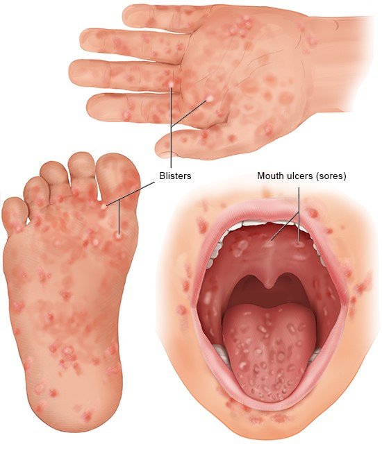
Hand, foot, and mouth disease (HFMD) is a contagious viral illness predominantly affecting young children but can occur in individuals of all ages. The condition is caused by enteroviruses, with coxsackievirus A16 and enterovirus 71 being the most common culprits. Characterized by fever, painful sores in the mouth, and a rash on the hands and feet, HFMD is typically mild but may occasionally lead to complications. It spreads through respiratory droplets, direct contact with contaminated surfaces, or fecal matter.

Nursing Paper Example on HFMD

Causes

HFMD is caused by viruses belonging to the Enterovirus genus, primarily coxsackievirus A16 and enterovirus 71. These viruses thrive in the gastrointestinal tract and are highly contagious. Infections are more common in warm climates and during summer and fall.

The disease is spread through close contact, respiratory droplets, and fecal-oral routes, with outbreaks often occurring in schools and childcare settings. Viral shedding in stool can persist for weeks after symptoms resolve, increasing the risk of transmission.

Signs and Symptoms

The incubation period for HFMD is typically three to six days. Early symptoms include fever, sore throat, reduced appetite, and general malaise. Painful ulcers may develop in the mouth, particularly on the tongue, gums, and inner cheeks, leading to difficulty swallowing. A characteristic rash appears on the hands, feet, and sometimes the buttocks, progressing to vesicular lesions.

In some cases, individuals may experience irritability, excessive drooling, or vomiting. Severe cases involving enterovirus 71 can lead to neurological complications such as meningitis, encephalitis, or polio-like paralysis.

Etiology

The etiology of HFMD involves infection with enteroviruses, with different strains causing variations in disease severity. Enterovirus 71 has been associated with more severe cases and complications compared to coxsackievirus A16.

Children under the age of five are most vulnerable due to their developing immune systems and frequent exposure in communal settings. Factors such as poor hygiene, crowded environments, and close physical contact contribute to the rapid spread of the virus.

Pathophysiology

The virus enters the body through the oral or respiratory tract, replicating in the mucosa and lymphoid tissue. From there, it disseminates to the bloodstream, targeting specific organs. The development of oral ulcers and rashes is due to viral replication and immune-mediated responses.

The virus’s affinity for neural and cardiac tissues explains the potential for severe complications, including aseptic meningitis, encephalitis, or myocarditis. Host immune responses, including inflammation and cytokine production, contribute to symptom development and resolution.

(Nursing Paper Example on HFMD)

Diagnosis

HFMD diagnosis is primarily clinical, based on characteristic symptoms such as fever, oral ulcers, and rash distribution. A detailed history of exposure and symptom onset aids diagnosis. Laboratory confirmation may involve polymerase chain reaction (PCR) testing of throat swabs, stool samples, or vesicular fluid to identify the specific enterovirus. Serological testing is less commonly used but may support diagnosis in complex cases or outbreaks.

Treatment Regimens

There is no specific antiviral treatment for HFMD. Management focuses on symptom relief and supportive care. Over-the-counter pain relievers such as acetaminophen or ibuprofen alleviate fever and discomfort. Topical oral gels or rinses can reduce pain from mouth sores.

Maintaining hydration is crucial, as painful swallowing may lead to reduced fluid intake. Severe cases with complications like meningitis or encephalitis may require hospitalization, intravenous fluids, or antiviral therapy under specific circumstances. Strict hygiene practices help prevent further transmission.

Patient Education

Patient education is vital in controlling the spread and managing HFMD. Caregivers should be informed about the importance of handwashing, disinfecting surfaces, and avoiding close contact during active illness. Children with HFMD should stay home from school or daycare until fever resolves and sores heal.

Encouraging hydration and soft, cool foods minimizes discomfort from mouth sores. Parents should seek medical attention if symptoms worsen or neurological signs such as confusion, neck stiffness, or seizures develop.

(Nursing Paper Example on HFMD)

Prevention

Preventive measures include maintaining good hygiene, avoiding close contact with infected individuals, and frequent cleaning of shared surfaces. In outbreak settings, public health interventions like temporary closures of childcare facilities or schools may be necessary.

Vaccination programs targeting enterovirus 71 have been implemented in some countries, reducing severe HFMD cases.

Conclusion

Hand, foot, and mouth disease is a common but usually mild illness that predominantly affects young children. While most cases resolve without complications, severe forms can lead to life-threatening neurological or cardiac issues. Prompt diagnosis, supportive care, and public health measures are essential in managing HFMD and preventing outbreaks. Ongoing research into vaccines and antiviral therapies offers hope for improved prevention and treatment strategies in the future.

References

American Academy of Pediatrics. (2022). Hand, foot, and mouth disease clinical guidelines. Pediatrics Journal. https://www.aap.org
Xu, W., & Liu, C. (2021). Epidemiology of hand, foot, and mouth disease in Asia. Infectious Disease Reports. https://www.infectiousdiseasereports.org
Shen, L., & Ma, H. (2020). Pathogenesis of severe HFMD caused by enterovirus 71. Virology Journal. https://www.virologyjournal.com
Centers for Disease Control and Prevention. (2023). Hand, foot, and mouth disease overview. https://www.cdc.gov
World Health Organization. (2023). Prevention of hand, foot, and mouth disease in community settings. https://www.who.int

 
Do you need a similar assignment done for you from scratch? Order now!
Use Discount Code "Newclient" for a 15% Discount!

Nursing Paper Example on Graves’ Disease

Nursing Paper Example on Graves’ Disease

Graves’ disease is an autoimmune disorder and the most common cause of hyperthyroidism. It occurs when the immune system produces autoantibodies that stimulate the thyroid gland, leading to excessive production of thyroid hormones. This hormonal imbalance affects various bodily functions, including metabolism, heart rate, and thermoregulation. Graves’ disease can present at any age but is most prevalent in women aged 30 to 50. Early diagnosis and treatment are essential to manage symptoms and prevent complications.

Nursing Paper Example on Graves' Disease

Causes

Graves’ disease is caused by the production of thyroid-stimulating immunoglobulins (TSI), which mimic thyroid-stimulating hormone (TSH). These autoantibodies bind to TSH receptors on thyroid cells, prompting the gland to overproduce thyroid hormones.

Although the exact trigger remains unknown, genetic predisposition and environmental factors play significant roles. Stress, infections, and pregnancy are known to exacerbate immune dysregulation, contributing to the onset of Graves’ disease. A family history of thyroid disorders or autoimmune conditions also increases the likelihood of developing the disease.

Signs and Symptoms

Graves’ disease presents with various symptoms due to hyperthyroidism. Common signs include unexplained weight loss, heat intolerance, and increased appetite. Patients may experience palpitations, sweating, and fatigue due to the overactive metabolism. Neurological symptoms such as tremors, anxiety, and irritability are frequent.

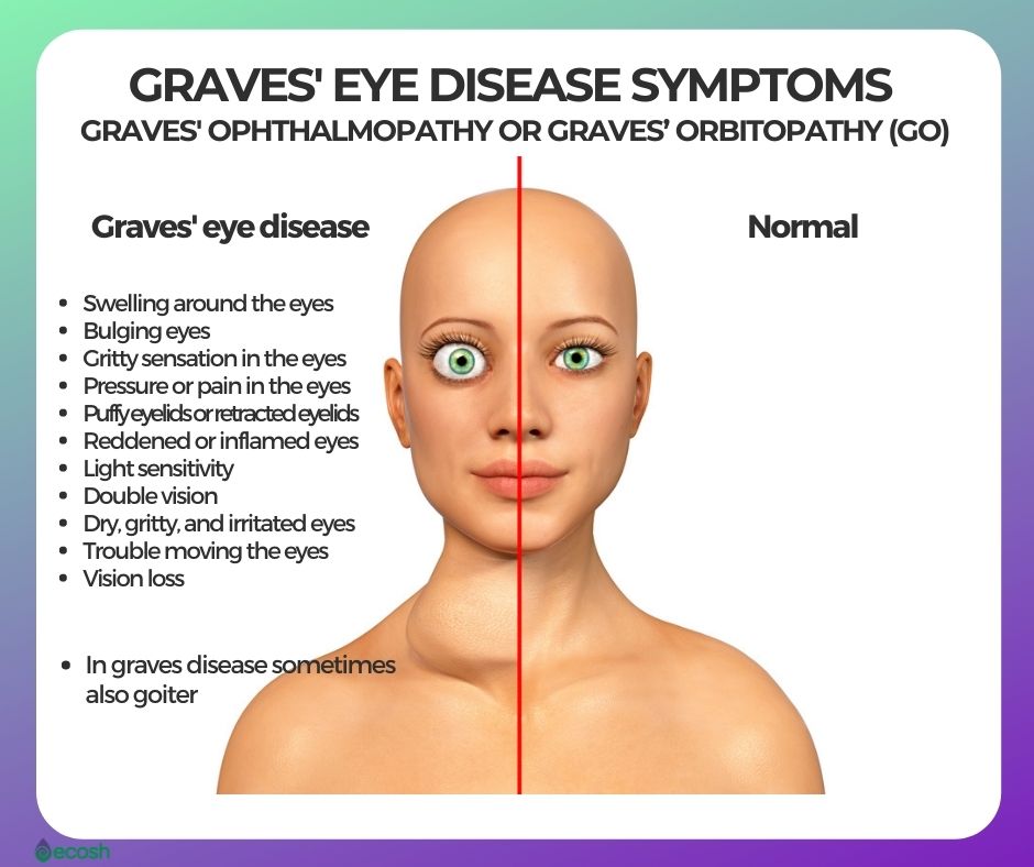
Ocular manifestations, known as Graves’ ophthalmopathy, include eye bulging, redness, and double vision. In severe cases, skin thickening on the lower legs, termed pretibial myxedema, may occur. The intensity of symptoms varies among individuals, with some presenting mildly and others severely.

Etiology

The etiology of Graves’ disease involves immune system dysfunction. The primary autoantibody implicated is thyroid-stimulating immunoglobulin, which disrupts normal thyroid regulation. Genetic factors, including specific human leukocyte antigen (HLA) types and other immune-regulatory genes, predispose individuals to the disease.

Environmental triggers such as smoking, infections, and high iodine intake have been linked to the onset of Graves’ disease. Hormonal fluctuations, particularly in women, are also believed to influence disease development and severity.

Pathophysiology

Graves’ disease results from the interplay between genetic susceptibility and environmental triggers, leading to autoimmune activation. The thyroid-stimulating immunoglobulin activates the TSH receptor on thyroid cells, causing unregulated thyroid hormone synthesis and release. Excess thyroid hormones amplify metabolic activity and enhance sympathetic nervous system sensitivity. Graves’ ophthalmopathy arises from the autoimmune attack on orbital tissues, leading to inflammation and fibrosis. Pretibial myxedema occurs due to the deposition of glycosaminoglycans in the dermis.

Diagnosis

Graves’ disease diagnosis combines clinical evaluation, laboratory tests, and imaging studies. Blood tests reveal elevated free thyroxine (T4) and triiodothyronine (T3) levels, with suppressed TSH levels. Thyroid-stimulating immunoglobulin or TSH receptor antibody tests confirm autoimmune activity.

A radioactive iodine uptake scan demonstrates diffuse thyroid gland overactivity. Ultrasound may identify thyroid enlargement and increased vascularity. A comprehensive assessment helps distinguish Graves’ disease from other causes of hyperthyroidism, such as toxic nodular goiter.

Treatment Regimens

Treatment options for Graves’ disease aim to control hyperthyroidism, alleviate symptoms, and address autoimmune activity. Antithyroid medications such as methimazole and propylthiouracil reduce thyroid hormone production. Beta-blockers like propranolol manage cardiovascular symptoms, including palpitations and tachycardia. Radioactive iodine therapy destroys overactive thyroid tissue, leading to a reduction in hormone production.

In some cases, thyroidectomy, the surgical removal of the thyroid gland, is performed. For Graves’ ophthalmopathy, glucocorticoids and orbital decompression surgery may be necessary in severe cases.

(Nursing Paper Example on Graves’ Disease)

Patient Education

Patient education is crucial for effective management of Graves’ disease. Individuals should understand the chronic nature of the condition and the importance of adhering to treatment plans. Regular follow-up visits are essential for monitoring thyroid hormone levels and adjusting therapy. Patients should be informed about potential side effects of treatments, such as hypothyroidism following radioactive iodine therapy.

Lifestyle modifications, including stress reduction, avoiding smoking, and maintaining a balanced diet, support overall health and may reduce symptom severity.

Research and Future Directions

Ongoing research on Graves’ disease focuses on understanding its genetic basis and immune mechanisms to develop targeted therapies. Advances in biologics targeting specific immune pathways, such as monoclonal antibodies, show promise in reducing autoimmune activity.

Studies exploring the role of gut microbiota in immune regulation may provide new insights into disease prevention and management. Improved diagnostic tools for early detection and risk assessment are also under investigation.

Conclusion

Graves’ disease is a complex autoimmune disorder with diverse clinical manifestations. Its pathogenesis involves genetic and environmental factors, leading to unregulated thyroid hormone production. Early diagnosis and tailored treatment strategies are essential for managing symptoms and preventing complications. Advances in research continue to enhance understanding and provide hope for improved therapeutic options in the future.

References

Davies, T. F., & Larsen, P. R. (2022). Thyroid hormone synthesis and secretion. New England Journal of Medicine. https://www.nejm.org
Smith, T. J., & Hegedüs, L. (2023). Graves’ disease: Pathophysiology and treatment approaches. Lancet Diabetes & Endocrinology. https://www.thelancet.com
Bahn, R. S. (2020). Graves’ ophthalmopathy. New England Journal of Medicine. https://www.nejm.org
Antonelli, A., Ferrari, S. M., & Fallahi, P. (2021). Environmental triggers in Graves’ disease. Autoimmunity Reviews. https://www.sciencedirect.com
Cooper, D. S., & Ross, D. S. (2023). Antithyroid drugs in the treatment of Graves’ disease. Journal of Clinical Endocrinology & Metabolism. https://academic.oup.com

 
Do you need a similar assignment done for you from scratch? Order now!
Use Discount Code "Newclient" for a 15% Discount!

Nursing Paper Example on Granulomatosis with Polyangiitis

Nursing Paper Example on Granulomatosis with Polyangiitis

Granulomatosis with polyangiitis (GPA), formerly known as Wegener’s granulomatosis, is a rare, systemic vasculitis affecting small to medium-sized blood vessels. It is characterized by necrotizing granulomas primarily in the respiratory tract and kidneys. GPA often presents with a wide range of symptoms, making diagnosis challenging. Early detection and treatment are crucial to preventing severe organ damage and improving prognosis. This autoimmune disease typically occurs in adults, with a slight predominance in males, and has no clear genetic or environmental cause.

Nursing Paper Example on Granulomatosis with Polyangiitis

Causes

The exact cause of GPA remains unknown. However, it is believed to result from an abnormal immune response triggered by environmental or infectious agents. Research suggests that bacterial or viral infections may initiate the autoimmune response.

Genetic predisposition may also play a role, particularly variants in human leukocyte antigen (HLA) genes that regulate immune function. Environmental factors such as exposure to silica and other occupational hazards have been loosely associated with GPA but require further study.

Signs and Symptoms

The clinical presentation of GPA is variable, but it typically involves the respiratory tract, kidneys, and other organ systems. Common symptoms include persistent nasal congestion, nosebleeds, and sinus infections due to inflammation in the upper respiratory tract. Lung involvement may lead to coughing, shortness of breath, and hemoptysis.  Kidney involvement often manifests as rapidly progressive glomerulonephritis, which can cause hematuria and proteinuria.

Generalized symptoms such as fever, weight loss, and fatigue are also common. Skin rashes, joint pain, and eye inflammation may occur in some patients, reflecting systemic vasculitis.

Etiology

GPA is an autoimmune disease primarily driven by the production of antineutrophil cytoplasmic antibodies (ANCAs). These autoantibodies target proteins in neutrophils, particularly proteinase-3, leading to neutrophil activation and subsequent vascular inflammation.

The underlying etiology involves complex interactions between genetic susceptibility, environmental triggers, and immune system dysregulation. Although the precise mechanism remains unclear, studies suggest that activated neutrophils release inflammatory mediators, causing endothelial injury and granuloma formation.

Pathophysiology

The pathophysiology of GPA involves immune-mediated inflammation of blood vessels and surrounding tissues. ANCAs play a central role by activating neutrophils, which adhere to vascular endothelium and release reactive oxygen species and proteolytic enzymes. This process damages blood vessel walls, leading to necrosis and granuloma formation.

Granulomas, a hallmark of GPA, consist of macrophages, multinucleated giant cells, and lymphocytes and are particularly prominent in the respiratory tract. Kidney involvement occurs when immune complexes deposit in glomeruli, causing glomerulonephritis. The systemic nature of GPA reflects widespread vascular inflammation.

(Nursing Paper Example on Granulomatosis with Polyangiitis)

Diagnosis

The diagnosis of GPA relies on clinical evaluation, laboratory tests, and imaging studies. Blood tests often reveal elevated inflammatory markers such as erythrocyte sedimentation rate (ESR) and C-reactive protein (CRP). The presence of ANCAs, specifically proteinase-3 ANCA, is highly suggestive of GPA but not definitive.

Imaging studies, including chest X-rays and computed tomography (CT), can identify granulomas and other abnormalities in the lungs and sinuses. Tissue biopsy remains the gold standard for diagnosis, with findings of necrotizing granulomas and vasculitis confirming the disease.

Treatment Regimens

Treatment for GPA aims to suppress inflammation, prevent disease progression, and manage organ damage. Initial therapy typically involves high-dose corticosteroids combined with immunosuppressive agents such as cyclophosphamide or rituximab. Corticosteroids rapidly reduce inflammation, while immunosuppressive drugs target the underlying autoimmune process.

For maintenance therapy, methotrexate or azathioprine is often used to reduce the risk of relapse. In severe cases, plasma exchange may be employed to remove ANCAs from the bloodstream. Patients with kidney involvement may require dialysis or, in rare cases, kidney transplantation.

Patient Education

Education is a critical component of GPA management. Patients should understand the chronic nature of the disease and the importance of adherence to treatment regimens to prevent relapses. Awareness of potential medication side effects, including infections and bone loss due to long-term corticosteroid use, is essential.

Regular follow-ups with healthcare providers are necessary to monitor disease activity and organ function. Patients should also be encouraged to maintain a healthy lifestyle, including smoking cessation, to reduce additional cardiovascular risks.

Research and Future Directions

Advances in understanding GPA are leading to new therapeutic approaches. Ongoing research focuses on identifying specific genetic markers and immune pathways involved in the disease. Monoclonal antibodies targeting inflammatory cytokines and immune cells, such as rituximab, are being studied for their efficacy and safety in long-term disease control.

Additionally, biomarkers for early detection and monitoring disease activity are under investigation. These developments hold promise for improving outcomes and reducing the burden of GPA.

Conclusion

Granulomatosis with polyangiitis is a complex autoimmune disease characterized by systemic vasculitis and granuloma formation. Although its exact cause is unknown, it is closely associated with ANCAs and immune dysregulation. Early diagnosis and aggressive treatment are vital to preventing irreversible organ damage and improving quality of life. Advances in immunotherapy and ongoing research provide hope for better management and outcomes for patients with this challenging condition.

References

Jennette, J. C., & Falk, R. J. (2020). Pathogenesis of ANCA-associated vasculitis: The role of autoantibodies and neutrophils. Annual Review of Pathology: Mechanisms of Disease. https://www.annualreviews.org
Falk, R. J., & Jennette, J. C. (2023). Clinical features and diagnosis of granulomatosis with polyangiitis. New England Journal of Medicine. https://www.nejm.org
Yates, M., Watts, R. A., & Scott, D. G. (2021). Advances in the treatment of ANCA-associated vasculitis. Lancet Rheumatology. https://www.thelancet.com
Niles, J. L., & Mejia-Vilet, J. M. (2023). Immunosuppressive therapy in granulomatosis with polyangiitis: Current strategies. Kidney International Reports. https://www.kidney-international.org
Luqmani, R. A., & Suppiah, R. (2020). Long-term management and prognosis of granulomatosis with polyangiitis. Rheumatology (Oxford). https://academic.oup.com/rheumatology

 
Do you need a similar assignment done for you from scratch? Order now!
Use Discount Code "Newclient" for a 15% Discount!