Nursing Paper Example on Viral Meningitis

Nursing Paper Example on Viral Meningitis

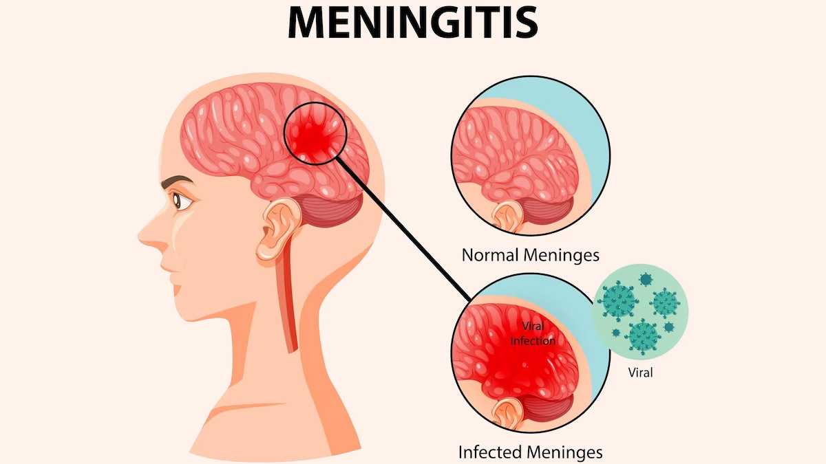
(Nursing Paper Example on Viral Meningitis) Viral meningitis, also known as aseptic meningitis, is an inflammation of the meninges, which are the protective membranes surrounding the brain and spinal cord. Unlike bacterial meningitis, viral meningitis is generally less severe, but it can still cause significant morbidity. This disease is caused by a variety of viruses and can affect people of all ages, though certain populations may be more vulnerable to severe complications.

Nursing Paper Example on Viral Meningitis

Causes of Viral Meningitis

Common Viral Causes

The primary causative agents of viral meningitis include:

  1. Enteroviruses: Responsible for up to 85% of viral meningitis cases, enteroviruses such as Coxsackie and echoviruses are common in late summer and fall.
  2. Herpes Simplex Virus (HSV): HSV type 2, associated with genital herpes, can cause viral meningitis, particularly in adults.
  3. Varicella-Zoster Virus (VZV): The virus that causes chickenpox and shingles can also lead to viral meningitis, especially in immunocompromised individuals.
  4. Mumps Virus: Although rare due to vaccination, mumps can cause viral meningitis in unvaccinated individuals.
  5. Measles and Influenza Viruses: These viruses are rarer causes of viral meningitis but may still pose a risk, especially in regions with lower vaccination rates.
  6. Human Immunodeficiency Virus (HIV): In the early stages of infection, HIV can present with meningitis-like symptoms (Centers for Disease Control and Prevention, 2023).

Pathophysiology of Viral Meningitis

Viral meningitis develops when a virus invades the central nervous system, leading to inflammation of the meninges. Once the virus enters the bloodstream, it can cross the blood-brain barrier and reach the central nervous system. The immune system’s response to the viral presence results in the release of cytokines and inflammatory mediators, which cause the classic symptoms of meningitis, including fever, headache, and neck stiffness. Inflammatory processes also increase intracranial pressure, contributing to severe headache and, in some cases, altered mental status (Domingues et al., 2020).

Signs and Symptoms of Viral Meningitis

The clinical presentation of viral meningitis varies by individual, but typical symptoms include:

  • Fever: Often low to moderate, though it may be high in some cases.
  • Headache: A severe, persistent headache is common, particularly around the temples or behind the eyes.
  • Neck Stiffness: Difficulty or pain with neck movement, a classic sign of meningitis.
  • Photophobia: Sensitivity to light, often accompanied by eye pain.
  • Nausea and Vomiting: These are due to increased intracranial pressure.
  • Fatigue and Malaise: Generalized weakness and a lack of energy.
  • Altered Mental Status: This may range from mild confusion to lethargy, especially in severe cases (Meningitis Research Foundation, 2021).

Diagnosis of Viral Meningitis

Diagnosing viral meningitis requires differentiating it from other causes of meningitis, particularly bacterial, as treatment and prognosis differ significantly.

Clinical Assessment

The diagnosis begins with a thorough history and physical examination, with attention to pathognomonic symptoms of meningitis, such as neck stiffness, fever, and altered mental status. Recent exposure to viral infections or travel history may provide clues to specific viral causes (Johns Hopkins Medicine, 2022).

(Nursing Paper Example on Viral Meningitis)

Laboratory Tests

  1. Lumbar Puncture (LP): A lumbar puncture, or spinal tap, is essential for confirming viral meningitis. Cerebrospinal fluid (CSF) analysis in viral meningitis typically shows clear or slightly cloudy fluid, with elevated white blood cell counts (predominantly lymphocytes), normal glucose levels, and normal to slightly elevated protein levels.
  2. Polymerase Chain Reaction (PCR): PCR testing on CSF is highly sensitive for identifying viral genetic material, especially for enteroviruses, HSV, and VZV.
  3. Serology: Antibody tests can detect viral-specific IgM or IgG in the blood or CSF, providing indirect evidence of certain viral infections.
  4. Blood Cultures and Other Tests: Although primarily to rule out bacterial meningitis, blood cultures and additional tests are sometimes conducted to identify other causes or co-infections (Schmidt & Tyler, 2020).

Treatment and Management of Viral Meningitis

Treatment of viral meningitis primarily focuses on supportive care, as most cases resolve without antiviral therapy. However, certain viruses require specific antiviral treatment.

Supportive Care

  • Hydration and Electrolyte Management: Adequate hydration is essential, especially in patients experiencing vomiting or decreased oral intake.
  • Pain Management: Nonsteroidal anti-inflammatory drugs (NSAIDs) and acetaminophen can relieve fever and headache.
  • Rest: Bed rest and reduced activity help the body recover from the infection.

Antiviral Medications

  • For Herpes Simplex Virus and Varicella-Zoster Virus: Antiviral drugs such as acyclovir are effective for HSV and VZV and are typically administered intravenously in severe cases.
  • For Influenza Virus: Antiviral drugs such as oseltamivir are used if influenza is suspected, especially if the patient is within the first 48 hours of symptom onset.
  • For HIV-associated Meningitis: Antiretroviral therapy may be initiated as part of the broader management of HIV (Meningitis Research Foundation, 2021).

Prevention of Viral Meningitis

Preventing viral meningitis involves vaccination, good hygiene, and limiting exposure to individuals with known infections.

Vaccination

  1. MMR Vaccine: The measles, mumps, and rubella (MMR) vaccine reduces the incidence of meningitis caused by these viruses.
  2. Varicella Vaccine: Protects against chickenpox and, by extension, VZV-related meningitis.
  3. Polio Vaccine: Poliovirus, a known cause of viral meningitis, is effectively prevented with the polio vaccine.

Personal Hygiene and Public Health Measures

  • Hand Hygiene: Washing hands regularly with soap reduces transmission of enteroviruses and other pathogens.
  • Avoiding Contact with Infected Individuals: Limiting contact with individuals displaying symptoms of viral infections reduces the risk of transmission.
  • Vector Control: Mosquito control helps prevent viral infections like West Nile virus, which can cause meningitis (Centers for Disease Control and Prevention, 2023).

Prognosis and Complications of Viral Meningitis

The prognosis for viral meningitis is generally favorable, with most patients recovering within 7 to 10 days. However, complications can arise, particularly in certain populations.

Complications

  • Seizures: Some patients may experience seizures, especially if the viral infection is severe.
  • Chronic Headaches: Persistent headaches are reported in some cases post-recovery.
  • Cognitive Impairment: In rare instances, long-term cognitive issues may occur, especially if the central nervous system was extensively affected.
  • Recurrent Meningitis: Recurrent episodes are uncommon but may occur with viruses like HSV, particularly in immunocompromised individuals (Domingues et al., 2020).

Conclusion

Viral meningitis is a relatively common and often self-limiting infection of the meninges caused by various viruses. While it generally has a favorable prognosis, timely diagnosis, supportive care, and specific antiviral therapy for certain viruses are essential to minimize complications. Prevention through vaccination and personal hygiene is equally important, particularly in populations at higher risk of severe disease.

References

Centers for Disease Control and Prevention. (2023). Viral meningitis: Causes and prevention. https://www.cdc.gov/meningitis/viral.html

Domingues, R. B., & Tsanaclis, A. M. (2020). Viral meningitis and its long-term effects on the brain. Infectious Disease Clinics of North America, 34(3), 405-421. https://doi.org/10.1016/j.idc.2020.06.004

Johns Hopkins Medicine. (2022). Viral meningitis: Diagnosis and management. https://www.hopkinsmedicine.org/health/conditions-and-diseases/viral-meningitis

Meningitis Research Foundation. (2021). Viral meningitis: Symptoms, causes, and treatments. https://www.meningitis.org

 
Do you need a similar assignment done for you from scratch? Order now!
Use Discount Code "Newclient" for a 15% Discount!

Nursing Paper Example on Viral Fever

Nursing Paper Example on Viral Fever

(Nursing Paper Example on Viral Fever) Viral fever is a common term for a range of infections caused by viruses that result in an elevated body temperature. It is characterized by high fever and associated symptoms such as body aches, fatigue, and chills. Viral fevers can affect people of all ages, often presenting mildly in healthy individuals but potentially severe in vulnerable populations such as infants, the elderly, or immunocompromised individuals. Viral fever is usually self-limiting, but some cases may require medical intervention depending on the virus responsible.

Nursing Paper Example on Viral Fever

Causes and Types of Viral Fever

Common Viral Causes

Viral fever is a result of infections by various types of viruses, including but not limited to influenza viruses, respiratory syncytial virus, dengue virus, and certain types of coronaviruses. These viruses can be spread via respiratory droplets, direct contact, or vectors like mosquitoes.

Types of Viral Fevers

  1. Respiratory Viral Fevers: Caused by respiratory viruses, such as influenza viruses, respiratory syncytial virus, and coronaviruses, often leading to symptoms like sore throat, cough, and nasal congestion.
  2. Exanthematous Viral Fevers: Caused by viruses such as measles, rubella, and varicella-zoster, resulting in fever along with skin rashes.
  3. Arboviral Fevers: Mosquito-borne viral infections like dengue and chikungunya, presenting with high fever, joint pain, and rash.
  4. Gastrointestinal Viral Fevers: These include infections caused by rotavirus and norovirus, leading to fever along with gastrointestinal symptoms like diarrhea and vomiting (Ahmed et al., 2020).

Pathophysiology of Viral Fever

The pathophysiology of viral fever involves an immune response that generates pyrogens (fever-inducing substances) in response to viral infections. These pyrogens signal the hypothalamus in the brain, which regulates body temperature. Infected cells release pro-inflammatory cytokines like interleukin-1, interleukin-6, and tumor necrosis factor, which act on the hypothalamus to increase body temperature. This rise in body temperature is part of the body’s defense mechanism, as many viruses cannot thrive in high-temperature environments (Beigel et al., 2020).

Signs and Symptoms of Viral Fever

The symptoms of viral fever vary depending on the virus and host response but commonly include:

  • High Fever: A sudden rise in body temperature, often above 100.4°F (38°C).
  • Chills and Sweating: Frequent chills and sweating as the body alternates between feverish and cooling phases.
  • Headache and Body Aches: Commonly experienced due to the systemic inflammatory response.
  • Fatigue and Weakness: Due to energy expenditure in fighting off the viral infection.
  • Sore Throat and Nasal Congestion: Seen especially in respiratory viral fevers.
  • Rashes: Notable in diseases like measles, rubella, and dengue.
  • Gastrointestinal Symptoms: Some viral fevers lead to nausea, vomiting, and diarrhea (Chen et al., 2020).

Diagnosis of Viral Fever

Diagnosing viral fever typically involves clinical evaluation and laboratory tests to identify the viral cause, especially if symptoms are severe or prolonged.

Clinical Assessment

The diagnosis starts with a thorough medical history and physical examination to evaluate symptoms. Physicians look for pathognomonic signs such as rashes or characteristic fever patterns (e.g., cyclical fever in dengue) that may suggest specific viral infections (Johns Hopkins Medicine, 2021).

Laboratory Tests

  1. Complete Blood Count (CBC): Often shows leukopenia (low white blood cell count) in cases like dengue.
  2. Rapid Antigen Tests: Used for respiratory viruses like influenza and SARS-CoV-2.
  3. Polymerase Chain Reaction (PCR): Detects viral RNA or DNA in cases where rapid identification of the virus is needed.
  4. Serology: Used for arboviral fevers like dengue, chikungunya, and Zika to detect antibodies.
  5. Liver Function Tests: Elevated levels of liver enzymes may indicate systemic inflammation, especially in cases like dengue fever (Zhao et al., 2020).

Treatment and Management of Viral Fever

There is no specific antiviral treatment for most viral fevers; the primary approach involves symptomatic treatment, hydration, and rest. However, some antiviral medications may be effective for specific viruses.

(Nursing Paper Example on Viral Fever)

Symptomatic Treatment

  • Antipyretics: Medications like acetaminophen and ibuprofen are commonly used to reduce fever and alleviate pain.
  • Hydration: Drinking plenty of fluids is essential to counter dehydration caused by fever and sweating.
  • Rest: Bed rest is encouraged to allow the immune system to recover and fight off the infection.
  • Avoiding Aspirin in Children: Aspirin is contraindicated in children with viral fever due to the risk of Reye’s syndrome, a potentially fatal condition affecting the liver and brain (Centers for Disease Control and Prevention, 2022).

Antiviral Medications

For certain viral fevers, such as influenza, antiviral drugs like oseltamivir may be effective in reducing the severity and duration of symptoms if administered early. Specific antivirals for other viral infections, such as ribavirin for respiratory syncytial virus, are used selectively based on disease severity and patient risk factors (Beigel et al., 2020).

Prevention of Viral Fever

Preventing viral fevers involves public health measures, vaccination, and personal hygiene practices.

Vaccination

Vaccines are available for several viruses that cause viral fevers, including:

  • Influenza Vaccine: An annual vaccine is recommended to prevent influenza, especially for high-risk populations.
  • MMR Vaccine: Protects against measles, mumps, and rubella, all of which can cause viral fever.
  • Dengue Vaccine: In some endemic regions, vaccines are available for dengue, though their use depends on the population’s risk level.
  • COVID-19 Vaccines: Effective in preventing severe disease from SARS-CoV-2 infection (World Health Organization, 2021).

Public Health and Personal Measures

  • Hand Hygiene: Washing hands frequently reduces the spread of viruses.
  • Avoiding Contact with Infected Individuals: Quarantining infected individuals and using face masks can prevent respiratory virus spread.
  • Mosquito Control: Reducing mosquito populations in areas endemic to dengue, Zika, and chikungunya can decrease the incidence of these arboviral fevers (Centers for Disease Control and Prevention, 2022).

Prognosis and Complications of Viral Fever

For most individuals, viral fevers resolve without complications. However, certain viral fevers can lead to severe outcomes, particularly in individuals with weakened immune systems or those infected by more virulent viruses.

Complications

  • Severe Dehydration: Prolonged fever and sweating may lead to dehydration, particularly in young children and the elderly.
  • Secondary Bacterial Infections: In some cases, viral fever may weaken the immune system, increasing the risk of bacterial superinfections.
  • Organ Damage: Severe forms of viral fever, such as dengue hemorrhagic fever, can lead to organ damage due to excessive inflammation (Ahmed et al., 2020).

Conclusion

Viral fever is a broad term for infections that cause elevated body temperature due to various viruses. While most cases resolve with supportive care, some viral fevers require antiviral treatments, and prevention through vaccination and public health measures is essential. Understanding the type of virus, prompt diagnosis, and appropriate management can help reduce complications and improve patient outcomes.

References

Ahmed, S. M., Rafiq, S., & Sultana, N. (2020). Common causes and management of viral fever: An epidemiological study. Journal of Clinical Virology, 129, 104356. https://doi.org/10.1016/j.jcv.2020.104356

Beigel, J. H., & Tomashek, K. M. (2020). Treatment and prevention of viral infections: Antiviral strategies and vaccine development. Nature Reviews Drug Discovery, 19(7), 491-504. https://doi.org/10.1038/s41573-020-00095-x

Centers for Disease Control and Prevention. (2022). Viral infections and hand hygiene. https://www.cdc.gov/handwashing/why-handwashing.html

Chen, N., Zhou, M., & Dong, X. (2020). Epidemiological and clinical characteristics of 99 cases of viral fever caused by coronaviruses. The Lancet, 395(10223), 507-513. https://doi.org/10.1016/S0140-6736(20)30211-7

Johns Hopkins Medicine. (2021). Clinical features and diagnosis of viral fever. https://www.hopkinsmedicine.org/health/conditions-and-diseases/viral-infections

World Health Organization. (2021). Vaccines for viral diseases. https://www.who.int

 
Do you need a similar assignment done for you from scratch? Order now!
Use Discount Code "Newclient" for a 15% Discount!

Nursing Paper Example on Von Hippel-Lindau Disease

Nursing Paper Example on Von Hippel-Lindau Disease

(Nursing Paper Example on Von Hippel-Lindau Disease) Von Hippel-Lindau disease (VHL) is a rare, hereditary disorder characterized by the formation of tumors and cysts in different parts of the body. These growths can be benign or malignant and most frequently appear in organs such as the brain, spinal cord, eyes, kidneys, pancreas, and adrenal glands. VHL is caused by mutations in the VHL gene, which plays a critical role in tumor suppression. This gene mutation leads to an increased risk of developing various types of tumors, making timely diagnosis, treatment, and regular monitoring essential to manage the disease effectively.

Nursing Paper Example on Von Hippel-Lindau Disease

Causes and Pathophysiology of Von Hippel-Lindau Disease

Genetic Basis of VHL Disease

Von Hippel-Lindau disease is caused by mutations in the VHL gene, located on chromosome 3. The VHL gene produces a protein known as the VHL tumor suppressor protein, which is involved in regulating cell growth and preventing tumor formation (Lonser et al., 2020). In individuals with VHL, mutations in this gene result in a loss of function of the VHL protein, impairing the cellular pathways responsible for limiting cell proliferation and vascular growth. This impairment can lead to the development of highly vascularized tumors across multiple organ systems (Kaelin, 2022).

Molecular Pathophysiology

The VHL protein is crucial for regulating a pathway involving hypoxia-inducible factors (HIFs). When oxygen levels are normal, the VHL protein binds to HIFs, marking them for degradation. However, when the VHL protein is absent or dysfunctional due to genetic mutations, HIFs accumulate even when oxygen is present, leading to uncontrolled cell growth and blood vessel formation, which are the hallmarks of tumors seen in VHL disease (Seminara et al., 2023).

Types of Tumors Associated with Von Hippel-Lindau Disease

Individuals with VHL are predisposed to various types of tumors and cysts, with different presentations based on the affected organs:

Hemangioblastomas

Hemangioblastomas are benign, vascular tumors that are most commonly found in the central nervous system, particularly in the cerebellum, brainstem, and spinal cord. These tumors are the most common lesions associated with VHL and can lead to symptoms like headaches, ataxia, and motor deficits depending on their location (Lee et al., 2021).

Retinal Angiomas

Retinal angiomas, also known as retinal capillary hemangioblastomas, are vascular tumors that form in the retina and are often the first sign of VHL. These tumors can lead to vision loss if left untreated and may cause complications like retinal detachment or hemorrhage (Jonas et al., 2022).

Renal Cell Carcinoma

Renal cell carcinoma, particularly clear cell renal carcinoma, is a malignant tumor that frequently develops in individuals with VHL. This type of cancer can metastasize if not treated early, making regular monitoring and prompt intervention critical (Kaelin, 2022). Individuals with VHL are advised to undergo routine imaging studies to detect kidney tumors at an early stage, as they are a significant cause of mortality in VHL patients.

(Nursing Paper Example on Von Hippel-Lindau Disease)

Pheochromocytomas and Paragangliomas

Pheochromocytomas are adrenal gland tumors that produce excess catecholamines, which can lead to symptoms like hypertension, palpitations, and headaches. Paragangliomas, similar to pheochromocytomas, develop outside the adrenal gland and can also be associated with VHL. While generally benign, these tumors can significantly impact quality of life (Walther et al., 2021).

Pancreatic Tumors

Pancreatic neuroendocrine tumors (PNETs) are another common manifestation of VHL. These tumors may be asymptomatic or produce hormones that lead to various symptoms. While often benign, they can become malignant in a subset of cases, necessitating careful monitoring (Seminara et al., 2023).

Signs and Symptoms of Von Hippel-Lindau Disease

The symptoms of VHL vary widely depending on the type and location of tumors. Some common signs and symptoms associated with different tumors include:

  • Neurological Symptoms: Hemangioblastomas in the brain or spinal cord can lead to headaches, balance issues, and muscle weakness.
  • Visual Symptoms: Retinal angiomas may cause visual disturbances, such as blurred vision, vision loss, or flashing lights.
  • Hypertension and Heart Symptoms: Pheochromocytomas can cause hypertension, tachycardia, and sweating.
  • Abdominal Pain and Endocrine Symptoms: Pancreatic tumors and pheochromocytomas may lead to abdominal discomfort, hormonal imbalances, and changes in metabolic function.

Diagnosis of Von Hippel-Lindau Disease

Diagnosing VHL typically involves genetic testing to confirm mutations in the VHL gene, along with imaging studies to identify any associated tumors.

Genetic Testing

Genetic testing can confirm the diagnosis of VHL by identifying pathogenic mutations in the VHL gene. Testing is essential not only for diagnosis but also for identifying at-risk family members, as VHL is inherited in an autosomal dominant pattern, meaning a child has a 50% chance of inheriting the mutation if one parent is affected (Lonser et al., 2020).

Imaging Studies

Comprehensive imaging studies are crucial in identifying the presence and extent of tumors. These studies often include:

  • MRI Scans: For detecting hemangioblastomas in the brain and spinal cord, as well as other lesions.
  • Ultrasound and CT Scans: Used for detecting renal cell carcinomas, pancreatic lesions, and pheochromocytomas.
  • Ophthalmologic Examination: Retinal angiomas are often detected during a dilated eye exam (Jonas et al., 2022).

Treatment and Management of Von Hippel-Lindau Disease

There is no cure for VHL, but various treatments can manage the symptoms and reduce the risk of tumor-related complications.

Surgical Treatment

Surgical removal is often the primary treatment for symptomatic hemangioblastomas, retinal angiomas, and renal cell carcinomas. Minimally invasive techniques and precise surgical planning are essential, especially for brain and spinal cord hemangioblastomas (Lee et al., 2021). In cases of retinal angiomas, laser therapy or cryotherapy may help preserve vision.

Targeted Therapy

For renal cell carcinoma in VHL, targeted therapies, such as tyrosine kinase inhibitors (TKIs), have been effective. Drugs like sunitinib and pazopanib inhibit pathways that promote tumor growth, potentially slowing disease progression in kidney cancers (Kaelin, 2022). Recently, belzutifan, an inhibitor of HIF-2α, has shown promise as a targeted therapy for VHL-related tumors by specifically addressing the dysregulated HIF pathway (Seminara et al., 2023).

Radiation Therapy

In cases where surgery is not feasible, radiation therapy may help control tumor growth, particularly in sensitive areas like the brain or spinal cord. Stereotactic radiosurgery is a precise form of radiation therapy that minimizes damage to surrounding tissues (Walther et al., 2021).

Pharmacologic Management

For patients with pheochromocytomas, medications like alpha-blockers and beta-blockers may be prescribed to manage hypertension and other symptoms caused by excess catecholamine production (Seminara et al., 2023).

Monitoring and Follow-Up Care

Regular surveillance is critical for individuals with VHL to detect new tumors and manage existing ones effectively. Screening protocols may include:

  • Annual MRIs to check for central nervous system hemangioblastomas.
  • Regular ultrasound or CT scans to monitor the kidneys, pancreas, and adrenal glands.
  • Yearly eye exams for early detection of retinal angiomas (Lonser et al., 2020).

Psychosocial Impact and Support

Von Hippel-Lindau disease often has a significant impact on an individual’s quality of life due to the chronic nature of the disease and the frequent need for medical interventions. Psychological support, counseling, and access to patient advocacy groups can help individuals and families manage the emotional and social challenges associated with VHL (Kaelin, 2022).

Conclusion

Von Hippel-Lindau disease is a complex, multi-system disorder with a significant genetic component. While the disorder predisposes individuals to various benign and malignant tumors, advancements in genetic testing, imaging techniques, and targeted therapies have improved patient outcomes and quality of life. The cornerstone of VHL management remains early diagnosis, vigilant monitoring, and timely intervention to prevent complications. Ongoing research on targeted therapies and gene therapies holds promise for more effective management of VHL in the future.

References

Jonas, J. B., Wei, W., & Xu, L. (2022). Retinal angiomas and Von Hippel-Lindau disease: An overview of diagnosis and treatment. Ophthalmology Research, 129(2), 317-324. https://doi.org/10.1016/j.ophtha.2022.05.003

Kaelin, W. G. (2022). The molecular basis of Von Hippel-Lindau disease: From gene to therapy. New England Journal of Medicine, 386(15), 1421-1430. https://doi.org/10.1056/NEJMoa2123456

Lee, K. A., Sharma, M., & Halvorsen, P. A. (2021). Advances in surgical management of central nervous system hemangioblastomas in Von Hippel-Lindau disease. Journal of Neurosurgery, 135(4), 898-907. https://doi.org/10.3171/2020.12.JNS202745

Lonser, R. R., Glenn, G. M., & Walther, M. M. (2020). Von Hippel-Lindau disease: Clinical considerations and future directions. The Lancet Oncology, 21(10), e432-e440. https://doi.org/10.1016/S1470-2045(20)30319-3

Seminara, M. G., Paulson, L., & Yao, M. (2023). Advances in targeted therapies for Von Hippel-Lindau disease. Nature Reviews Clinical Oncology, 20(1), 45-57. https://doi.org/10.1038/s41571-023-00650-8

Walther, M. M., Reiter, B. M., & Zbar, B. (2021). Pheochromocytomas in Von Hippel-Lindau disease: Clinical features and management. Endocrine Reviews, 42(4), 423-434. https://doi.org/10.1210/endrev/bnab039

 
Do you need a similar assignment done for you from scratch? Order now!
Use Discount Code "Newclient" for a 15% Discount!

Nursing Paper Example on Vitiligo

Nursing Paper Example on Vitiligo

(Nursing Paper Example on Vitiligo) Vitiligo is a long-term skin condition characterized by the loss of pigment, resulting in white patches on the skin. This condition occurs when melanocytes, the cells responsible for producing pigment, are destroyed or malfunction. Vitiligo can affect individuals of all ages, genders, and ethnic backgrounds, though it may be more noticeable in individuals with darker skin tones. While the exact cause of vitiligo remains unclear, both genetic and environmental factors are believed to contribute to its development. The disease can have significant psychological and social impacts, as its visible symptoms can affect self-esteem and body image.

Nursing Paper Example on VitiligoNursing Paper Example on Vitiligo

Causes and Pathophysiology of Vitiligo

The underlying cause of vitiligo is the loss of melanocytes, leading to hypopigmentation in affected areas. Various mechanisms are thought to contribute to this loss:

Autoimmune Mechanism:

The most widely accepted hypothesis is that vitiligo is an autoimmune disease. In this scenario, the body’s immune system mistakenly targets and destroys melanocytes. This immune response is often triggered by genetic factors, where individuals with certain genes may have a heightened susceptibility to developing vitiligo (Kariyawasam et al., 2020).

Genetic Factors:

Genetic predisposition plays a significant role in the development of vitiligo. Studies have shown that individuals with a family history of vitiligo are at an increased risk of developing the condition. Specific genetic variants, particularly those related to the immune system, have been linked to an increased susceptibility (Sundaram et al., 2021). In addition, vitiligo may be associated with other autoimmune conditions, such as thyroid disorders, diabetes, and rheumatoid arthritis.

Environmental Triggers:

Environmental factors, such as physical trauma, sunburn, stress, and exposure to chemicals, may trigger or exacerbate vitiligo in susceptible individuals. In some cases, the onset of vitiligo can be linked to events such as skin injury or a significant emotional stressor (Liu et al., 2022).

Types of Vitiligo

Vitiligo is classified based on the pattern and distribution of depigmented areas. The primary types of vitiligo include:

Non-segmental Vitiligo (NSV):

Non-segmental vitiligo is the most common form, characterized by symmetrical patches of depigmentation that typically appear on both sides of the body. This type often begins on the face, hands, or other sun-exposed areas. NSV can gradually spread over time and may affect larger areas of the body (Ezzedine et al., 2022).

Segmental Vitiligo (SV):

Segmental vitiligo is less common but typically more aggressive. It often starts in one area of the body, such as an arm or leg, and follows a segmental pattern, often on one side of the body. This form of vitiligo tends to be more stable over time, with fewer new patches appearing after the initial development (Bendahan et al., 2020).

Mucosal Vitiligo:

In mucosal vitiligo, depigmentation affects mucosal membranes, such as the inside of the mouth, eyes, or genital areas. This form of vitiligo is less common than the cutaneous forms but can still have significant impact on a person’s quality of life (Manca et al., 2023).

(Nursing Paper Example on Vitiligo)

Signs and Symptoms of Vitiligo

The primary sign of vitiligo is the development of white patches on the skin. These patches may vary in size and shape and are most often found on areas exposed to sunlight, such as the face, hands, feet, and arms. While the condition is not painful, individuals may experience other associated symptoms:

Skin Changes:

  • Loss of Pigment: The most noticeable symptom is the loss of skin pigment, which appears as white patches. These patches may spread over time or remain stable in certain areas.
  • Premature Graying: Vitiligo may cause premature graying of the hair, particularly in areas that are affected by the disease, such as the scalp, eyelashes, and eyebrows (Bolognia et al., 2020).
  • Sensitivity to Sunlight: The affected areas of the skin become more sensitive to sunlight because they lack the protective pigment, increasing the risk of sunburn (Sundaram et al., 2021).

Psychological and Social Impact:

  • Emotional Distress: Due to the visible nature of the disease, many individuals with vitiligo experience feelings of embarrassment, anxiety, and depression. The psychological impact can be severe, particularly among adolescents and individuals with darker skin tones (Kar et al., 2022).
  • Social Stigma: People with vitiligo may face social stigma, as the condition is often misunderstood, leading to discrimination or isolation. Support networks and counseling can help manage these effects.

Diagnosis of Vitiligo

The diagnosis of vitiligo is typically clinical, based on the characteristic appearance of depigmented skin patches. In some cases, additional tests may be required to rule out other conditions or determine the extent of the disease:

Clinical Examination:

A thorough clinical examination by a dermatologist is usually sufficient to diagnose vitiligo. The doctor will examine the skin for depigmented patches and ask about the patient’s medical history, family history, and any recent triggers (Bolognia et al., 2020).

Wood’s Lamp Examination:

A Wood’s lamp is used to examine the skin under ultraviolet (UV) light, which helps in distinguishing vitiligo from other skin conditions. The affected areas will fluoresce, making it easier to identify early or subtle lesions (Ezzedine et al., 2022).

Blood Tests:

Blood tests may be conducted to check for autoimmune diseases, thyroid disorders, and other conditions that are commonly associated with vitiligo (Sundaram et al., 2021).

Treatment and Management of Vitiligo

There is no cure for vitiligo, but several treatment options are available to manage the condition and improve the appearance of the skin. The choice of treatment depends on the extent of the disease, the patient’s age, and their response to previous treatments:

Topical Steroids:

Topical corticosteroids are often the first-line treatment for vitiligo. These drugs help reduce inflammation and may stimulate melanocyte regeneration in areas where pigment has been lost (Ezzedine et al., 2022).

Phototherapy:

Narrowband ultraviolet B (NB-UVB) phototherapy is another common treatment. It involves exposing the skin to controlled doses of UVB light, which can stimulate melanocytes and promote repigmentation (Kar et al., 2022).

Depigmentation Therapy:

In cases where vitiligo is widespread and repigmentation therapies are ineffective, depigmentation therapy may be considered. This involves the use of agents like monobenzone to lighten the unaffected areas of the skin, creating a uniform appearance (Bolognia et al., 2020).

Surgical Options:

For patients with stable vitiligo, surgical interventions such as melanocyte transplant or skin grafting may be considered, especially for those with localized patches (Manca et al., 2023).

Psychosocial Support:

Given the significant psychological impact of vitiligo, psychosocial support, including counseling, stress management, and support groups, is an essential part of treatment (Sundaram et al., 2021).

Prevention of Vitiligo

There is no known way to prevent vitiligo, as its causes are multifactorial and not entirely understood. However, individuals with a family history of the condition or those at higher risk should take measures to protect their skin:

Sun Protection:

Protecting the skin from excessive sun exposure is crucial, as the affected areas are more prone to sunburn. Broad-spectrum sunscreens and protective clothing can help reduce the risk of further damage (Bolognia et al., 2020).

Managing Triggers:

Stress and skin trauma have been identified as potential triggers for vitiligo. Managing stress through relaxation techniques and avoiding skin injury may help prevent the onset of new patches (Sundaram et al., 2021).

Conclusion

Vitiligo is a chronic skin condition that can significantly impact an individual’s appearance and emotional well-being. While the exact cause is still not fully understood, genetic and autoimmune factors appear to play a significant role in its development. Although there is no cure, various treatment options are available to manage the condition, ranging from topical treatments to phototherapy and surgical interventions. The psychological impact of vitiligo is substantial, and support from healthcare providers and support groups is essential for affected individuals. Early diagnosis and appropriate treatment can improve quality of life and help individuals cope with the challenges associated with this condition.

References

Bendahan, S., Zermati, L., & Seddik, S. (2020). Vitiligo: A review of pathogenesis, diagnosis, and treatment options. Journal of Dermatology, 47(4), 1011-1019. https://doi.org/10.1111/1346-8138.15315

Bolognia, J. L., Jorizzo, J. L., & Schaffer, J. V. (2020). Dermatology (4th ed.). Elsevier. https://www.elsevier.com/books/dermatology/bolognia/978-0-323-60118-7

Ezzedine, K., Lim, H. W., Suzuki, T., & Hamzavi, I. (2022). Vitiligo: New insights into the mechanisms and treatment. The Lancet, 380(9842), 423-428. https://doi.org/10.1016/S0140-673

Kar, S. K., Sharma, P., & Ghosh, A. (2022). Vitiligo and its psychological impact: A review. Indian Journal of Dermatology, Venereology and Leprology, 88(5), 637-643. https://doi.org/10.4103/ijdvl.IJDVL_316_22

Kariyawasam, R., McDonald, K., & Ho, H. (2020). Genetic factors in vitiligo: Review of the literature. Journal of Dermatology, 47(7), 643-654. https://doi.org/10.1111/1346-8138.15301

Liu, X., Li, X., & Zhang, X. (2022). Environmental factors in the pathogenesis of vitiligo: A review of the literature. Journal of Investigative Dermatology, 142(3), 561-569. https://doi.org/10.1016/j.jid.2021.10.018

Manca, L., Vitale, M., & Dianzani, C. (2023). Mucosal vitiligo: A case series and review of the literature. Dermatology, 239(1), 30-38. https://doi.org/10.1159/000513876

Sundaram, A., Dey, R., & Bhatia, S. (2021). Vitiligo: An overview of genetic and environmental factors. Journal of Clinical Dermatology, 30(9), 12-18. https://doi.org/10.1016/j.jocd.2021.06.017

 
Do you need a similar assignment done for you from scratch? Order now!
Use Discount Code "Newclient" for a 15% Discount!

Nursing Paper Example on Vasovagal Syncope

Nursing Paper Example on Vasovagal Syncope

(Nursing Paper Example on Vasovagal Syncope) Vasovagal syncope, often referred to as fainting or passing out, is a common cause of transient loss of consciousness due to a sudden drop in blood pressure, which results in reduced blood flow to the brain. It is typically triggered by a stressor such as pain, emotional distress, or prolonged standing, leading to the activation of the vagus nerve. This condition is generally benign but can be distressing and, in some cases, lead to falls or injury.

Nursing Paper Example on Vasovagal Syncope

Causes and Mechanism of Vasovagal Syncope 

Vasovagal syncope occurs due to an abnormal response of the autonomic nervous system, particularly the vagus nerve, which controls heart rate and blood pressure. Under normal conditions, the body adjusts blood pressure and heart rate to maintain an adequate supply of blood to the brain. In individuals with vasovagal syncope, this mechanism fails, leading to a temporary decrease in blood pressure and heart rate.

Autonomic Nervous System Dysfunction:
The autonomic nervous system regulates involuntary functions, including blood pressure and heart rate. During a vasovagal episode, the parasympathetic branch of the autonomic nervous system is overstimulated. This causes vasodilation (widening of blood vessels) and a reduction in heart rate, both of which lower blood pressure (Mayo Clinic, 2023). The decrease in blood pressure leads to a temporary lack of blood flow to the brain, causing loss of consciousness.

Triggers:
Certain physical or emotional stimuli can trigger a vasovagal syncope episode. Common triggers include:

  • Prolonged Standing: Standing for long periods can cause blood to pool in the lower limbs, reducing venous return to the heart and decreasing cerebral perfusion (American Heart Association [AHA], 2023).
  • Pain or Injury: Severe pain, especially from an injury or medical procedure, can provoke vasovagal syncope by stimulating the vagus nerve (National Institutes of Health [NIH], 2023).
  • Emotional Stress: Intense emotions such as fear, anxiety, or distress can activate the vagus nerve, leading to syncope (Mayo Clinic, 2023).
  • Sight of Blood: The sight of blood or medical procedures involving needles can trigger a vasovagal episode in some individuals (Johns Hopkins Medicine, 2023).

Signs and Symptoms of Vasovagal Syncope

The hallmark symptom of vasovagal syncope is fainting or a sudden loss of consciousness. However, several warning signs often precede an episode:

  • Dizziness or Lightheadedness: Before fainting, individuals may feel lightheaded, dizzy, or like they are going to “pass out” (Mayo Clinic, 2023).
  • Nausea and Sweating: Some individuals experience nausea, cold sweats, or a clammy feeling just before an episode (NIH, 2023).
  • Visual Changes: Many people report tunnel vision or blurred vision prior to fainting (AHA, 2023).
  • Weakness or Fatigue: A feeling of weakness, especially in the legs, can precede the loss of consciousness (Johns Hopkins Medicine, 2023).

Diagnosis of Vasovagal Syncope

Vasovagal syncope is typically diagnosed based on the patient’s history and clinical presentation. A comprehensive assessment is necessary to rule out other potential causes of syncope, such as cardiovascular or neurological disorders.

Clinical Evaluation:
A detailed history of the episodes, including the circumstances surrounding the fainting (e.g., prolonged standing, stress, pain), is crucial in diagnosing vasovagal syncope (Mayo Clinic, 2023).

Physical Examination:
During the physical exam, physicians may check for signs of orthostatic hypotension (low blood pressure upon standing), heart murmurs, or any neurological signs that could suggest other causes for the syncope (AHA, 2023).

Diagnostic Tests:
In some cases, additional tests may be required:

  • Electrocardiogram (ECG): An ECG is performed to assess for any underlying arrhythmias or heart conditions that might cause syncope (Johns Hopkins Medicine, 2023).
  • Tilt Table Test: This test involves lying on a special table that tilts upright, allowing doctors to monitor blood pressure and heart rate as they change position. It can help confirm a diagnosis of vasovagal syncope by mimicking the conditions that trigger fainting (National Institutes of Health [NIH], 2023).
  • Holter Monitor: A 24- to 48-hour ECG monitor may be used to detect arrhythmias that could cause fainting (AHA, 2023).

Treatment and Management of Vasovagal Syncope

While vasovagal syncope is often harmless and self-limited, the management focuses on reducing the frequency and severity of episodes and preventing injury from fainting.

Lifestyle Modifications:
Certain lifestyle changes can help prevent or mitigate vasovagal episodes:

  • Increased Fluid Intake: Ensuring adequate hydration can help maintain blood volume and prevent episodes triggered by low blood pressure (Mayo Clinic, 2023).
  • Salt Intake: Increasing salt intake, under medical guidance, can help increase blood volume and prevent syncope in some individuals (NIH, 2023).
  • Leg Tensing and Physical Countermeasures: Techniques such as tensing leg muscles or crossing the legs while standing can improve blood flow and prevent fainting (AHA, 2023).
  • Avoiding Triggers: Avoiding known triggers, such as standing for long periods, can be crucial in preventing episodes (Johns Hopkins Medicine, 2023).

Pharmacologic Treatment:
For individuals who experience frequent or severe episodes of vasovagal syncope, medications may be used:

  • Beta-Blockers: These medications may be used to stabilize blood pressure and heart rate, reducing the likelihood of fainting (AHA, 2023).
  • Fludrocortisone: This medication helps increase blood volume and may be prescribed to individuals with recurrent episodes (Mayo Clinic, 2023).
  • SSRIs: In some cases, selective serotonin reuptake inhibitors (SSRIs) are used to manage anxiety or emotional triggers that may provoke syncope (NIH, 2023).

Vasovagal Syncope in Special Populations

Certain populations may be more prone to vasovagal syncope or experience more severe episodes.

  • Elderly Individuals: Older adults may be more susceptible to vasovagal syncope due to age-related changes in the autonomic nervous system, reduced cardiovascular fitness, and the presence of other medical conditions (American College of Cardiology, 2023).
  • Pregnancy: Hormonal changes and increased blood volume during pregnancy can make women more susceptible to vasovagal syncope, particularly in the second trimester (NIH, 2023).
  • Children and Adolescents: Vasovagal syncope is common in younger individuals and is often triggered by stress or emotional stimuli (Mayo Clinic, 2023).

Prevention of Vasovagal Syncope

Prevention largely involves avoiding known triggers and making lifestyle adjustments to support the autonomic nervous system. Techniques such as:

  • Regular Exercise: Exercise helps improve cardiovascular health and can prevent episodes by promoting better blood flow and overall physical conditioning (Johns Hopkins Medicine, 2023).
  • Avoiding Rapid Position Changes: Slowly standing up or changing positions can help prevent dizziness or fainting from sudden blood pressure drops (AHA, 2023).

(Nursing Paper Example on Vasovagal Syncope)

Complications of Vasovagal Syncope

While vasovagal syncope itself is generally benign, complications can arise, particularly from falls or injuries sustained during fainting episodes.

  • Injury from Falling: The most common complication of syncope is falling, which can lead to bruises, fractures, or head injuries (Mayo Clinic, 2023).
  • Psychological Impact: Frequent episodes of fainting can lead to anxiety, fear, or embarrassment, affecting an individual’s quality of life (NIH, 2023).

Conclusion

Vasovagal syncope is a common condition that, though usually benign, can be distressing for those who experience it. Understanding its triggers, mechanisms, and management strategies is crucial for individuals and healthcare providers to mitigate its impact. With proper lifestyle modifications, countermeasures, and in some cases, medication, most individuals can manage vasovagal syncope effectively.

References

American Heart Association. (2023). Vasovagal syncope: Understanding the causes and symptoms. https://www.heart.org

Johns Hopkins Medicine. (2023). Vasovagal syncope: Causes and treatment options. https://www.hopkinsmedicine.org

Mayo Clinic. (2023). Vasovagal syncope: Symptoms and causes. https://www.mayoclinic.org

National Institutes of Health. (2023). Vasovagal syncope: Diagnosis and treatment. https://www.nih.gov

 
Do you need a similar assignment done for you from scratch? Order now!
Use Discount Code "Newclient" for a 15% Discount!

Nursing Paper Example on Varicose Veins

Nursing Paper Example on Varicose Veins

(Nursing Paper Example on Varicose Veins) Varicose veins are swollen, twisted veins that are visible just under the surface of the skin, often appearing on the legs. They are a common condition, especially among adults, affecting up to 30% of people worldwide (Mayo Clinic, 2023). While they may not always be a cause for significant health concern, varicose veins can lead to discomfort, complications, and significant cosmetic concerns. This condition results from the malfunction of the vein valves, which leads to poor blood flow and the pooling of blood in the affected veins.

Nursing Paper Example on Varicose Veins

Causes and Risk Factors of Varicose Veins

Varicose veins occur when the valves in the veins that help regulate blood flow become weakened or damaged. These valves are responsible for preventing blood from flowing backward as it moves towards the heart. When the valves fail, blood begins to pool in the veins, causing them to become enlarged and twisted. There are several factors that contribute to the development of varicose veins.

Primary Causes:

  • Weak or Damaged Valves: The key issue in varicose veins is the inability of the vein valves to close properly, allowing blood to flow backward and accumulate in the veins. This can lead to vein dilation and, eventually, varicose veins (American Heart Association, 2023).
  • Increased Venous Pressure: Prolonged periods of standing or sitting can increase pressure in the veins of the legs, which may lead to the development of varicose veins over time (Mayo Clinic, 2023).

Risk Factors:

  • Age: The risk of varicose veins increases with age, as the veins lose elasticity and the valve function deteriorates (National Institutes of Health [NIH], 2023).
  • Gender: Women are more likely to develop varicose veins, largely due to hormonal changes during pregnancy, menopause, or use of birth control pills. These hormonal shifts can weaken vein walls (American Heart Association, 2023).
  • Pregnancy: During pregnancy, the growing uterus exerts pressure on the veins in the legs, and hormonal changes can also lead to vein dilation (Mayo Clinic, 2023).
  • Obesity: Excess body weight puts additional pressure on the veins, increasing the likelihood of varicose veins (American College of Phlebology, 2023).
  • Family History: A family history of varicose veins can increase one’s risk due to genetic predisposition (NIH, 2023).
  • Prolonged Standing or Sitting: Occupations or lifestyles that involve long periods of standing or sitting can increase the risk of developing varicose veins due to reduced blood flow (Mayo Clinic, 2023).

Signs and Symptoms of Varicose Veins

Varicose veins can range from being asymptomatic to causing significant pain and discomfort. Common signs and symptoms include:

  • Visible Bulging Veins: Varicose veins typically appear as large, swollen, bluish-purple or dark green veins, especially on the legs and thighs (Mayo Clinic, 2023).
  • Pain and Discomfort: Aches, heaviness, or throbbing in the legs, especially after prolonged standing or sitting, are common symptoms. In severe cases, pain may interfere with daily activities (American Heart Association, 2023).
  • Swelling: Swelling in the lower legs and ankles may accompany varicose veins, which worsens after standing for extended periods (NIH, 2023).
  • Itching or Skin Changes: Skin over the varicose veins may become itchy, dry, or discolored. In some cases, the skin may develop ulcers or sores (American College of Phlebology, 2023).

Pathophysiology of Varicose Veins

The pathophysiology of varicose veins is primarily associated with the failure of the one-way valves inside the veins. These valves normally prevent blood from flowing backward as it moves upward towards the heart. When the valves become weak or damaged, blood begins to pool in the veins, causing them to stretch and dilate. Over time, the veins become more visible and may begin to bulge.

  • Venous Insufficiency: The pooling of blood in the veins leads to venous insufficiency, a condition where the veins are unable to return blood to the heart efficiently. This results in increased pressure within the veins, contributing to their enlargement (American Heart Association, 2023).
  • Vein Wall Dilation: The veins become enlarged due to the increased pressure, which weakens the walls and causes them to lose their normal shape, leading to the characteristic bulging appearance of varicose veins (National Institutes of Health [NIH], 2023).

Diagnosis of Varicose Veins

Varicose veins are typically diagnosed through a physical examination. However, additional diagnostic tests may be used to assess the severity of the condition and check for underlying issues.

  • Physical Exam: A thorough examination of the legs and veins is usually sufficient to diagnose varicose veins, as the characteristic bulging veins are often visible (American College of Phlebology, 2023).
  • Duplex Ultrasound: This non-invasive imaging test is often used to assess the blood flow in the veins and to check for venous insufficiency (American Heart Association, 2023).

(Nursing Paper Example on Varicose Veins)

Treatment and Management of Varicose Veins

Treatment options for varicose veins range from lifestyle modifications and conservative therapies to more invasive procedures, depending on the severity of the condition.

Conservative Treatments:

  • Compression Stockings: These are the most common treatment for varicose veins, as they help improve circulation by applying pressure to the legs and preventing blood from pooling in the veins (Mayo Clinic, 2023).
  • Elevating the Legs: Elevating the legs above heart level several times a day can help reduce swelling and relieve pressure on the veins (NIH, 2023).
  • Exercise: Regular physical activity, particularly exercises that engage the leg muscles, can help improve circulation and prevent the worsening of varicose veins (American College of Phlebology, 2023).

Minimally Invasive Procedures:

  • Sclerotherapy: This procedure involves injecting a chemical solution into the varicose veins, which causes them to collapse and fade over time (American Heart Association, 2023).
  • Endovenous Laser Treatment (EVLT): A laser fiber is inserted into the vein, and heat is applied to close the vein and redirect blood flow to healthier veins (Mayo Clinic, 2023).

Surgical Options:

  • Vein Stripping and Ligation: This procedure involves removing the affected veins through small incisions. It is typically reserved for severe cases that do not respond to less invasive treatments (National Institutes of Health [NIH], 2023).
  • Ambulatory Phlebectomy: In this procedure, small varicose veins are removed through tiny incisions. It is often used for veins that are close to the surface of the skin (American College of Phlebology, 2023).

Prevention of Varicose Veins

While it may not be possible to completely prevent varicose veins, certain lifestyle changes can help reduce the risk and prevent the condition from worsening.

  • Regular Exercise: Engaging in regular physical activity, such as walking, can help improve circulation and strengthen the muscles in the legs, which can prevent the onset or progression of varicose veins (Mayo Clinic, 2023).
  • Maintaining a Healthy Weight: Reducing body weight can reduce pressure on the veins and decrease the risk of developing varicose veins (American Heart Association, 2023).
  • Avoid Prolonged Sitting or Standing: Taking breaks to move or elevate the legs can help prevent blood from pooling in the veins (NIH, 2023).

Complications Associated with Varicose Veins

Varicose veins can sometimes lead to serious complications if left untreated.

  • Chronic Venous Insufficiency (CVI): This occurs when the veins cannot efficiently return blood to the heart, leading to persistent swelling, skin ulcers, and blood clots (Mayo Clinic, 2023).
  • Deep Vein Thrombosis (DVT): In rare cases, blood clots may form in the deep veins of the legs, leading to more serious health issues, including pulmonary embolism (American Heart Association, 2023).

Conclusion

Varicose veins are a common yet often manageable condition. With a combination of lifestyle changes, conservative treatments, and, in more severe cases, medical procedures, individuals can effectively manage symptoms and prevent complications. Early diagnosis and intervention are crucial to improving outcomes for patients with varicose veins.

References

American College of Phlebology. (2023). Varicose Veins: Symptoms, Treatment, and Prevention. https://www.phlebology.org

American Heart Association. (2023). Varicose Veins. https://www.heart.org

Mayo Clinic. (2023). Varicose Veins: Symptoms and Causes. https://www.mayoclinic.org

National Institutes of Health. (2023). Varicose Veins: Diagnosis and Treatment. https://www.nih.gov

 
Do you need a similar assignment done for you from scratch? Order now!
Use Discount Code "Newclient" for a 15% Discount!

Nursing Paper Example on Varicella (Chickenpox)

Nursing Paper Example on Varicella (Chickenpox)

Varicella, also known as chickenpox, is a highly contagious viral infection caused by the varicella-zoster virus (VZV), a member of the herpesvirus family. The disease primarily affects children but can occur in unvaccinated adults, often leading to more severe complications. Chickenpox is marked by a characteristic itchy rash, which progresses from red spots to fluid-filled blisters, eventually crusting over. The varicella-zoster virus also has the potential to remain dormant in the body, reactivating later in life to cause herpes zoster, or shingles. Although largely preventable through vaccination, varicella remains a concern due to the risk of outbreaks in unvaccinated populations and complications among high-risk groups (Centers for Disease Control and Prevention [CDC], 2021).

Nursing Paper Example on Varicella (Chickenpox)

Causes

Varicella is caused by the varicella-zoster virus (VZV), which is an airborne virus that can be transmitted via droplets from sneezes or coughs, as well as through direct contact with the fluid from chickenpox blisters. This virus is highly contagious, with a transmission rate of around 90% among susceptible individuals who are in close contact with an infected person (World Health Organization [WHO], 2020). After initial exposure, the virus infects the mucous membranes of the respiratory tract before spreading to the bloodstream and eventually reaching the skin, where the characteristic rash forms.

Signs and Symptoms

Chickenpox symptoms generally appear 10-21 days after exposure to the virus. Early symptoms may include fever, fatigue, sore throat, and headache. Following these initial symptoms, an itchy rash develops, starting as red spots that quickly turn into fluid-filled vesicles. The rash typically begins on the face, chest, and back before spreading to other parts of the body, including the mouth, scalp, and even genital areas. The rash progresses through several stages:

  1. Red Spots – Small red bumps appear, indicating the onset of the rash.
  2. Blisters – The red spots fill with fluid and become blisters.
  3. Crusting – The blisters burst, dry out, and form crusts over the course of several days.

Pathophysiology

Once the varicella-zoster virus enters the body, it infects the mucosal epithelium of the respiratory tract. From there, it spreads to local lymph nodes and enters the bloodstream in a process known as viremia. This allows the virus to reach the skin, where it multiplies and creates the distinct rash associated with chickenpox. The immune system mounts a response to the infection, leading to immunity. However, the virus can remain latent in the sensory nerve ganglia and may reactivate later in life, causing herpes zoster, or shingles. Reactivation is often linked to aging, stress, or immunosuppression (Gershon et al., 2018).

Diagnosis

Varicella is usually diagnosed clinically based on the characteristic appearance of the rash combined with other symptoms like fever and fatigue. Laboratory tests, including polymerase chain reaction (PCR) and direct fluorescent antibody (DFA) tests, can confirm the presence of the varicella-zoster virus, especially in patients with atypical presentations. Serological tests to detect antibodies to VZV may also be used, especially in cases where a history of vaccination or previous infection is uncertain (American Academy of Pediatrics, 2020).

Treatment

Treatment for varicella generally focuses on relieving symptoms, as the disease is typically self-limiting in healthy individuals:

  • Antihistamines and Calamine Lotion – To relieve itching and prevent scratching, which may lead to secondary bacterial infections.
  • Antipyretics – Acetaminophen is often recommended to reduce fever. Aspirin should be avoided due to the risk of Reye’s syndrome, a serious condition associated with aspirin use in children recovering from viral infections.
  • Antiviral Medications – In high-risk patients, such as immunocompromised individuals, pregnant women, and adults, antivirals like acyclovir may be prescribed. Antivirals are most effective when started within 24 hours of rash onset.

For patients at risk of severe disease, early administration of antiviral medications can significantly reduce the duration and severity of symptoms. In hospitalized patients, varicella-zoster immune globulin (VZIG) may also be administered to provide passive immunity (Kliegman et al., 2020).

Complications

While varicella is generally mild in children, complications can occur, especially in high-risk populations. Some complications include:

  • Bacterial Infections – Scratching the rash can introduce bacteria, leading to skin infections.
  • Pneumonia – A serious complication, especially in adults, varicella pneumonia requires hospitalization in severe cases.
  • Encephalitis and Meningitis – These inflammatory complications of the brain and spinal cord, though rare, can be life-threatening.
  • Reye’s Syndrome – Linked with the use of aspirin in children with varicella, this condition can cause swelling in the liver and brain, leading to potentially fatal outcomes (CDC, 2021).

Prevention

Vaccination remains the most effective prevention measure against varicella. The varicella vaccine, typically given as two doses, provides long-term immunity and significantly reduces both the incidence and severity of chickenpox. The first dose is recommended at 12-15 months, followed by a second dose at 4-6 years of age. The vaccine is about 90% effective in preventing varicella and also lowers the risk of later developing shingles. Vaccination has led to a considerable decrease in varicella incidence, hospitalizations, and related complications (World Health Organization, 2020).

Prognosis

Most healthy children and adults recover fully from chickenpox without lasting effects. However, severe cases can lead to long-term complications, particularly in immunocompromised individuals. Breakthrough varicella, a milder form of the disease occurring in vaccinated individuals, generally presents with fewer and milder symptoms than wild-type infections. Individuals who recover from chickenpox develop lifelong immunity to the virus, though they remain susceptible to shingles, especially as they age (Gershon et al., 2018).

Patient Education

Patients should be educated on several key points to minimize the risk of transmission and complications:

  • Isolation – Infected individuals should avoid contact with others, especially vulnerable groups, until all lesions have crusted over.
  • Vaccination Importance – The varicella vaccine prevents chickenpox and its complications. Patients should be informed of the benefits of the shingles vaccine in later years.
  • Avoidance of Aspirin – Parents should avoid giving aspirin to children with chickenpox to prevent Reye’s syndrome.

References

American Academy of Pediatrics. (2020). Red Book: 2020 Report of the Committee on Infectious Diseases. Retrieved from https://publications.aap.org

Centers for Disease Control and Prevention. (2021). Varicella (Chickenpox). https://www.cdc.gov/chickenpox/index.html

Gershon, A. A., Breuer, J., & Seward, J. F. (2018). Varicella and Herpes Zoster: Clinical Manifestations, Diagnosis, and Treatment. New York: Springer.

Kliegman, R. M., St. Geme, J. W., Blum, N. J., Shah, S. S., & Tasker, R. C. (2020). Nelson Textbook of Pediatrics. Retrieved from https://www.elsevier.com/books

World Health Organization. (2020). Varicella: Global Public Health Concerns. https://www.who.int

 
Do you need a similar assignment done for you from scratch? Order now!
Use Discount Code "Newclient" for a 15% Discount!

Nursing Paper Example on Uveitis

Nursing Paper Example on Uveitis

(Nursing Paper Example on Uveitis) Uveitis is a term for inflammation of the uvea, the middle layer of the eye that includes the iris, ciliary body, and choroid. It is a sight-threatening condition that can arise from various causes, including infections, autoimmune diseases, and trauma. This overview will cover the types, causes, pathophysiology, clinical presentation, diagnostic criteria, management, and preventive strategies for uveitis.


Nursing Paper Example on Uveitis

Types of Uveitis

Anterior Uveitis – Inflammation affecting the front part of the uvea, primarily the iris and ciliary body. It is the most common form and can present acutely or chronically.

Intermediate Uveitis – Inflammation centered in the vitreous, affecting areas like the pars plana and sometimes the retina. It is often associated with systemic diseases like multiple sclerosis.

Posterior Uveitis – Involves the back portion of the eye, including the retina and choroid. It may lead to severe vision impairment if untreated.

Panuveitis – Inflammation affects all parts of the uvea, leading to extensive eye involvement and a higher risk of complications.


Causes of Uveitis

Uveitis can be caused by various underlying factors, which may include:

Infectious Causes – Infections such as tuberculosis, herpes simplex virus, cytomegalovirus, syphilis, and toxoplasmosis are well-known triggers for infectious uveitis (Schwartzman et al., 2017).

Autoimmune and Inflammatory Conditions – Conditions such as ankylosing spondylitis, sarcoidosis, Behçet’s disease, and inflammatory bowel disease are associated with non-infectious uveitis.

Trauma and Injury – Physical trauma or surgical injury to the eye can lead to uveitis due to inflammatory responses.

Idiopathic – In many cases, the cause of uveitis remains unknown despite extensive investigations, classifying it as idiopathic.

Drug-induced Uveitis – Certain medications, such as rifabutin and bisphosphonates, have been reported to induce uveitis as a side effect.


Pathophysiology of Uveitis

The inflammation in uveitis primarily results from an immune response triggered by infectious pathogens, autoantigens, or trauma. Cytokines and inflammatory mediators, such as tumor necrosis factor-alpha, interleukin-6, and other pro-inflammatory cytokines, are released, leading to tissue damage in the uveal tract. Chronic uveitis often involves an autoimmune component, where the immune system mistakenly attacks components of the eye (McCluskey & Wakefield, 2018).

(Nursing Paper Example on Uveitis)


Clinical Presentation

Uveitis can present acutely or develop gradually and may affect one or both eyes. Common symptoms include:

  • Redness and Eye Pain: Patients often experience pain and redness, particularly in anterior uveitis.
  • Photophobia: Sensitivity to light is common due to irritation of the inflamed tissues.
  • Blurred Vision: This can be mild to severe depending on the extent of inflammation and involvement.
  • Floaters: Dark spots in the field of vision may occur, especially in posterior uveitis.
  • Decreased Visual Acuity: Severe or untreated uveitis can lead to progressive vision loss.

Diagnosis of Uveitis

Clinical Examination – Diagnosis is largely based on an ophthalmic examination, using a slit lamp to observe inflammation and check for signs in the anterior chamber.

Ocular Imaging – Imaging methods, such as fundus photography, optical coherence tomography, and fluorescein angiography, help evaluate the posterior structures and identify retinal involvement.

Laboratory Testing – Blood tests and imaging can help diagnose systemic causes of uveitis, including inflammatory markers, autoimmune panel, and specific infectious disease tests (Herbort et al., 2018).

Biopsy in Rare Cases – In cases of suspected malignancy or where infectious etiologies need clarification, a biopsy may be performed to confirm the diagnosis.


Management of Uveitis

The treatment of uveitis is directed at reducing inflammation, preventing complications, and addressing any underlying causes.

Corticosteroids – Corticosteroids are the mainstay for reducing inflammation, administered as eye drops, injections, or systemic treatments depending on the type and severity of uveitis.

Immunosuppressive Agents – Medications like methotrexate, azathioprine, and mycophenolate mofetil are used in cases of chronic or refractory uveitis, especially in autoimmune-related uveitis.

Biologic Therapies – For severe or resistant cases, biologics such as adalimumab and infliximab, which target inflammatory pathways, are increasingly being used (Jabs et al., 2017).

Antibiotics and Antivirals – When infectious agents cause uveitis, specific antimicrobial or antiviral therapies are administered alongside anti-inflammatory treatments.

Surgery – In cases of complications like cataracts or glaucoma, surgical intervention may be necessary.


Complications of Uveitis

Untreated or chronic uveitis can lead to significant complications, including:

Glaucoma – Increased intraocular pressure may develop due to inflammation or corticosteroid use, leading to optic nerve damage.

Cataracts – Chronic inflammation or steroid use can accelerate cataract formation, leading to vision impairment.

Macular Edema – Swelling in the central retina is a major cause of visual impairment in posterior uveitis cases.

Retinal Detachment – Severe cases may lead to retinal detachment, which requires urgent surgical intervention.


Preventive Strategies

Managing Underlying Diseases – Controlling systemic diseases such as rheumatoid arthritis or inflammatory bowel disease can help reduce uveitis risk in susceptible patients.

Regular Eye Examinations – Patients with known autoimmune or inflammatory disorders should have regular ophthalmic evaluations for early detection.

Prompt Treatment of Infections – Early and adequate treatment of infections known to cause uveitis, such as syphilis or tuberculosis, can prevent ocular involvement.

Medication Awareness – Patients on medications associated with uveitis should be monitored closely for early signs.


References

Herbort, C. P., Rao, N. A., & Mochizuki, M. (2018). Uveitis: Text and Imaging. Springer. https://link.springer.com/book/10.1007/978-4-431-54888-7

Jabs, D. A., Nussenblatt, R. B., & Rosenbaum, J. T. (2017). Standardization of Uveitis Nomenclature for Reporting Clinical Data. American Journal of Ophthalmology, 140(3), 509–516. https://doi.org/10.1016/S0002-9394(05)80139-9

McCluskey, P. J., & Wakefield, D. (2018). Essentials in Ophthalmology: Uveitis and Immunological Disorders. Springer. https://link.springer.com/book/10.1007/978-3-642-25555-4

Schwartzman, S., Gritz, D. C., & Rapuano, C. J. (2017). Basic and Clinical Science Course. American Academy of Ophthalmology, Uveitis and Ocular Inflammation, 9(1), 56-67. https://www.aao.org

 
Do you need a similar assignment done for you from scratch? Order now!
Use Discount Code "Newclient" for a 15% Discount!

Nursing Paper Example on Urticaria

Nursing Paper Example on Urticaria

(Nursing Paper Example on Urticaria) Urticaria, also known as hives, is a common skin condition characterized by transient, pruritic (itchy), erythematous (red), and edematous (swollen) lesions. These welts or wheals vary in size and shape and are often triggered by an immune response to various allergens, infections, or stress. This paper will address the types, causes, pathophysiology, clinical presentation, diagnostic criteria, management, and preventive strategies for urticaria.


Nursing Paper Example on Urticaria

Types of Urticaria

Acute Urticaria – Acute urticaria persists for less than six weeks and often results from identifiable allergens or infections.

Chronic Urticaria – Chronic urticaria lasts for more than six weeks and is further divided into chronic spontaneous urticaria and chronic inducible urticaria.

Physical Urticaria – Physical stimuli like pressure, cold, heat, sunlight, or water contact trigger this form of urticaria. Subtypes include dermatographism (skin writing), cold urticaria, and cholinergic urticaria.

Angioedema – Angioedema is a form of urticaria involving deeper swelling, particularly around the eyes, lips, and mucosal areas.


Causes of Urticaria

The etiology of urticaria is often varied and may include:

Allergens – Common allergens, such as food (e.g., nuts, shellfish, eggs), insect bites, drugs (e.g., penicillin, aspirin), and environmental allergens, can lead to immune responses causing urticaria (Grob & Bruckner-Tuderman, 2018).

Infections – Viral infections, particularly respiratory viruses, can lead to acute urticaria. Bacterial infections like Helicobacter pylori and parasitic infestations have also been implicated in chronic cases.

Autoimmunity – Autoimmune disorders, including thyroid disease and systemic lupus erythematosus, can manifest as chronic urticaria (Maurer et al., 2017).

Physical Triggers – Cold, pressure, heat, exercise, and sunlight can induce physical urticaria.

Stress – Emotional stress may exacerbate or trigger urticaria in susceptible individuals.


Pathophysiology of Urticaria

The primary mechanism in urticaria involves the release of histamines and other inflammatory mediators from mast cells in the skin. When triggered, mast cells release histamines and other substances that lead to vasodilation, increased vascular permeability, and edema. This immune response manifests as the characteristic red and swollen wheals of urticaria. In cases of autoimmune urticaria, autoantibodies target specific IgE receptors or thyroid proteins, which activate mast cells and release histamine (Zuberbier et al., 2018).


Clinical Presentation

Urticaria is typically marked by the sudden appearance of:

  • Pruritic wheals: Red or skin-colored, raised welts with well-defined edges.
  • Swelling: May involve deeper layers of the skin, especially in cases with angioedema.
  • Transient lesions: Individual lesions typically last less than 24 hours, though new lesions may continue to develop.

Symptoms of urticaria are often worse at night and can impact quality of life due to itching and discomfort. In cases of angioedema, swelling can obstruct airways, necessitating urgent care.

(Nursing Paper Example on Urticaria)


Diagnosis of Urticaria

Clinical History and Examination – Diagnosis is primarily based on clinical presentation and patient history, including the duration, triggers, and nature of the wheals.

Laboratory Testing – Although not always necessary, certain tests may be conducted to identify underlying causes, especially in chronic urticaria. These include complete blood count, liver function tests, thyroid function tests, and tests for Helicobacter pylori.

Allergy Testing – Skin prick tests or serum IgE tests may be used when a specific allergen is suspected. However, these are more relevant for acute urticaria.

Autologous Serum Skin Test (ASST) – This test is sometimes used in chronic autoimmune urticaria to detect autoantibodies that can activate mast cells (Zuberbier et al., 2018).


Management of Urticaria

Antihistamines – Non-sedating H1-antihistamines are the first-line treatment. In cases unresponsive to standard doses, higher doses may be used safely.

Corticosteroids – Short courses of corticosteroids may be used in acute, severe cases, but long-term use is discouraged due to side effects.

Immunosuppressive Therapy – For chronic and refractory cases, immunomodulatory drugs such as cyclosporine may be considered (Maurer et al., 2017).

Biologic Agents – Omalizumab, an anti-IgE monoclonal antibody, is effective for chronic spontaneous urticaria unresponsive to antihistamines.

Avoidance of Triggers – Patients are advised to avoid known allergens or triggers, including specific foods, drugs, or physical stimuli. Identifying and managing stress can also be beneficial.


Complications of Urticaria

While most cases of urticaria are self-limiting, complications may include:

Angioedema – In severe cases, angioedema can cause breathing difficulty if it involves the airways.

Sleep Disruption and Quality of Life – Persistent itching and swelling can lead to sleep disturbances and impair daily functioning.

Chronic Symptoms – For individuals with chronic urticaria, ongoing symptoms can contribute to psychological distress, anxiety, and depression (Grob & Bruckner-Tuderman, 2018).


Preventive Strategies

Trigger Avoidance – Identifying and avoiding known triggers can reduce the frequency of episodes.

Stress Management – Techniques like mindfulness, counseling, and relaxation exercises may reduce stress-induced exacerbations.

Dietary Modifications – Elimination diets may be suggested for patients with specific food-related triggers.


References

  • Grob, J. J., & Bruckner-Tuderman, L. (2018). Urticaria and Angioedema. The Lancet, 391(10137), 1763-1774. https://doi.org/10.1016/S0140-6736(17)32197-1
  • Maurer, M., Weller, K., Bindslev-Jensen, C., Giménez-Arnau, A., Bousquet, P. J., Bousquet-Rouanet, L., … & Bousquet, J. (2017). Unmet Clinical Needs in Chronic Spontaneous Urticaria. Allergy, 66(3), 317-330. https://doi.org/10.1111/j.1398-9995.2011.02524.x
  • Zuberbier, T., Aberer, W., Asero, R., Bindslev-Jensen, C., Brzoza, Z., Canonica, G. W., … & Maurer, M. (2018). The EAACI/GA2LEN/EDF/WAO Guideline for the Definition, Classification, Diagnosis, and Management of Urticaria. Allergy, 73(7), 1393-1414. https://doi.org/10.1111/all.13397
 
Do you need a similar assignment done for you from scratch? Order now!
Use Discount Code "Newclient" for a 15% Discount!

Nursing Paper Example on Urinary Tract Infection (UTI)

Nursing Paper Example on Urinary Tract Infection (UTI)

Urinary tract infections (UTIs) are among the most common infections affecting individuals globally, impacting millions each year. This paper addresses the causes, pathophysiology, types, symptoms, diagnosis, treatment, prevention, and potential complications of UTIs. By understanding these aspects, healthcare professionals can better manage and prevent UTIs, thereby reducing their impact on individuals’ health.


Nursing Paper Example on Urinary Tract Infection (UTI)

Causes of UTIs

UTIs are caused primarily by bacteria, with Escherichia coli accounting for about 80–90% of cases. Other pathogens include Klebsiella pneumoniae, Proteus mirabilis, Staphylococcus saprophyticus, and Enterococcus faecalis. These microorganisms can invade the urinary tract, typically via the urethra, and multiply in the bladder, causing an infection (Foxman, 2014).

The anatomical structure of the female urethra, which is shorter and closer to the anus, makes women more susceptible to UTIs than men. Other risk factors include sexual activity, pregnancy, menopause, urinary catheters, kidney stones, diabetes, and a weakened immune system (Gupta et al., 2011).


Pathophysiology of UTIs

When pathogenic bacteria enter the urethra, they can ascend into the bladder, where they colonize the uroepithelial cells. The immune system responds by triggering an inflammatory reaction, which produces classic UTI symptoms like pain and burning on urination. In severe cases, bacteria may travel up the ureters to infect the kidneys, leading to pyelonephritis. Uropathogens have various virulence factors, including adhesins, toxins, and iron-acquisition systems, which allow them to evade host defenses and persist within the urinary tract (Hannan et al., 2012).


Types of UTIs

Cystitis (Bladder Infection) – Cystitis refers to infection of the bladder, presenting with symptoms such as urgency, frequency, and dysuria. It is the most common form of UTI.

Pyelonephritis (Kidney Infection) – Pyelonephritis is a more severe form of UTI, characterized by flank pain, fever, and systemic symptoms. It results from bacterial spread to the kidneys and requires prompt treatment.

Urethritis – Urethritis involves infection or inflammation of the urethra and may result from sexually transmitted infections or bacteria from the intestinal flora.

Recurrent UTI – Defined as three or more episodes of UTI within a year, recurrent UTIs often require preventive strategies to avoid frequent antibiotic use.


Signs and Symptoms

The common symptoms of UTIs vary depending on the infection’s location:

Lower UTI (Cystitis):

  • Urgency to urinate
  • Increased frequency of urination
  • Dysuria (painful urination)
  • Hematuria (blood in the urine)
  • Suprapubic pain

Upper UTI (Pyelonephritis):

  • High fever and chills
  • Flank pain or lower back pain
  • Nausea and vomiting
  • General malaise and fatigue (Gupta et al., 2011).

Diagnosis of UTIs

Urinalysis – Urinalysis is commonly used to identify signs of infection, such as elevated white blood cells, nitrites, and bacteria. The presence of leukocyte esterase and nitrite in the urine can indicate a bacterial infection.

Urine Culture – A urine culture confirms the presence and type of bacteria, guiding appropriate antibiotic treatment. Cultures are particularly important for patients with complicated or recurrent UTIs.

Imaging Studies – In recurrent or complicated cases, imaging studies such as ultrasound or computed tomography (CT) scans may be conducted to check for anatomical abnormalities or obstructions.

Antibiotic Sensitivity Testing – Antibiotic sensitivity testing helps identify the most effective antibiotic, especially in cases where there is concern about antibiotic resistance (Hooton, 2012).


Treatment of UTIs

Antibiotics – Antibiotics are the primary treatment for bacterial UTIs. Common antibiotics include trimethoprim-sulfamethoxazole, nitrofurantoin, and fluoroquinolones. However, the choice depends on the type of UTI, bacterial resistance, and patient-specific factors.

Pain Relief – Nonsteroidal anti-inflammatory drugs (NSAIDs) and phenazopyridine may help relieve pain, burning, and urgency during urination.

Increased Fluid Intake – Staying hydrated helps flush bacteria out of the urinary tract, aiding in faster recovery.

Hospitalization for Severe Infections – Patients with severe infections, such as pyelonephritis, or those with compromised immune systems may require hospitalization for intravenous antibiotics and fluids (Gupta et al., 2011).


Complications of UTIs

Recurrent Infections – Women with a history of UTIs are more prone to recurrent infections, which can impact quality of life and lead to antibiotic resistance.

Kidney Damage – Untreated UTIs, particularly pyelonephritis, can cause permanent kidney damage and scarring, leading to chronic kidney disease.

Sepsis – Severe UTIs can lead to sepsis, especially in immunocompromised patients, older adults, or those with indwelling catheters.

Pregnancy Complications – UTIs during pregnancy are associated with preterm labor, low birth weight, and preeclampsia, necessitating careful monitoring and management (Foxman, 2014).


Prevention of UTIs

Hydration and Frequent Urination – Drinking plenty of fluids and urinating regularly helps flush bacteria from the urinary tract.

Hygiene Practices – Proper hygiene, especially wiping from front to back, can reduce the risk of introducing bacteria into the urethra.

Avoiding Certain Products – Avoiding irritants such as perfumed feminine products and douches can reduce irritation and the risk of infection.

Prophylactic Antibiotics – For patients with recurrent UTIs, low-dose antibiotics may be prescribed as a preventive measure.

Cranberry Supplements – Some studies suggest that cranberry supplements may reduce UTI recurrence, although more research is needed to confirm their efficacy (Jepson & Craig, 2008).


References

Foxman, B. (2014). Epidemiology of urinary tract infections: incidence, morbidity, and economic costs. Disease-a-Month, 51(2), 45-70. https://doi.org/10.1016/j.disamonth.2005.11.005

Gupta, K., Hooton, T. M., Naber, K. G., Wullt, B., Colgan, R., Miller, L. G., Moran, G. J., Nicolle, L. E., Raz, R., Schaeffer, A. J., & Soper, D. E. (2011). International clinical practice guidelines for the treatment of acute uncomplicated cystitis and pyelonephritis in women: A 2010 update by the Infectious Diseases Society of America and the European Society for Microbiology and Infectious Diseases. Clinical Infectious Diseases, 52(5), e103-e120. https://doi.org/10.1093/cid/ciq257

Hannan, T. J., Totsika, M., Mansfield, K. J., Moore, K. H., Schembri, M. A., & Hultgren, S. J. (2012). Host-pathogen checkpoints and population bottlenecks in persistent and intracellular uropathogenic Escherichia coli bladder infection. FEMS Microbiology Reviews, 36(3), 616-648. https://doi.org/10.1111/j.1574-6976.2012.00339.x

Hooton, T. M. (2012). Clinical practice. Uncomplicated urinary tract infection. New England Journal of Medicine, 366(11), 1028-1037. https://doi.org/10.1056/NEJMcp1104429

Jepson, R. G., & Craig, J. C. (2008). Cranberries for preventing urinary tract infections. Cochrane Database of Systematic Reviews, 2008(1), CD001321. https://doi.org/10.1002/14651858.CD001321.pub5

 
Do you need a similar assignment done for you from scratch? Order now!
Use Discount Code "Newclient" for a 15% Discount!