Nursing Paper Example on Ichthyosis

Nursing Paper Example on Ichthyosis

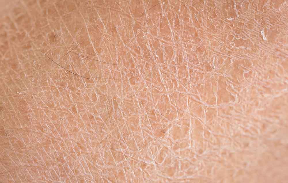
Ichthyosis refers to a group of skin disorders characterized by dry, thickened, and scaly skin. The condition can range from mild to severe, with varying degrees of impact on the patient’s quality of life. There are several forms of ichthyosis, including inherited types, which can manifest from birth, and acquired types, which develop later in life. The severity of symptoms varies widely, and treatment is focused on managing symptoms and improving skin appearance.

Nursing Paper Example on Ichthyosis

Causes

Ichthyosis is primarily caused by genetic mutations that affect the skin’s ability to retain moisture and regulate cell turnover. Inherited forms of ichthyosis are typically caused by mutations in specific genes, such as the keratin 1 and keratin 10 genes in ichthyosis vulgaris. These mutations lead to abnormal keratin production, which causes the skin cells to pile up, forming scales. Environmental triggers can also contribute to the condition, although the primary cause is genetic.

In some acquired forms, factors such as infections, metabolic disorders, and other systemic diseases may play a role. Inherited ichthyosis is often passed down in an autosomal dominant or recessive manner, depending on the specific form.

Signs and Symptoms

The most prominent symptom of ichthyosis is the presence of thickened, scaly skin that may cover large areas of the body. The scales can range from fine, powdery flakes to large, thick plaques that are often dry and cracked. Common sites of involvement include the limbs, torso, and scalp, although the condition can affect any part of the body. The severity of scaling varies and can worsen in cold weather or with dry skin conditions. In some forms of ichthyosis, the skin may be red, inflamed, or painful, especially during flare-ups.

Other symptoms may include itching, increased sensitivity to temperature changes, and skin fissures that can lead to secondary infections.

Etiology

Ichthyosis is caused by mutations in genes responsible for skin cell function. For example, ichthyosis vulgaris, the most common form, results from a mutation in the filaggrin gene. This protein is important for maintaining the skin’s barrier function, and its absence or dysfunction leads to skin dryness and scaling. X-linked recessive ichthyosis is another inherited form caused by mutations in the steroid sulfatase gene, which leads to a buildup of certain substances that impair skin turnover.

Other rarer forms of ichthyosis are caused by defects in proteins responsible for skin development or lipid metabolism. Acquired ichthyosis, while less common, can be associated with systemic conditions such as HIV, cancer, hypothyroidism, and sarcoidosis, all of which may lead to abnormal skin changes resembling ichthyosis.

Pathophysiology

The pathophysiology of ichthyosis involves a disruption in the normal processes of skin cell production, differentiation, and desquamation. In healthy skin, the stratum corneum, the outermost layer, is composed of dead skin cells that are continuously shed and replaced. In individuals with ichthyosis, abnormal keratinization leads to the accumulation of thickened skin cells, resulting in the formation of scales.

The defective genes responsible for ichthyosis disrupt the structure and function of the skin’s outer layer, impairing its ability to retain moisture and protect against environmental stressors. As a result, the skin becomes dry, rough, and prone to cracking. In some forms of ichthyosis, there may be an overproduction of lipids or altered lipid composition in the skin, further contributing to the formation of scales. This process not only affects the appearance of the skin but also its ability to function as a barrier, leaving individuals more vulnerable to infections and other complications.

DSM-5 Diagnosis

There is no specific diagnostic category for ichthyosis in the Diagnostic and Statistical Manual of Mental Disorders (DSM-5) since it is a dermatological condition rather than a psychiatric one. Diagnosis is based on clinical evaluation, including a thorough patient history and physical examination. In most cases, the characteristic appearance of the skin lesions is enough for diagnosis, although genetic testing can confirm the specific form of ichthyosis. In some cases, a skin biopsy may be performed to assess the thickness and composition of the skin layers.

Family history is also an important factor, as many forms of ichthyosis are inherited in an autosomal dominant or recessive pattern. Genetic counseling may be recommended for families affected by hereditary forms of ichthyosis to understand the inheritance pattern and potential risks for future generations.

Treatment Regimens

Management of ichthyosis focuses on alleviating symptoms and preventing complications, as there is currently no cure for the condition. Topical treatments, such as moisturizers, emollients, and keratolytics (e.g., urea, salicylic acid), are commonly used to hydrate the skin and reduce scaling. For more severe cases, prescription-strength creams containing tretinoin or corticosteroids may be used to help normalize skin cell turnover and reduce inflammation.

Oral retinoids such as acitretin may be prescribed for more widespread or severe cases, particularly in conditions like lamellar ichthyosis. In cases where ichthyosis is associated with an underlying systemic condition, treating the primary disease may help improve the skin symptoms.

It is also essential for patients to avoid triggers such as harsh soaps, hot water, and environmental irritants that can worsen dryness and scaling. Regular bathing with lukewarm water and the use of soap substitutes are often recommended to help maintain skin hydration.

Patient Education

Educating patients with ichthyosis about proper skin care is critical to managing the condition effectively. Regular moisturizing is essential, as it helps prevent the skin from becoming dry and cracked. Patients should be advised to apply moisturizers immediately after bathing to lock in moisture. Gentle skin care products, including non-soap cleansers and lukewarm water, should be used to avoid further irritation.

In addition, patients should avoid tight clothing and friction in areas where scaling is prominent, as this can worsen the condition. Sunscreen should be applied regularly to protect the skin from UV damage, as patients with ichthyosis may have increased sensitivity to the sun. Genetic counseling is recommended for patients with hereditary forms of ichthyosis to help them understand the potential risks for family members. Regular follow-up with a dermatologist is important to monitor disease progression and adjust treatment as needed.

Conclusion

Ichthyosis is a group of skin disorders characterized by dry, scaly skin due to defective skin cell production and differentiation. While the condition is often genetic in origin, environmental factors and systemic conditions can also contribute to the disease. Treatment focuses on symptom management, including moisturizing, keratolytics, and oral retinoids for severe cases. Patient education plays a key role in managing the condition, with advice on skin care, avoiding triggers, and protecting the skin from environmental damage. Despite the chronic nature of the disease, early intervention and appropriate treatment can significantly improve the quality of life for individuals affected by ichthyosis.

References

Tobin, D. J., & Gawkrodger, D. J. (2020). Ichthyosis: Clinical presentation and management. American Journal of Clinical Dermatology, 21(3), 347-357. https://link.springer.com/article/10.1007/s40257-020-00507-2

Zouboulis, C. C., & Bechara, F. G. (2021). Ichthyosis: Diagnosis, pathophysiology, and treatment. Dermatologic Clinics, 39(2), 167-183. https://doi.org/10.1016/j.det.2020.11.007

Bialynicki-Birula, R., & Wróblewska, A. (2020). The genetics of ichthyosis: Pathogenesis and clinical presentation. Journal of the European Academy of Dermatology and Venereology, 34(5), 1015-1024. https://doi.org/10.1111/jdv.16456

 
Do you need a similar assignment done for you from scratch? Order now!
Use Discount Code "Newclient" for a 15% Discount!

Nursing Paper Example on Hidradenitis Suppurativa

Nursing Paper Example on Hidradenitis Suppurativa

Hidradenitis suppurativa (HS) is a chronic skin condition characterized by the formation of painful, inflamed lumps and abscesses in areas where skin rubs together, such as the armpits, groin, buttocks, and under the breasts. This condition primarily affects hair follicles and sweat glands, leading to the development of lesions that may drain pus or blood. Over time, repeated flare-ups can result in scar tissue and tunnels under the skin. HS is often misdiagnosed or dismissed as simple acne or boils, making timely treatment critical to reducing complications.

Nursing Paper Example on Hidradenitis Suppurativa

Causes

The exact cause of HS is not fully understood, but genetic and environmental factors are believed to play a role. A major contributing factor is inflammation within the hair follicles and sweat glands. The condition has a strong genetic component, as it is often seen in families, suggesting an inherited predisposition.

Environmental triggers such as obesity, smoking, hormonal changes, and friction from tight clothing can exacerbate symptoms. Lifestyle factors, particularly smoking, are significant risk factors for the disease, worsening the severity and frequency of flare-ups. Additionally, alterations in immune system functioning are thought to play a role in the abnormal inflammatory response associated with HS.

While it is not classified as an autoimmune disorder, the body’s immune system appears to mistakenly attack the skin’s hair follicles, causing the inflammation and abscesses that define the disease. However, not all cases of HS are genetically determined, and environmental triggers can also play a key role in the development and exacerbation of the condition.

Signs and Symptoms

The hallmark symptoms of hidradenitis suppurativa are painful, inflamed lumps or abscesses in the skin’s folds, often leading to abscess formation and pus drainage. These lumps may rupture over time, releasing foul-smelling fluid. These painful lumps can recur in the same areas, causing the formation of tunnel-like structures under the skin called sinus tracts. Over time, repeated flare-ups lead to the thickening of the skin and the formation of scar tissue. The affected areas typically include the armpits, groin, buttocks, and under the breasts, but any skin fold can be involved.

Early-stage symptoms may present as small, pimple-like lumps that progress into larger abscesses. As the disease progresses, these areas may become swollen, red, and tender. Individuals may also experience an increased frequency of flare-ups, leading to chronic discomfort and even disability in severe cases. In addition to visible skin changes, patients with hidradenitis suppurativa may experience systemic symptoms such as fever and fatigue during active flare-ups.

Etiology

The precise etiology of hidradenitis suppurativa remains unclear, though it is thought to involve a combination of genetic, environmental, and immunological factors. A family history of the condition is a strong indicator of a genetic predisposition, suggesting that certain genetic mutations may contribute to its onset. Mutations in the NF-kB pathway, which regulates immune responses and inflammation, have been linked to HS. This pathway’s dysfunction can result in an exaggerated inflammatory response in the skin’s hair follicles and sweat glands.

Environmental factors, particularly smoking, obesity, and excessive friction or sweating, are known to increase the risk of HS flare-ups. Hormonal changes, especially those occurring during puberty or menstruation, are also considered important factors in the development of the disease, with many individuals noticing a worsening of symptoms during these times. The inflammation in HS is not caused by bacterial infection, though infected abscesses can develop secondary to the primary condition.

Pathophysiology

Hidradenitis suppurativa is believed to arise from a combination of hyperkeratinization and inflammation of the hair follicles, particularly in areas of friction. This inflammation leads to the blockage of sweat glands, followed by the formation of painful lumps and abscesses. Over time, these abscesses may rupture, causing the release of pus or blood. Repeated flare-ups lead to the formation of scar tissue, and the development of sinus tracts and fistulas, which are abnormal connections between skin surfaces.

The process is thought to involve an abnormal immune response where the body’s immune system, instead of protecting the skin, triggers inflammation in the affected areas. This chronic inflammation results in the progressive damage to skin structures and the underlying tissue.

Over time, individuals with HS may experience significant scarring, thickened skin, and the development of painful, interconnected tunnels under the skin. In severe cases, extensive scarring can lead to functional and aesthetic impairments, and complications such as secondary infections and mobility limitations may occur.

DSM-5 Diagnosis

Hidradenitis suppurativa is not specifically classified in the DSM-5, as it is a dermatologic condition rather than a psychiatric disorder. Diagnosis is primarily clinical and involves evaluating the patient’s symptoms, medical history, and physical examination findings. No definitive laboratory test or imaging is required for the diagnosis, but it may be confirmed through the identification of characteristic skin lesions in areas of friction.

In some cases, biopsy or culture may be necessary to rule out other conditions or confirm the presence of infection. The diagnosis is typically made based on the presence of recurrent, painful abscesses, sinus tracts, and scarring, often in the armpits, groin, buttocks, and under the breasts. In some cases, associated systemic symptoms such as fever or malaise may also aid in diagnosis.

Treatment Regimens

Management of hidradenitis suppurativa focuses on controlling inflammation, preventing infection, and reducing the frequency and severity of flare-ups. Mild cases may be treated with topical antibiotics or corticosteroids to reduce inflammation and prevent infection. In more severe cases, oral antibiotics such as tetracycline or doxycycline may be prescribed to reduce bacterial colonization and inflammation.

Additionally, biologic agents targeting the immune system, such as adalimumab, have shown promise in reducing flare-ups and managing the condition long-term. Surgical intervention may be necessary in advanced cases to remove abscesses or to drain cysts and sinus tracts. In some instances, patients may require skin grafts to restore the affected areas after significant scarring.

Lifestyle changes, including weight management and smoking cessation, are essential components of treatment to reduce flare-up frequency and severity. Pain management may also be part of the treatment regimen, with nonsteroidal anti-inflammatory drugs (NSAIDs) or opioids being used in severe cases. Early intervention is key to preventing long-term complications and improving quality of life.

Patient Education

Patient education is an integral part of managing hidradenitis suppurativa. Educating patients on the nature of the disease, its triggers, and the importance of early intervention can help reduce the severity of symptoms and improve outcomes.

Smoking cessation is strongly recommended, as tobacco use is a major risk factor for flare-ups. Patients should also be educated on the role of weight management in controlling the condition, as obesity can exacerbate symptoms. In addition, patients should be instructed on proper skin hygiene to reduce irritation and the risk of infection in affected areas. For those with recurrent flare-ups, it is important to avoid tight clothing or friction in areas prone to the condition.

Medications prescribed for HS, including antibiotics or biologics, should be taken as directed, and patients should be aware of potential side effects. In cases of severe disease, patients should be informed about the potential need for surgery, and psychological support may be necessary to address the emotional and social challenges associated with living with a chronic skin condition.

Conclusion

Hidradenitis suppurativa is a chronic and often painful condition that primarily affects areas where skin rubs together, such as the armpits and groin. Although the exact cause remains unclear, it is believed to involve a combination of genetic, environmental, and immunological factors. Early intervention with lifestyle modifications, medications, and, when necessary, surgery, can help manage the disease and improve the quality of life for affected individuals. Educating patients on the importance of avoiding triggers, maintaining proper hygiene, and adhering to treatment plans is essential to managing the condition effectively.

References

Ting, T. Y., & Tey, H. L. (2021). Hidradenitis suppurativa: Epidemiology, pathogenesis, and diagnosis. Journal of Clinical Medicine, 10(11), 2345. https://www.ncbi.nlm.nih.gov/pmc/articles/PMC8002679/

Zouboulis, C. C., & Bechara, F. G. (2020). Hidradenitis suppurativa: Current concepts in pathogenesis and treatment. Journal of Dermatology, 47(7), 712-725. https://doi.org/10.1111/1346-8138.15431

Saunte, D. M. L., & Jemec, G. B. E. (2020). Hidradenitis suppurativa: Advances in diagnosis and treatment. Journal of the American Academy of Dermatology, 82(2), 323-333. https://doi.org/10.1016/j.jaad.2019.06.036

 
Do you need a similar assignment done for you from scratch? Order now!
Use Discount Code "Newclient" for a 15% Discount!

Nursing Paper Example on Henoch-Schönlein purpura

Nursing Paper Example on Henoch-Schönlein purpura

Henoch-Schönlein purpura (HSP) is a form of small-vessel vasculitis that most commonly affects children but can also occur in adults. The disease is characterized by the deposition of immunoglobulin A (IgA) complexes in the blood vessels, which leads to inflammation. HSP typically presents with a purpuric rash, joint pain, abdominal pain, and kidney involvement. The pathophysiology of the disease is complex and not fully understood, though it is often triggered by infections, medications, or other environmental factors.

Nursing Paper Example on Henoch-Schönlein purpura

Causes

Henoch-Schönlein purpura is often triggered by a variety of factors, with infections being the most common cause. Upper respiratory infections, particularly those caused by viral agents such as the adenovirus, parvovirus, and influenza, are frequently linked to the onset of HSP. Streptococcus infections, often in the form of strep throat, are another common trigger.

In addition to infections, certain medications have been implicated in triggering the disease. These include nonsteroidal anti-inflammatory drugs (NSAIDs), antibiotics, and vaccinations, which can induce immune responses that lead to the deposition of immunoglobulin A (IgA) in blood vessel walls.

Genetic factors also play a role in the development of HSP. Individuals with a family history of autoimmune diseases or vasculitis may be more susceptible to the condition. Research indicates that abnormalities in immune system regulation, particularly involving IgA, contribute to the pathogenesis of the disease.

Environmental factors, such as exposure to pollutants or changes in climate, may further contribute to the development of Henoch-Schönlein purpura in genetically predisposed individuals. However, the interplay between genetics, environmental factors, and immune dysregulation remains complex and not fully understood.

Signs and Symptoms

The primary symptom of Henoch-Schönlein purpura is the presence of purpura, which refers to purple spots on the skin caused by bleeding underneath. These spots typically appear on the lower extremities and buttocks, and may also be seen in the arms. The skin lesions can range from small, red dots to larger bruises.

Abdominal pain is another hallmark symptom of HSP, affecting many patients. This pain is often cramp-like and can be intense, sometimes accompanied by nausea and vomiting. It is important to note that abdominal pain may mimic other conditions like appendicitis, making diagnosis challenging.

Joint pain and swelling are also common, particularly affecting the knees and ankles. In some cases, arthritis-like symptoms may occur, leading to discomfort and limited mobility.

Additionally, some individuals with HSP experience kidney involvement, which can manifest as hematuria (blood in the urine) or proteinuria (excess protein in the urine). If left untreated, kidney damage may occur, potentially leading to renal failure.

Fever is present in some patients, and systemic symptoms like fatigue and malaise can contribute to the overall discomfort. These symptoms may occur suddenly and often follow an upper respiratory infection or other triggering event.

Etiology

The exact cause of Henoch-Schönlein purpura (HSP) remains unclear, but it is thought to be triggered by a variety of factors that lead to an immune system response. In many cases, HSP follows a preceding infection, most commonly a respiratory infection caused by viruses like adenovirus or streptococcus. These infections seem to prompt the immune system to produce antibodies, which then form immune complexes that deposit in blood vessels, leading to inflammation and the hallmark symptoms of HSP, such as purpura.

Genetics may also play a role in the development of HSP, as certain family members may have an increased risk of the disease, although no specific gene has been definitively linked to the disorder. HSP is more common in children than adults, with male children being more affected than females.

Other factors that might contribute to HSP include drug reactions, such as to antibiotics or nonsteroidal anti-inflammatory drugs (NSAIDs), and autoimmune conditions. The inflammatory response in HSP is thought to result in vasculitis, which affects small blood vessels and causes leakage of blood and fluid into the surrounding tissue, leading to symptoms like purpura and organ involvement.

Despite these associations, the precise etiology remains complex and likely involves both genetic predisposition and environmental factors.

(Nursing Paper Example on Henoch-Schönlein purpura)

Pathophysiology

The pathophysiology of Henoch-Schönlein purpura (HSP) is primarily characterized by the formation of immune complexes that deposit in small blood vessels, causing vasculitis. The immune complexes, typically consisting of immunoglobulin A (IgA), are triggered by an initial infection or other stimuli. Once these complexes accumulate in the walls of blood vessels, especially in the skin, kidneys, joints, and gastrointestinal tract, they lead to inflammation.

The deposition of these complexes activates the complement system, which amplifies the inflammatory response. This results in the damage and leakage of blood vessels, causing the characteristic symptoms of HSP. The skin manifestations, such as purpura, occur due to hemorrhage from the ruptured blood vessels, while the renal involvement can lead to glomerulonephritis. This can sometimes progress to kidney damage if left untreated.

In the gastrointestinal tract, inflammation may result in abdominal pain, nausea, and vomiting, while joint involvement leads to swelling and pain. The overall inflammation seen in HSP is a result of both the immune complexes and the subsequent activation of inflammatory pathways, including T-cell-mediated responses, further promoting vasculitis. This disorder primarily affects small vessels but can involve medium-sized vessels in severe cases, leading to more widespread tissue damage.

DSM-5 Diagnosis

Henoch-Schönlein purpura (HSP) is not explicitly listed as a separate disorder in the DSM-5, as it is primarily considered a physical, autoimmune condition rather than a psychiatric disorder. Therefore, HSP does not have specific diagnostic criteria outlined in the DSM-5. Instead, its diagnosis is based on clinical features and laboratory findings, including the presence of palpable purpura, abdominal pain, kidney involvement (hematuria or proteinuria), and arthritis or arthralgia.

The clinical diagnosis of HSP is often confirmed by identifying characteristic skin lesions and the presence of IgA deposits in affected tissues. In addition, a thorough history and clinical presentation are essential for differentiating HSP from other vasculitides or systemic diseases that may present with similar symptoms.

While the DSM-5 does not address HSP, clinicians can use it to rule out other psychiatric or psychosomatic disorders if the patient presents with associated anxiety, depression, or other psychological symptoms secondary to the physical condition. It is important to note that managing HSP often requires a multidisciplinary approach, including pediatricians, rheumatologists, and nephrologists, rather than a focus on psychiatric diagnosis alone.

Treatment Regimens

The treatment of Henoch-Schönlein purpura (HSP) primarily focuses on managing symptoms, preventing complications, and addressing underlying causes when possible. Most cases are self-limiting, requiring only supportive care. However, in severe cases, especially those involving kidney dysfunction or gastrointestinal symptoms, more intensive interventions may be necessary.

For mild cases, the use of nonsteroidal anti-inflammatory drugs (NSAIDs) can help alleviate joint pain and inflammation. Corticosteroids, such as prednisone, are commonly used for moderate to severe cases, particularly when there is significant kidney involvement, gastrointestinal bleeding, or severe purpura. These medications help to reduce inflammation and prevent further damage to organs.

In cases where renal involvement progresses to nephrotic syndrome or significant proteinuria, immunosuppressive therapies such as cyclophosphamide or azathioprine may be considered to control the inflammatory process. Rituximab has also been explored as a treatment option in resistant or recurrent cases of HSP.

For patients with severe gastrointestinal manifestations or life-threatening bleeding, intravenous immunoglobulin (IVIG) may be used to reduce inflammation and support immune function. Additionally, ongoing monitoring of renal function is crucial to detect any potential complications early, especially in children and those with long-term symptoms.

The treatment plan for HSP should always be tailored to the individual, taking into account the severity of the disease and the presence of systemic complications.

(Nursing Paper Example on Henoch-Schönlein purpura)

Patient Education

For patients diagnosed with Henoch-Schönlein purpura (HSP), education about the disease and its management is crucial. HSP is often self-limiting, but patients need to understand the importance of monitoring symptoms and managing potential complications.

Patients should be advised to rest and avoid activities that might worsen joint pain or skin lesions. It is essential to keep the skin clean and dry to prevent infection in areas affected by purpura. As the disease can cause abdominal pain and gastrointestinal symptoms, a low-fat, easy-to-digest diet may help reduce discomfort.

Managing pain and inflammation with over-the-counter medications, such as acetaminophen or ibuprofen, should be discussed. However, nonsteroidal anti-inflammatory drugs should be used cautiously in patients with kidney involvement. In more severe cases, corticosteroids or immunosuppressive treatments may be prescribed, and patients must follow the prescribed medication regimen closely.

Close monitoring of kidney function is necessary, as HSP can affect the kidneys, leading to proteinuria or even nephritis. Regular check-ups with a healthcare provider to assess kidney health, as well as blood pressure monitoring, should be part of ongoing care.

Patients should be informed about the possible recurrence of the disease and when to seek medical attention if symptoms worsen, particularly if there are signs of kidney problems or gastrointestinal bleeding.

(Nursing Paper Example on Henoch-Schönlein purpura)

Conclusion

Henoch-Schönlein purpura is a small-vessel vasculitis primarily affecting children, though it can occur in adults. The disease involves immune-mediated damage caused by the deposition of IgA in the blood vessels. Common symptoms include purpura, arthritis, abdominal pain, and kidney involvement. The etiology of HSP is multifactorial, with infections and immune system dysregulation being key contributors. Although the disease is typically self-limiting, it can lead to long-term complications, particularly involving the kidneys. Treatment focuses on symptom management with NSAIDs, corticosteroids, and immunosuppressive medications when necessary. Patient education on recognizing symptoms and monitoring kidney function is crucial for managing the disease and preventing long-term consequences. Regular follow-up care is important, especially for individuals with significant kidney involvement, as early intervention can improve outcomes.

References

Ozen, S., & Pistorio, A. (2010). Epidemiology of Henoch-Schönlein purpura. Rheumatology, 49(6), 1035-1042. https://doi.org/10.1093/rheumatology/keq340

Lee, T. H., & So, K. M. (2014). Henoch-Schönlein purpura: Pathophysiology and diagnosis. The Journal of Pediatric Pharmacology and Therapeutics, 19(2), 75-81. https://www.jpedpharm.org/doi/10.5863/1551-6776-19.2.75

Kuo, C. F., & See, L. C. (2011). Henoch-Schönlein purpura in children: Epidemiology, clinical features, and complications. Pediatrics, 128(3), e675-e683. https://doi.org/10.1542/peds.2010-3734

 
Do you need a similar assignment done for you from scratch? Order now!
Use Discount Code "Newclient" for a 15% Discount!

Nursing Paper Example on Hashimoto’s Thyroiditis

Nursing Paper Example on Hashimoto’s Thyroiditis

Hashimoto’s thyroiditis, also known as chronic lymphocytic thyroiditis, is an autoimmune disorder that causes inflammation and gradual destruction of the thyroid gland. It is the most common cause of hypothyroidism in iodine-sufficient regions and predominantly affects women, particularly between the ages of 30 and 50. This condition results from an immune system malfunction where antibodies attack thyroid tissue, leading to reduced thyroid hormone production. Hashimoto’s thyroiditis often progresses slowly, with symptoms developing over months or years.

Nursing Paper Example on Hashimoto's Thyroiditis

Causes

The exact causes of Hashimoto’s thyroiditis involve a combination of genetic, environmental, and immune system factors. Genetics play a significant role, as individuals with a family history of autoimmune diseases are more likely to develop the condition. Specific genes, such as those associated with immune regulation, including the human leukocyte antigen (HLA) system, are linked to increased susceptibility.

Environmental factors also contribute to the development of Hashimoto’s thyroiditis. Excessive iodine intake, either from supplements or dietary sources, can trigger or worsen the autoimmune response in susceptible individuals. Infections, particularly viral infections, may initiate an immune reaction through molecular mimicry, where the immune system mistakenly attacks thyroid tissue. Chronic stress has been associated with the onset or exacerbation of autoimmune conditions, including Hashimoto’s thyroiditis.

The immune system’s malfunction underpins the disease process. In Hashimoto’s thyroiditis, the immune system generates antibodies, including thyroid peroxidase and thyroglobulin antibodies, which target the thyroid gland. This autoimmune response leads to inflammation, tissue destruction, and a gradual decline in thyroid hormone production. Coexisting autoimmune disorders, such as rheumatoid arthritis or lupus, further increase the likelihood of developing Hashimoto’s thyroiditis.

Understanding these causes helps clinicians identify at-risk individuals and implement preventive or early intervention strategies to manage the disease effectively.

Signs and Symptoms

Hashimoto’s thyroiditis often progresses slowly, with subtle symptoms that develop as thyroid function declines. Early signs may include fatigue, sensitivity to cold, and unexplained weight gain due to a slowed metabolism. A feeling of fullness or discomfort in the neck may occur if the thyroid gland becomes enlarged, forming a painless goiter.

As the condition progresses, other symptoms may arise, including dry skin, hair thinning, and brittle nails, reflecting the body’s reduced thyroid hormone levels. Cognitive symptoms, such as difficulty concentrating or memory issues often described as “brain fog,” are common. Additionally, individuals may experience mood changes, including depression or irritability.

Women with Hashimoto’s thyroiditis often report menstrual irregularities, such as heavy or prolonged periods, or difficulties with fertility. Muscle weakness, joint stiffness, or generalized body aches can develop, further affecting daily life. Advanced cases may cause hoarseness, puffiness in the face, or swelling in the extremities due to fluid retention.

These diverse symptoms often overlap with other conditions, complicating early diagnosis. Recognizing this broad spectrum is crucial for timely identification and management to prevent complications such as severe hypothyroidism or myxedema, a rare but life-threatening condition. Regular medical evaluation helps monitor symptoms and thyroid function effectively.

Etiology

Hashimoto’s thyroiditis arises due to a combination of genetic, environmental, and immune factors. It is classified as an autoimmune disorder, where the immune system mistakenly attacks thyroid tissue. Genetic predisposition plays a key role, as individuals with family histories of autoimmune conditions are at greater risk. Specific genes linked to immune regulation, such as HLA-DR, are implicated.

Environmental triggers also contribute to the development of Hashimoto’s thyroiditis. Exposure to high levels of iodine, selenium deficiencies, and certain infections are known to initiate the autoimmune response in genetically predisposed individuals. Viral infections like hepatitis C or Epstein-Barr virus may also disrupt immune tolerance and trigger thyroid autoimmunity.

Hormonal factors are significant, especially in women, who are more commonly affected. Fluctuations in estrogen levels during pregnancy or menopause may enhance susceptibility. Stressful life events or psychological stress can also exacerbate immune dysregulation.

The disease is mediated by the production of autoantibodies, particularly against thyroid peroxidase and thyroglobulin. These antibodies target and gradually destroy thyroid cells, impairing hormone synthesis. Over time, this immune-mediated damage results in hypothyroidism. Understanding this multifactorial etiology is critical for developing preventive strategies and targeted therapies.

Pathophysiology

Hashimoto’s thyroiditis is an autoimmune condition in which the immune system attacks the thyroid gland, impairing its function. The process begins with genetic susceptibility and environmental triggers, leading to immune dysregulation. Key mechanisms involve the activation of CD4+ T helper cells that recognize thyroid antigens as foreign. This immune response stimulates the production of autoantibodies, including anti-thyroid peroxidase and anti-thyroglobulin antibodies.

These autoantibodies bind to thyroid cells, marking them for destruction. Concurrently, cytotoxic T lymphocytes infiltrate the thyroid gland, releasing inflammatory cytokines like interleukin-2 and tumor necrosis factor-alpha. This inflammatory cascade damages thyroid follicles, disrupting hormone synthesis and leading to reduced levels of thyroxine (T4) and triiodothyronine (T3).

As the gland is progressively destroyed, compensatory mechanisms such as increased thyroid-stimulating hormone production from the pituitary gland attempt to restore hormone levels. However, the ongoing immune-mediated destruction eventually surpasses the gland’s capacity to compensate, resulting in hypothyroidism.

The chronic inflammation can also lead to fibrosis and thyroid gland enlargement, forming a characteristic goiter. The interplay between immune dysregulation, genetic factors, and environmental triggers drives this progressive and multifaceted disease process.

DSM-5 Diagnosis

Hashimoto’s thyroiditis is not classified as a distinct diagnosis in the Diagnostic and Statistical Manual of Mental Disorders, Fifth Edition (DSM-5). However, its associated psychiatric and neuropsychological symptoms may warrant evaluation under DSM-5 criteria. Mental health issues such as depression, anxiety, and cognitive dysfunction can emerge due to hypothyroidism or autoimmune processes.

To assess mood disorders in Hashimoto’s thyroiditis, clinicians evaluate depressive symptoms using DSM-5 criteria for major depressive disorder. Symptoms like persistent sadness, fatigue, and sleep disturbances are common in individuals with hypothyroidism. Anxiety disorders, including generalized anxiety disorder, may also align with DSM-5 diagnostic standards, particularly when patients experience heightened worry or physical tension.

Cognitive impairments related to Hashimoto’s thyroiditis, often described as “brain fog,” may fall under neurocognitive disorders in DSM-5 if they interfere significantly with daily functioning. These issues highlight the interplay between thyroid function and mental health.

While DSM-5 is not specific to thyroid conditions, its framework assists in diagnosing coexisting psychiatric conditions. A comprehensive clinical assessment of physical and mental symptoms, combined with laboratory testing of thyroid hormones and antibodies, provides a holistic understanding of Hashimoto’s thyroiditis and its impact on mental health.

Diagnosis

Diagnosing Hashimoto’s thyroiditis involves a combination of clinical evaluation, laboratory tests, and imaging studies. Physicians assess the patient’s symptoms, including fatigue, weight gain, and cold intolerance, which suggest hypothyroidism. A detailed medical history, including family history of autoimmune diseases, provides critical diagnostic clues.

Laboratory evaluation focuses on thyroid hormone levels and antibodies. Low serum levels of free thyroxine (T4) and elevated thyroid-stimulating hormone (TSH) indicate hypothyroidism. The presence of antithyroid peroxidase (anti-TPO) antibodies confirms the autoimmune nature of the condition. In some cases, antithyroglobulin antibodies may also be detected.

Ultrasound imaging helps assess the thyroid gland’s structure. Common findings include reduced echogenicity, heterogeneity, and nodules, indicating inflammation or fibrosis. These imaging features support the diagnosis and help exclude malignancy.

Fine-needle aspiration biopsy may be performed in cases of suspicious thyroid nodules. This technique rules out thyroid cancer and confirms the inflammatory nature of the glandular tissue.

Accurate diagnosis integrates clinical features, laboratory findings, and imaging results. Early identification of Hashimoto’s thyroiditis is crucial for initiating treatment to prevent complications like severe hypothyroidism, cardiovascular issues, or goiter. Comprehensive evaluation ensures effective management and symptom relief.

Treatment Regimens

The primary treatment for Hashimoto’s thyroiditis is thyroid hormone replacement therapy, typically with levothyroxine, a synthetic form of thyroxine (T4). This therapy is designed to normalize thyroid hormone levels, alleviating symptoms of hypothyroidism such as fatigue, weight gain, and cold intolerance. The dosage is adjusted based on regular blood tests that measure TSH and free T4 levels.

Levothyroxine is usually taken orally once a day, and the dose is individualized for each patient. Regular monitoring of thyroid function is essential, especially during the first few months of treatment, to prevent over- or under-treatment. If a patient has difficulty swallowing pills, liquid forms of levothyroxine can be prescribed.

In cases where a goiter (enlarged thyroid) is present, treatment with levothyroxine may help reduce its size. However, if the goiter is large and causing difficulty in swallowing or breathing, surgery may be considered.

Although there is no cure for Hashimoto’s thyroiditis, thyroid hormone replacement is highly effective in managing the disease and preventing complications related to hypothyroidism. In rare cases, patients may require additional therapies if complications arise, such as in those with severe hypothyroidism or who are pregnant. In these cases, close monitoring and adjustments in treatment are crucial.

Patient Education

For individuals diagnosed with Hashimoto’s thyroiditis, patient education is vital to ensure proper management of the condition. The most important aspect is the adherence to thyroid hormone replacement therapy, typically with levothyroxine, to regulate thyroid hormone levels. Patients must understand the importance of taking their medication consistently, usually once daily on an empty stomach, to maximize absorption.

Patients should be informed about potential side effects of thyroid medication, such as heart palpitations or nervousness, which may indicate over-replacement. Regular blood tests will be required to monitor thyroid function and adjust the dosage as needed. It is essential for patients to attend follow-up appointments for ongoing management and to discuss any concerns with their healthcare provider.

Patients should also be educated about the symptoms of hypothyroidism, including fatigue, weight gain, and cold intolerance, so they can monitor their condition and seek adjustments in treatment when necessary. Additionally, lifestyle modifications such as a healthy diet, regular exercise, and stress management can help improve overall well-being.

Pregnant women with Hashimoto’s thyroiditis must be closely monitored as proper thyroid function is crucial for fetal development. With appropriate treatment and care, most individuals with Hashimoto’s thyroiditis can lead normal, healthy lives.

Conclusion

Hashimoto’s thyroiditis is a common autoimmune disorder that leads to hypothyroidism and affects thyroid function. The causes include genetic predisposition, environmental factors, and immune system dysfunction. Early diagnosis, based on clinical symptoms, laboratory tests, and imaging, is critical for effective management. Patients require thyroid hormone replacement therapy, most commonly levothyroxine, with regular monitoring of thyroid levels. Lifestyle modifications and adherence to medication play an important role in managing symptoms and preventing complications. Pregnant women and those with comorbid conditions need additional monitoring. Overall, with timely treatment and patient education, individuals can manage the condition and maintain a good quality of life.

References

American Thyroid Association. (2023). Hashimoto’s thyroiditis: Diagnosis and treatment. https://www.thyroid.org
Leung, A. M., & Braverman, L. E. (2022). Thyroid dysfunction in autoimmune disease. Journal of Clinical Endocrinology. https://www.jcem.org
McLeod, D. S. A., & Cooper, D. S. (2021). The pathogenesis of Hashimoto’s thyroiditis. Endocrine Reviews. https://www.endocrinereviews.org
National Institute of Diabetes and Digestive and Kidney Diseases. (2023). Autoimmune thyroid disorders. https://www.niddk.nih.gov
Roberts, C. G., & Ladenson, P. W. (2020). Hypothyroidism management in primary care. Annals of Internal Medicine. https://www.acpjournals.org

 
Do you need a similar assignment done for you from scratch? Order now!
Use Discount Code "Newclient" for a 15% Discount!

Nursing Paper Example on HFMD

Nursing Paper Example on HFMD

Hand, foot, and mouth disease (HFMD) is a contagious viral illness predominantly affecting young children but can occur in individuals of all ages. The condition is caused by enteroviruses, with coxsackievirus A16 and enterovirus 71 being the most common culprits. Characterized by fever, painful sores in the mouth, and a rash on the hands and feet, HFMD is typically mild but may occasionally lead to complications. It spreads through respiratory droplets, direct contact with contaminated surfaces, or fecal matter.

Nursing Paper Example on HFMD

Causes

HFMD is caused by viruses belonging to the Enterovirus genus, primarily coxsackievirus A16 and enterovirus 71. These viruses thrive in the gastrointestinal tract and are highly contagious. Infections are more common in warm climates and during summer and fall.

The disease is spread through close contact, respiratory droplets, and fecal-oral routes, with outbreaks often occurring in schools and childcare settings. Viral shedding in stool can persist for weeks after symptoms resolve, increasing the risk of transmission.

Signs and Symptoms

The incubation period for HFMD is typically three to six days. Early symptoms include fever, sore throat, reduced appetite, and general malaise. Painful ulcers may develop in the mouth, particularly on the tongue, gums, and inner cheeks, leading to difficulty swallowing. A characteristic rash appears on the hands, feet, and sometimes the buttocks, progressing to vesicular lesions.

In some cases, individuals may experience irritability, excessive drooling, or vomiting. Severe cases involving enterovirus 71 can lead to neurological complications such as meningitis, encephalitis, or polio-like paralysis.

Etiology

The etiology of HFMD involves infection with enteroviruses, with different strains causing variations in disease severity. Enterovirus 71 has been associated with more severe cases and complications compared to coxsackievirus A16.

Children under the age of five are most vulnerable due to their developing immune systems and frequent exposure in communal settings. Factors such as poor hygiene, crowded environments, and close physical contact contribute to the rapid spread of the virus.

Pathophysiology

The virus enters the body through the oral or respiratory tract, replicating in the mucosa and lymphoid tissue. From there, it disseminates to the bloodstream, targeting specific organs. The development of oral ulcers and rashes is due to viral replication and immune-mediated responses.

The virus’s affinity for neural and cardiac tissues explains the potential for severe complications, including aseptic meningitis, encephalitis, or myocarditis. Host immune responses, including inflammation and cytokine production, contribute to symptom development and resolution.

(Nursing Paper Example on HFMD)

Diagnosis

HFMD diagnosis is primarily clinical, based on characteristic symptoms such as fever, oral ulcers, and rash distribution. A detailed history of exposure and symptom onset aids diagnosis. Laboratory confirmation may involve polymerase chain reaction (PCR) testing of throat swabs, stool samples, or vesicular fluid to identify the specific enterovirus. Serological testing is less commonly used but may support diagnosis in complex cases or outbreaks.

Treatment Regimens

There is no specific antiviral treatment for HFMD. Management focuses on symptom relief and supportive care. Over-the-counter pain relievers such as acetaminophen or ibuprofen alleviate fever and discomfort. Topical oral gels or rinses can reduce pain from mouth sores.

Maintaining hydration is crucial, as painful swallowing may lead to reduced fluid intake. Severe cases with complications like meningitis or encephalitis may require hospitalization, intravenous fluids, or antiviral therapy under specific circumstances. Strict hygiene practices help prevent further transmission.

Patient Education

Patient education is vital in controlling the spread and managing HFMD. Caregivers should be informed about the importance of handwashing, disinfecting surfaces, and avoiding close contact during active illness. Children with HFMD should stay home from school or daycare until fever resolves and sores heal.

Encouraging hydration and soft, cool foods minimizes discomfort from mouth sores. Parents should seek medical attention if symptoms worsen or neurological signs such as confusion, neck stiffness, or seizures develop.

(Nursing Paper Example on HFMD)

Prevention

Preventive measures include maintaining good hygiene, avoiding close contact with infected individuals, and frequent cleaning of shared surfaces. In outbreak settings, public health interventions like temporary closures of childcare facilities or schools may be necessary.

Vaccination programs targeting enterovirus 71 have been implemented in some countries, reducing severe HFMD cases.

Conclusion

Hand, foot, and mouth disease is a common but usually mild illness that predominantly affects young children. While most cases resolve without complications, severe forms can lead to life-threatening neurological or cardiac issues. Prompt diagnosis, supportive care, and public health measures are essential in managing HFMD and preventing outbreaks. Ongoing research into vaccines and antiviral therapies offers hope for improved prevention and treatment strategies in the future.

References

American Academy of Pediatrics. (2022). Hand, foot, and mouth disease clinical guidelines. Pediatrics Journal. https://www.aap.org
Xu, W., & Liu, C. (2021). Epidemiology of hand, foot, and mouth disease in Asia. Infectious Disease Reports. https://www.infectiousdiseasereports.org
Shen, L., & Ma, H. (2020). Pathogenesis of severe HFMD caused by enterovirus 71. Virology Journal. https://www.virologyjournal.com
Centers for Disease Control and Prevention. (2023). Hand, foot, and mouth disease overview. https://www.cdc.gov
World Health Organization. (2023). Prevention of hand, foot, and mouth disease in community settings. https://www.who.int

 
Do you need a similar assignment done for you from scratch? Order now!
Use Discount Code "Newclient" for a 15% Discount!

Nursing Paper Example on Graves’ Disease

Nursing Paper Example on Graves’ Disease

Graves’ disease is an autoimmune disorder and the most common cause of hyperthyroidism. It occurs when the immune system produces autoantibodies that stimulate the thyroid gland, leading to excessive production of thyroid hormones. This hormonal imbalance affects various bodily functions, including metabolism, heart rate, and thermoregulation. Graves’ disease can present at any age but is most prevalent in women aged 30 to 50. Early diagnosis and treatment are essential to manage symptoms and prevent complications.

Nursing Paper Example on Graves' Disease

Causes

Graves’ disease is caused by the production of thyroid-stimulating immunoglobulins (TSI), which mimic thyroid-stimulating hormone (TSH). These autoantibodies bind to TSH receptors on thyroid cells, prompting the gland to overproduce thyroid hormones.

Although the exact trigger remains unknown, genetic predisposition and environmental factors play significant roles. Stress, infections, and pregnancy are known to exacerbate immune dysregulation, contributing to the onset of Graves’ disease. A family history of thyroid disorders or autoimmune conditions also increases the likelihood of developing the disease.

Signs and Symptoms

Graves’ disease presents with various symptoms due to hyperthyroidism. Common signs include unexplained weight loss, heat intolerance, and increased appetite. Patients may experience palpitations, sweating, and fatigue due to the overactive metabolism. Neurological symptoms such as tremors, anxiety, and irritability are frequent.

Ocular manifestations, known as Graves’ ophthalmopathy, include eye bulging, redness, and double vision. In severe cases, skin thickening on the lower legs, termed pretibial myxedema, may occur. The intensity of symptoms varies among individuals, with some presenting mildly and others severely.

Etiology

The etiology of Graves’ disease involves immune system dysfunction. The primary autoantibody implicated is thyroid-stimulating immunoglobulin, which disrupts normal thyroid regulation. Genetic factors, including specific human leukocyte antigen (HLA) types and other immune-regulatory genes, predispose individuals to the disease.

Environmental triggers such as smoking, infections, and high iodine intake have been linked to the onset of Graves’ disease. Hormonal fluctuations, particularly in women, are also believed to influence disease development and severity.

Pathophysiology

Graves’ disease results from the interplay between genetic susceptibility and environmental triggers, leading to autoimmune activation. The thyroid-stimulating immunoglobulin activates the TSH receptor on thyroid cells, causing unregulated thyroid hormone synthesis and release. Excess thyroid hormones amplify metabolic activity and enhance sympathetic nervous system sensitivity. Graves’ ophthalmopathy arises from the autoimmune attack on orbital tissues, leading to inflammation and fibrosis. Pretibial myxedema occurs due to the deposition of glycosaminoglycans in the dermis.

Diagnosis

Graves’ disease diagnosis combines clinical evaluation, laboratory tests, and imaging studies. Blood tests reveal elevated free thyroxine (T4) and triiodothyronine (T3) levels, with suppressed TSH levels. Thyroid-stimulating immunoglobulin or TSH receptor antibody tests confirm autoimmune activity.

A radioactive iodine uptake scan demonstrates diffuse thyroid gland overactivity. Ultrasound may identify thyroid enlargement and increased vascularity. A comprehensive assessment helps distinguish Graves’ disease from other causes of hyperthyroidism, such as toxic nodular goiter.

Treatment Regimens

Treatment options for Graves’ disease aim to control hyperthyroidism, alleviate symptoms, and address autoimmune activity. Antithyroid medications such as methimazole and propylthiouracil reduce thyroid hormone production. Beta-blockers like propranolol manage cardiovascular symptoms, including palpitations and tachycardia. Radioactive iodine therapy destroys overactive thyroid tissue, leading to a reduction in hormone production.

In some cases, thyroidectomy, the surgical removal of the thyroid gland, is performed. For Graves’ ophthalmopathy, glucocorticoids and orbital decompression surgery may be necessary in severe cases.

(Nursing Paper Example on Graves’ Disease)

Patient Education

Patient education is crucial for effective management of Graves’ disease. Individuals should understand the chronic nature of the condition and the importance of adhering to treatment plans. Regular follow-up visits are essential for monitoring thyroid hormone levels and adjusting therapy. Patients should be informed about potential side effects of treatments, such as hypothyroidism following radioactive iodine therapy.

Lifestyle modifications, including stress reduction, avoiding smoking, and maintaining a balanced diet, support overall health and may reduce symptom severity.

Research and Future Directions

Ongoing research on Graves’ disease focuses on understanding its genetic basis and immune mechanisms to develop targeted therapies. Advances in biologics targeting specific immune pathways, such as monoclonal antibodies, show promise in reducing autoimmune activity.

Studies exploring the role of gut microbiota in immune regulation may provide new insights into disease prevention and management. Improved diagnostic tools for early detection and risk assessment are also under investigation.

Conclusion

Graves’ disease is a complex autoimmune disorder with diverse clinical manifestations. Its pathogenesis involves genetic and environmental factors, leading to unregulated thyroid hormone production. Early diagnosis and tailored treatment strategies are essential for managing symptoms and preventing complications. Advances in research continue to enhance understanding and provide hope for improved therapeutic options in the future.

References

Davies, T. F., & Larsen, P. R. (2022). Thyroid hormone synthesis and secretion. New England Journal of Medicine. https://www.nejm.org
Smith, T. J., & Hegedüs, L. (2023). Graves’ disease: Pathophysiology and treatment approaches. Lancet Diabetes & Endocrinology. https://www.thelancet.com
Bahn, R. S. (2020). Graves’ ophthalmopathy. New England Journal of Medicine. https://www.nejm.org
Antonelli, A., Ferrari, S. M., & Fallahi, P. (2021). Environmental triggers in Graves’ disease. Autoimmunity Reviews. https://www.sciencedirect.com
Cooper, D. S., & Ross, D. S. (2023). Antithyroid drugs in the treatment of Graves’ disease. Journal of Clinical Endocrinology & Metabolism. https://academic.oup.com

 
Do you need a similar assignment done for you from scratch? Order now!
Use Discount Code "Newclient" for a 15% Discount!

Nursing Paper Example on Granulomatosis with Polyangiitis

Nursing Paper Example on Granulomatosis with Polyangiitis

Granulomatosis with polyangiitis (GPA), formerly known as Wegener’s granulomatosis, is a rare, systemic vasculitis affecting small to medium-sized blood vessels. It is characterized by necrotizing granulomas primarily in the respiratory tract and kidneys. GPA often presents with a wide range of symptoms, making diagnosis challenging. Early detection and treatment are crucial to preventing severe organ damage and improving prognosis. This autoimmune disease typically occurs in adults, with a slight predominance in males, and has no clear genetic or environmental cause.

Nursing Paper Example on Granulomatosis with Polyangiitis

Causes

The exact cause of GPA remains unknown. However, it is believed to result from an abnormal immune response triggered by environmental or infectious agents. Research suggests that bacterial or viral infections may initiate the autoimmune response.

Genetic predisposition may also play a role, particularly variants in human leukocyte antigen (HLA) genes that regulate immune function. Environmental factors such as exposure to silica and other occupational hazards have been loosely associated with GPA but require further study.

Signs and Symptoms

The clinical presentation of GPA is variable, but it typically involves the respiratory tract, kidneys, and other organ systems. Common symptoms include persistent nasal congestion, nosebleeds, and sinus infections due to inflammation in the upper respiratory tract. Lung involvement may lead to coughing, shortness of breath, and hemoptysis.  Kidney involvement often manifests as rapidly progressive glomerulonephritis, which can cause hematuria and proteinuria.

Generalized symptoms such as fever, weight loss, and fatigue are also common. Skin rashes, joint pain, and eye inflammation may occur in some patients, reflecting systemic vasculitis.

Etiology

GPA is an autoimmune disease primarily driven by the production of antineutrophil cytoplasmic antibodies (ANCAs). These autoantibodies target proteins in neutrophils, particularly proteinase-3, leading to neutrophil activation and subsequent vascular inflammation.

The underlying etiology involves complex interactions between genetic susceptibility, environmental triggers, and immune system dysregulation. Although the precise mechanism remains unclear, studies suggest that activated neutrophils release inflammatory mediators, causing endothelial injury and granuloma formation.

Pathophysiology

The pathophysiology of GPA involves immune-mediated inflammation of blood vessels and surrounding tissues. ANCAs play a central role by activating neutrophils, which adhere to vascular endothelium and release reactive oxygen species and proteolytic enzymes. This process damages blood vessel walls, leading to necrosis and granuloma formation.

Granulomas, a hallmark of GPA, consist of macrophages, multinucleated giant cells, and lymphocytes and are particularly prominent in the respiratory tract. Kidney involvement occurs when immune complexes deposit in glomeruli, causing glomerulonephritis. The systemic nature of GPA reflects widespread vascular inflammation.

(Nursing Paper Example on Granulomatosis with Polyangiitis)

Diagnosis

The diagnosis of GPA relies on clinical evaluation, laboratory tests, and imaging studies. Blood tests often reveal elevated inflammatory markers such as erythrocyte sedimentation rate (ESR) and C-reactive protein (CRP). The presence of ANCAs, specifically proteinase-3 ANCA, is highly suggestive of GPA but not definitive.

Imaging studies, including chest X-rays and computed tomography (CT), can identify granulomas and other abnormalities in the lungs and sinuses. Tissue biopsy remains the gold standard for diagnosis, with findings of necrotizing granulomas and vasculitis confirming the disease.

Treatment Regimens

Treatment for GPA aims to suppress inflammation, prevent disease progression, and manage organ damage. Initial therapy typically involves high-dose corticosteroids combined with immunosuppressive agents such as cyclophosphamide or rituximab. Corticosteroids rapidly reduce inflammation, while immunosuppressive drugs target the underlying autoimmune process.

For maintenance therapy, methotrexate or azathioprine is often used to reduce the risk of relapse. In severe cases, plasma exchange may be employed to remove ANCAs from the bloodstream. Patients with kidney involvement may require dialysis or, in rare cases, kidney transplantation.

Patient Education

Education is a critical component of GPA management. Patients should understand the chronic nature of the disease and the importance of adherence to treatment regimens to prevent relapses. Awareness of potential medication side effects, including infections and bone loss due to long-term corticosteroid use, is essential.

Regular follow-ups with healthcare providers are necessary to monitor disease activity and organ function. Patients should also be encouraged to maintain a healthy lifestyle, including smoking cessation, to reduce additional cardiovascular risks.

Research and Future Directions

Advances in understanding GPA are leading to new therapeutic approaches. Ongoing research focuses on identifying specific genetic markers and immune pathways involved in the disease. Monoclonal antibodies targeting inflammatory cytokines and immune cells, such as rituximab, are being studied for their efficacy and safety in long-term disease control.

Additionally, biomarkers for early detection and monitoring disease activity are under investigation. These developments hold promise for improving outcomes and reducing the burden of GPA.

Conclusion

Granulomatosis with polyangiitis is a complex autoimmune disease characterized by systemic vasculitis and granuloma formation. Although its exact cause is unknown, it is closely associated with ANCAs and immune dysregulation. Early diagnosis and aggressive treatment are vital to preventing irreversible organ damage and improving quality of life. Advances in immunotherapy and ongoing research provide hope for better management and outcomes for patients with this challenging condition.

References

Jennette, J. C., & Falk, R. J. (2020). Pathogenesis of ANCA-associated vasculitis: The role of autoantibodies and neutrophils. Annual Review of Pathology: Mechanisms of Disease. https://www.annualreviews.org
Falk, R. J., & Jennette, J. C. (2023). Clinical features and diagnosis of granulomatosis with polyangiitis. New England Journal of Medicine. https://www.nejm.org
Yates, M., Watts, R. A., & Scott, D. G. (2021). Advances in the treatment of ANCA-associated vasculitis. Lancet Rheumatology. https://www.thelancet.com
Niles, J. L., & Mejia-Vilet, J. M. (2023). Immunosuppressive therapy in granulomatosis with polyangiitis: Current strategies. Kidney International Reports. https://www.kidney-international.org
Luqmani, R. A., & Suppiah, R. (2020). Long-term management and prognosis of granulomatosis with polyangiitis. Rheumatology (Oxford). https://academic.oup.com/rheumatology

 
Do you need a similar assignment done for you from scratch? Order now!
Use Discount Code "Newclient" for a 15% Discount!

Nursing Paper Example on Guillain-Barré Syndrome

Nursing Paper Example on Guillain-Barré Syndrome

Guillain-Barré syndrome (GBS) is an acute autoimmune disorder that affects the peripheral nervous system, often triggered by an infection. It is characterized by the rapid onset of muscle weakness and paralysis, beginning in the lower extremities and potentially progressing to the upper body and respiratory muscles. Though rare, GBS is a serious condition that requires prompt diagnosis and treatment to prevent long-term disability or death. The condition is often self-limited, but the course can vary, with some individuals experiencing significant recovery while others may face lasting neurological deficits.

Nursing Paper Example on Guillain-Barré Syndrome

Causes

The primary trigger for GBS is an infection, with viral or bacterial pathogens being most commonly associated. The most frequently implicated virus is the cytomegalovirus, followed by Epstein-Barr virus, Zika virus, and the flu virus.

Bacterial infections, such as Campylobacter jejuni, are also a common precipitating factor. Vaccines, although rare, have also been linked to GBS, particularly after the administration of influenza or rabies vaccines. The exact mechanism of GBS involves molecular mimicry, where the immune system mistakenly attacks the myelin sheath of the nerves following an infection, leading to nerve damage.

Signs and Symptoms

GBS typically begins with a tingling sensation or weakness in the legs, often progressing to muscle paralysis. The symptoms can advance quickly, sometimes within hours or days, and affect the arms and facial muscles, leading to difficulty swallowing, speaking, and breathing. In severe cases, the paralysis can involve respiratory muscles, requiring mechanical ventilation.

Other signs include pain, particularly in the lower back, and autonomic dysfunction such as fluctuating blood pressure, heart rate abnormalities, and difficulties in temperature regulation. The severity and speed of symptom onset can vary from mild to life-threatening.

Etiology

The etiology of Guillain-Barré syndrome is autoimmune in nature. After an infection, the immune system produces antibodies that, in some cases, cross-react with the peripheral nervous system, leading to an inflammatory response. This response damages the myelin sheath that covers nerve fibers, impairing nerve signal transmission. The destruction of myelin results in the characteristic muscle weakness and sensory loss. The association between certain infections, particularly those caused by pathogens like Campylobacter, and the development of GBS underscores the importance of immune system dysfunction in its etiology.

Pathophysiology

Guillain-Barré syndrome is primarily a demyelinating condition, although some forms involve axonal damage. The immune system attacks peripheral nerves, initiating inflammation that damages the myelin sheath, which insulates nerve fibers. This damage leads to disrupted communication between nerves and muscles, resulting in the weakness, paralysis, and sensory disturbances that characterize the disorder.

In some variants of GBS, such as the acute motor axonal neuropathy, the axons themselves are damaged, causing more severe and longer-lasting disability. The pathophysiology involves both cellular immunity, particularly T cells, and humoral immunity, with antibodies playing a role in the damage to peripheral nerves.

Diagnosis

The diagnosis of Guillain-Barré syndrome is clinical and is supported by the patient’s history, symptoms, and diagnostic tests. The hallmark symptom is the progressive, symmetrical weakness starting in the legs. Diagnostic tests include nerve conduction studies, which reveal slowed nerve conduction due to demyelination, and cerebrospinal fluid analysis, which often shows elevated protein levels without a corresponding increase in white blood cells (albuminocytologic dissociation). Blood tests may help rule out other conditions, but no specific biomarker for GBS exists. In some cases, magnetic resonance imaging (MRI) can be used to assess the extent of nerve damage.

Treatment Regimens

The management of Guillain-Barré syndrome focuses on supportive care and immunotherapy. In the acute phase, hospitalization is usually required, especially if respiratory involvement is present. The main treatments are plasmapheresis (plasma exchange) and intravenous immunoglobulin (IVIg), both of which are used to modulate the immune response. Plasmapheresis involves removing harmful antibodies from the blood, while IVIg provides immune support by supplying healthy antibodies.

Pain management, physical therapy, and monitoring of autonomic functions are also key components of treatment. Early intervention is crucial for improving outcomes, although some patients may still experience prolonged weakness or residual effects.

Patient Education

Patient education is vital in the management of Guillain-Barré syndrome, both during the acute phase and throughout recovery. Patients should be informed about the potential for rapid deterioration of symptoms and the importance of early intervention. As recovery can be slow, patients and their families should be prepared for a gradual return to normal function, with some individuals experiencing long-term effects.

Rehabilitation therapies, including physical, occupational, and speech therapy, play an important role in recovery and should be initiated early. Support groups can help patients and their families manage the emotional challenges that often accompany the disease. Long-term monitoring may be necessary to address any ongoing complications.

Research and Future Directions

Ongoing research into Guillain-Barré syndrome is focused on better understanding its immune mechanisms, improving diagnostic techniques, and developing more effective treatments. Advances in immunotherapy, particularly monoclonal antibodies, are being investigated for their potential to more precisely target the autoimmune processes involved in GBS.

Researchers are also exploring the role of genetic predispositions and viral triggers in the development of the disease. The ultimate goal is to reduce the incidence of GBS, improve its prognosis, and provide better therapeutic options for patients with this potentially debilitating condition.

Conclusion

Guillain-Barré syndrome is a rare but potentially life-threatening condition that involves autoimmune damage to the peripheral nervous system. Though the disease has a variable course, with some individuals recovering fully and others facing lasting impairment, early diagnosis and prompt treatment significantly improve outcomes. Advances in immunotherapy and ongoing research provide hope for more effective treatments in the future. Awareness and education for both patients and healthcare providers are essential for managing this complex disease and ensuring the best possible recovery for those affected.

References

McGrogan, A., Leese, M., & Howell, J. (2023). Guillain-Barré syndrome: Epidemiology, clinical features, and diagnosis. Journal of Neurology, Neurosurgery, and Psychiatry. https://jnnp.bmj.com
Willison, H. J., & Yuki, N. (2023). Guillain-Barré syndrome: Mechanisms and management. The Lancet Neurology. https://www.thelancet.com/neurology
Van Doorn, P. A., & Hughes, R. A. C. (2023). Diagnosis and treatment of Guillain-Barré syndrome. British Medical Journal. https://www.bmj.com
Miller, R. G., & Haverkamp, M. H. (2023). Long-term outcomes in Guillain-Barré syndrome: A review of rehabilitation and management strategies. Journal of Neurologic Rehabilitation. https://www.journals.elsevier.com/journal-of-neurologic-rehabilitation
Willison, H. J., & Jacobs, B. C. (2023). Guillain-Barré syndrome and its infectious triggers. Journal of Clinical Investigation. https://www.jci.org

 
Do you need a similar assignment done for you from scratch? Order now!
Use Discount Code "Newclient" for a 15% Discount!

Nursing Paper Example on Glomerulonephritis

Nursing Paper Example on Glomerulonephritis

Glomerulonephritis refers to a group of kidney diseases characterized by inflammation of the glomeruli, the filtering units in the kidneys. This condition can occur as a primary kidney disease or secondary to systemic conditions like lupus or diabetes. The disease disrupts the kidneys’ ability to filter blood effectively, leading to complications such as proteinuria, hematuria, hypertension, and, in severe cases, kidney failure. The condition varies in severity and progression, requiring early detection and tailored treatment to prevent irreversible damage.

Nursing Paper Example on Glomerulonephritis

Causes

The causes of glomerulonephritis depend on its type. Primary glomerulonephritis occurs without an identifiable systemic disease, while secondary glomerulonephritis is associated with systemic conditions.

Common triggers include infections such as post-streptococcal infections, autoimmune diseases like systemic lupus erythematosus, and metabolic disorders such as diabetes mellitus.

Genetic predispositions and environmental factors, including toxins or drugs, may also play a role. The underlying pathogenesis involves immune system dysfunction, where immune complexes deposit in the glomeruli, triggering inflammation and damage.

Signs and Symptoms

The clinical presentation of glomerulonephritis varies. Early symptoms include swelling in the face, legs, or feet due to fluid retention, often accompanied by dark or pink-colored urine, indicating hematuria.

Patients may also experience frothy urine due to proteinuria. Hypertension and fatigue are common as the disease progresses.

In advanced cases, patients may develop uremia, characterized by nausea, confusion, and weight loss due to kidney failure. The variability in symptoms reflects the underlying cause and type of glomerulonephritis.

Etiology

The etiology of glomerulonephritis is linked to immune system dysregulation. In many cases, immune complexes formed in response to infections or autoantigens deposit in the glomeruli, leading to inflammation. For example, post-streptococcal glomerulonephritis follows a bacterial infection, while lupus nephritis is associated with autoantibody production. Genetic mutations affecting complement proteins can predispose individuals to glomerular injury, as seen in C3 glomerulopathy. Non-immune causes include hypertension and hyperglycemia, which damage the glomeruli over time.

Pathophysiology

Glomerulonephritis arises from immune-mediated or non-immune mechanisms. In immune-mediated types, immune complexes activate the complement system, resulting in recruitment of inflammatory cells that damage the glomerular structure. Non-immune forms involve direct injury to the glomerular cells, such as podocyte loss in diabetic nephropathy. This damage leads to proteinuria, reduced filtration capacity, and eventual glomerulosclerosis. Persistent inflammation and fibrosis contribute to chronic kidney disease, highlighting the importance of early intervention.

Diagnosis

Diagnosis begins with a thorough history, physical examination, and laboratory investigations. Urinalysis reveals hematuria and proteinuria, which are key indicators of glomerular damage.

Blood tests assess kidney function, including levels of serum creatinine and blood urea nitrogen. Complement levels and serologic tests help identify immune-mediated forms of the disease.

Imaging techniques, such as ultrasound, assess kidney size and structure. A kidney biopsy is often necessary for definitive diagnosis, providing insights into the type and extent of glomerular injury.

Treatment Regimens

Treatment for glomerulonephritis depends on the underlying cause and severity. For immune-mediated types, corticosteroids and immunosuppressive drugs like cyclophosphamide or rituximab are used to reduce inflammation. Infections causing glomerulonephritis are treated with antibiotics.

Angiotensin-converting enzyme inhibitors or angiotensin receptor blockers are prescribed to manage hypertension and proteinuria. In severe cases, plasmapheresis is employed to remove pathogenic antibodies from the blood. For patients with advanced kidney failure, dialysis or kidney transplantation may be necessary.

Patient Education

Patient education is critical for managing glomerulonephritis. Patients should understand the importance of adherence to prescribed medications to control inflammation and prevent disease progression. Dietary modifications, such as reducing salt and protein intake, help manage symptoms like swelling and hypertension.

Regular follow-ups are essential to monitor kidney function and adjust treatment as needed. Patients should be informed about the signs of disease exacerbation, such as worsening swelling or reduced urine output, to seek timely medical attention.

Research and Future Directions

Ongoing research aims to improve the understanding of glomerulonephritis and develop targeted therapies. Advances in genetic studies have identified novel mutations associated with the disease, opening avenues for personalized medicine. Biologic therapies targeting specific immune pathways, such as complement inhibitors, are being investigated for refractory cases. Improved biomarkers and imaging techniques are being developed to enhance early diagnosis and monitor treatment response. These efforts hold promise for better outcomes in patients with glomerulonephritis.

Conclusion

Glomerulonephritis encompasses a spectrum of kidney diseases with diverse causes, manifestations, and outcomes. Early recognition and a multidisciplinary approach are essential for effective management. Advances in diagnostics and treatment have improved patient outcomes, but challenges remain, particularly for severe or resistant cases. Continued research and patient education are crucial in combating the burden of this complex disease.

References

Jha, V., & Rathi, M. (2023). Glomerulonephritis: Overview and management. The Lancet. https://www.thelancet.com
Fervenza, F. C., & Appel, G. B. (2023). Advances in the treatment of glomerulonephritis. Kidney International. https://www.kidney-international.org
Bomback, A. S., & Canetta, P. A. (2023). Diagnosis of glomerular diseases: Role of biopsy and biomarkers. Clinical Journal of the American Society of Nephrology. https://cjasn.asnjournals.org
Turner, N., & Liyanage, T. (2023). Immune mechanisms in glomerular diseases. Nature Reviews Nephrology. https://www.nature.com/nrneph
Tang, S. C. W., & Lai, K. N. (2023). Innovations in glomerulonephritis treatment. Journal of Nephrology. https://www.jnephrology.org

 
Do you need a similar assignment done for you from scratch? Order now!
Use Discount Code "Newclient" for a 15% Discount!

Nursing Paper Example on Gaucher Disease

Nursing Paper Example on Gaucher Disease

Gaucher disease is a rare inherited lysosomal storage disorder caused by a deficiency in the enzyme beta-glucocerebrosidase. This enzyme’s dysfunction leads to the accumulation of a fatty substance called glucocerebroside in various cells and tissues, causing multi-organ dysfunction. The disease is most prevalent among Ashkenazi Jews, although it occurs worldwide. There are three primary subtypes of Gaucher disease, classified based on the presence and progression of neurological symptoms. Advances in therapeutic approaches have significantly enhanced disease management and patient outcomes, although challenges remain for more severe forms.

Nursing Paper Example on Gaucher Disease

Causes

The underlying cause of Gaucher disease is mutations in the GBA gene, which provides instructions for producing beta-glucocerebrosidase. This enzyme is essential for breaking down glucocerebroside into glucose and ceramide within lysosomes. When the enzyme is deficient or defective, glucocerebroside accumulates, particularly in macrophages, leading to the formation of Gaucher cells. These cells infiltrate organs such as the liver, spleen, and bone marrow, disrupting their function. The disease follows an autosomal recessive inheritance pattern, meaning two defective copies of the GBA gene—one from each parent—are required for the disease to manifest.

Signs and Symptoms

The symptoms of Gaucher disease vary by subtype. Type 1, the most common and non-neurological form, presents with hepatosplenomegaly, anemia, thrombocytopenia, bone pain, and fractures. Fatigue, growth delays, and a predisposition to infections are also observed. In type 2, the acute neuropathic form, symptoms manifest early in infancy and include severe neurological decline, spasticity, seizures, and difficulty swallowing. This form is typically fatal by age two. Type 3, the chronic neuropathic form, presents with systemic symptoms similar to type 1 but with progressive neurological impairment, such as ataxia, cognitive decline, and ocular abnormalities. Complications like pulmonary hypertension and an increased risk of malignancies, particularly multiple myeloma, are common across all forms.

Etiology

The etiology of Gaucher disease is directly linked to the biochemical and genetic defects caused by mutations in the GBA gene. These mutations hinder the normal degradation of glucocerebroside, leading to its pathological accumulation. The resultant Gaucher cells contribute to inflammation, organomegaly, and bone disease. Environmental and genetic factors, such as the specific type of mutation, influence the severity and type of Gaucher disease. Certain mutations, such as N370S, are associated with milder forms, whereas others like L444P are linked to severe neurological involvement.

Pathophysiology

The pathological hallmark of Gaucher disease is the presence of Gaucher cells, which are engorged macrophages containing undigested glucocerebroside. These cells infiltrate organs like the spleen, liver, bone marrow, and, in neuropathic types, the central nervous system. Their presence triggers inflammation and disrupts normal tissue architecture. In the bones, Gaucher cells interfere with vascularization and remodeling, leading to pain and fractures. In the nervous system, accumulated substrates disrupt lysosomal function, causing neuronal death and progressive neurodegeneration. The systemic inflammatory state induced by Gaucher cells further exacerbates organ dysfunction and contributes to disease complications.

Diagnosis

Gaucher disease diagnosis begins with clinical suspicion based on symptoms such as unexplained splenomegaly or bone pain. Enzyme assay testing is the gold standard, measuring beta-glucocerebrosidase activity in leukocytes or fibroblasts. Genetic testing identifies GBA mutations, confirming the diagnosis and assisting in subtype classification. Imaging studies, such as magnetic resonance imaging (MRI) or ultrasound, help evaluate organ involvement and monitor disease progression. Biomarkers like chitotriosidase and glucosylsphingosine levels are elevated in Gaucher disease and are valuable for tracking treatment response and disease activity.

Treatment Regimens

The management of Gaucher disease depends on its type and severity. Enzyme replacement therapy (ERT) is the cornerstone treatment for type 1 and some aspects of type 3 disease. Intravenous administration of recombinant beta-glucocerebrosidase, such as imiglucerase or velaglucerase, effectively reduces glucocerebroside accumulation, alleviating systemic symptoms. Substrate reduction therapy (SRT) with oral agents like eliglustat offers an alternative, especially for patients who cannot tolerate ERT. For neurological symptoms in type 3, interventions are largely supportive, as ERT cannot cross the blood-brain barrier. Palliative care is the mainstay for type 2, focusing on symptom management and quality of life. Additional therapies include blood transfusions for anemia, bisphosphonates for bone health, and pain management. Hematopoietic stem cell transplantation (HSCT) is considered in severe cases but carries significant risks.

Patient Education

Patient education plays a vital role in managing Gaucher disease. Patients and families must understand the genetic basis of the disease and the implications for family planning, including the importance of genetic counseling. Adherence to treatment regimens, whether ERT or SRT, is critical for symptom control and preventing complications. Patients should recognize early signs of disease progression, such as worsening fatigue or bone pain, to seek timely medical intervention. Lifestyle modifications, such as maintaining a balanced diet, regular exercise, and avoiding smoking, can help improve overall health and reduce disease complications.

Research and Future Directions

Ongoing research in Gaucher disease aims to address limitations in current therapies and uncover new treatment strategies. Gene therapy is being explored to provide a long-term cure by correcting the underlying genetic defect. Pharmacological chaperones, which stabilize misfolded beta-glucocerebrosidase, show promise in restoring enzyme function. Investigations into biomarkers and imaging techniques aim to improve disease monitoring and individualized treatment plans. Understanding the association between GBA mutations and Parkinson’s disease may provide insights into shared pathophysiological mechanisms, potentially benefiting both conditions.

Conclusion

Gaucher disease is a complex and multisystemic lysosomal storage disorder that poses significant challenges for patients and healthcare providers. While advancements in enzyme replacement and substrate reduction therapies have improved outcomes, severe neurological forms of the disease remain difficult to manage. Early diagnosis, personalized treatment strategies, and ongoing research are essential to optimize care for individuals with Gaucher disease. Continued education and multidisciplinary support are key to enhancing the quality of life for affected patients and their families.

References

Cox, T. M. (2023). Gaucher disease: Clinical features and current treatment. The Lancet. https://www.thelancet.com
Zimran, A., & Elstein, D. (2023). Enzyme replacement and substrate reduction in Gaucher disease. Orphanet Journal of Rare Diseases. https://ojrd.biomedcentral.com
Balwani, M., & Desnick, R. J. (2023). Genetic basis of Gaucher disease. Genetics in Medicine. https://geneticsinmedicine.org
Grabowski, G. A., & Mistry, P. K. (2023). Pathophysiology of Gaucher disease. Nature Reviews Endocrinology. https://nature.com/nrendocrin
de Fost, M., & Aerts, J. M. (2023). Advances in Gaucher biomarkers and therapies. Journal of Inherited Metabolic Disease. https://onlinelibrary.wiley.com

 
Do you need a similar assignment done for you from scratch? Order now!
Use Discount Code "Newclient" for a 15% Discount!