Nursing Paper Example on Cutaneous Lupus Erythematosus
Nursing Paper Example on Cutaneous Lupus Erythematosus
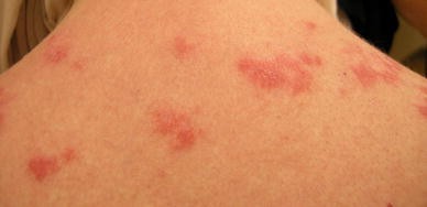
(Nursing Paper Example on Cutaneous Lupus Erythematosus) Cutaneous lupus erythematosus (CLE) is an autoimmune disorder characterized by skin manifestations associated with lupus erythematosus. It can occur independently or as part of systemic lupus erythematosus (SLE). CLE primarily affects the skin, causing disfigurement and impacting the quality of life. Early recognition and treatment are essential to manage symptoms and prevent complications.

Causes of Cutaneous Lupus Erythematosus
CLE arises from an interplay of genetic, environmental, and immunological factors.
Genetic Factors
Genetic predisposition plays a key role, with certain HLA alleles increasing susceptibility.
Family history of autoimmune diseases is a significant risk factor.
Environmental Triggers
Ultraviolet (UV) light: A major trigger causing photosensitivity and exacerbating skin lesions.
Infections: Viral infections such as Epstein-Barr virus may activate autoimmune responses.
Medications: Drug-induced CLE can result from hydralazine, procainamide, or isoniazid.
Smoking: Strongly associated with subacute cutaneous lupus erythematosus (SCLE).
Immunological Dysregulation
Abnormal activation of T and B lymphocytes leads to the production of autoantibodies.
Complement system activation contributes to tissue damage.
Signs and Symptoms
CLE manifests in distinct forms, each with unique features.
Acute Cutaneous Lupus Erythematosus (ACLE)
Butterfly-shaped rash (malar rash) across the cheeks and nose.
Associated with systemic lupus erythematosus.
Lesions may worsen with sun exposure.
Subacute Cutaneous Lupus Erythematosus (SCLE)
Annular or papulosquamous lesions on sun-exposed areas.
Often linked to anti-Ro/SSA antibodies.
Lesions heal without scarring but may cause pigment changes.
Chronic Cutaneous Lupus Erythematosus (CCLE)
Includes discoid lupus erythematosus (DLE), the most common form.
Plaques with central scarring, atrophy, and depigmentation.
Typically occurs on the face, scalp, and ears.
Can result in permanent scarring and hair loss (alopecia).
General Symptoms
Photosensitivity.
Itching or pain in affected areas.
Emotional distress due to cosmetic concerns.
Etiology
CLE is an autoimmune condition caused by an overactive immune system targeting healthy skin cells.
Genetic Susceptibility: Variants in genes related to immune regulation.
Environmental Triggers: UV radiation and smoking are significant external factors.
Immunological Mechanisms: Autoantibodies such as antinuclear antibodies (ANA) and anti-Ro/SSA are involved in pathogenesis.
Pathophysiology
CLE involves immune-mediated damage to the skin.
Role of Autoantibodies
Autoantibodies bind to nuclear antigens, forming immune complexes.
These complexes deposit in the skin, triggering inflammation.
T-Cell Activation
Dysregulated T-cells contribute to tissue damage.
Cytokines such as tumor necrosis factor-alpha (TNF-α) amplify inflammatory responses.
UV Radiation
UV light induces apoptosis in keratinocytes, exposing nuclear antigens.
This process exacerbates autoantibody production.
Diagnosis
The diagnosis of CLE involves clinical evaluation, laboratory tests, and sometimes skin biopsy.
Clinical Assessment
Detailed patient history, including sun exposure and medication use.
Physical examination of lesions for characteristic features.
Laboratory Tests
Antinuclear antibodies (ANA): Positive in most cases, especially ACLE.
Anti-Ro/SSA and Anti-La/SSB: Commonly associated with SCLE.
Skin Biopsy
Histopathological analysis shows vacuolar interface dermatitis and perivascular inflammation.
Direct immunofluorescence reveals immunoglobulin and complement deposition at the dermoepidermal junction.
Treatment Regimens
Treatment aims to control symptoms, reduce flares, and prevent scarring.
Topical Therapies
Corticosteroids: Reduce inflammation and control lesions.
Calcineurin Inhibitors: Tacrolimus and pimecrolimus for steroid-sparing effects.
Systemic Therapies
Antimalarials: Hydroxychloroquine is the first-line treatment for extensive disease.
Immunosuppressants: Methotrexate, mycophenolate mofetil, or azathioprine for severe or refractory cases.
Biologics: Belimumab, a B-cell inhibitor, may be beneficial in systemic involvement.
Photoprotection
Strict avoidance of UV exposure.
Broad-spectrum sunscreens with SPF ≥50.
Patient Education
Understanding CLE
Explain the nature of the disease and its triggers.
Emphasize the importance of adherence to treatment.
Lifestyle Modifications
Encourage wearing protective clothing and avoiding peak sunlight hours.
Stress the importance of smoking cessation to reduce disease activity.
Emotional Support
Address cosmetic concerns and provide resources for counseling.
Support groups can help patients cope with the emotional impact of the disease.
Additional Considerations
Complications
Scarring and permanent disfigurement from chronic lesions.
Progression to systemic lupus erythematosus in some cases.
Increased risk of secondary infections due to damaged skin.
Prognosis
Early treatment and effective management lead to favorable outcomes.
Chronic and recurrent cases require long-term follow-up.
Conclusion
Cutaneous lupus erythematosus is a challenging condition requiring a multidisciplinary approach. Its varied clinical presentations necessitate thorough evaluation for effective management. Educating patients on preventive measures and ensuring adherence to treatment are essential for improving outcomes.
References
Bolognia, J. L., Schaffer, J. V., & Cerroni, L. (2018). Dermatology (4th ed.). Elsevier. https://www.elsevier.com/books/dermatology/bolognia/978-0-7020-6285-8
Kuhn, A., & Sticherling, M. (2019). Cutaneous Lupus Erythematosus: Current Insights on Pathogenesis, Diagnosis, and Treatment. European Journal of Dermatology, 29(6), 535-551. https://www.journal-dermatology.com/article/S1167-1122(19)30583-2/fulltext
Werth, V. P. (2017). Clinical Manifestations of Cutaneous Lupus Erythematosus. UpToDate. https://www.uptodate.com/contents/cutaneous-lupus-erythematosus
Vasquez, R., & Isenberg, D. (2020). Current Concepts in the Management of Cutaneous Lupus Erythematosus. British Journal of Dermatology, 182(5), 1145-1153. https://onlinelibrary.wiley.com/doi/full/10.1111/bjd.18720
Mayo Clinic. (2023). Lupus. https://www.mayoclinic.org/diseases-conditions/lupus/symptoms-causes/syc-20365789


