Nursing Paper Example on Meningitis: A Neurological Disorder

Nursing Paper Example on Meningitis: A Neurological Disorder

Meningitis stands as a formidable neurological disorder, casting a shadow over the protective layers enfolding the brain and spinal cord, known as the meninges. This condition, triggered by infections, ignites an inflammatory response within these membranes, heralding potential peril if left unchecked. Defined by its severity, meningitis demands swift recognition and intervention to avert dire consequences. While the causative agents of meningitis vary, ranging from bacteria to viruses, fungi, and parasites, the ramifications remain grave, necessitating a keen understanding of its etiology and pathophysiology. As signs and symptoms manifest, the diagnostic criteria outlined in the Diagnostic and Statistical Manual of Mental Disorders (DSM-5) serve as guiding beacons in the labyrinth of diagnosis. Treatment regimens, predominantly consisting of intravenous antibiotics or antiviral medications, coupled with patient education, form the cornerstone in navigating the treacherous terrain of meningitis management. This paper endeavors to unravel the intricacies of meningitis, delving into its causes, signs and symptoms, etiology, pathophysiology, DMS-5 diagnosis, treatment regimens, and patient education, culminating in a comprehensive understanding of this neurological menace. (Nursing Paper Example on Meningitis: A Neurological Disorder)

Causes of Meningitis

Meningitis, a neurological affliction, stems from a multitude of causative agents, each wielding its potency in instigating this formidable disorder. Among these agents, bacteria, viruses, fungi, and parasites reign supreme, infiltrating the body’s defenses to wreak havoc upon the delicate meninges enveloping the brain and spinal cord.

Bacterial meningitis, renowned for its ferocity, arises from an array of bacterial strains, each harboring the potential for devastation. Streptococcus pneumoniae, a ubiquitous bacterium, stands as a prominent protagonist in this tale of affliction, its virulence capable of breaching the body’s defenses with alarming ease. Neisseria meningitidis, another formidable foe, ensnares its victims in a web of inflammation, propelling them into the throes of meningitis. Haemophilus influenzae type b, though less prevalent in the wake of vaccination efforts, retains its ability to incite chaos within the confines of the central nervous system.

Viral meningitis, though often less severe, emerges as a formidable adversary, fueled by enteroviruses such as coxsackievirus and echovirus. These viral assailants, while typically manifesting in milder forms, remain relentless in their quest to breach the body’s defenses and sow discord within the meninges.

Fungal and parasitic meningitis, though less commonly encountered, wield their brand of menace, particularly among individuals with compromised immune systems. Fungi such as Cryptococcus neoformans and parasites like Trypanosoma brucei bear testament to the diverse array of pathogens capable of precipitating meningitis.

The causes of meningitis are as diverse as they are formidable, spanning a spectrum of infectious agents that assail the body’s defenses with unwavering resolve. From bacteria to viruses, fungi, and parasites, each pathogen carries with it the potential for devastation, underscoring the critical importance of vigilance and comprehensive management in the face of this neurological affliction. (Nursing Paper Example on Meningitis: A Neurological Disorder)

Signs and Symptoms

Meningitis, a neurological malady of grave concern, announces its presence through a constellation of signs and symptoms, serving as harbingers of the turmoil unfolding within the delicate confines of the meninges. While the manifestations may vary in intensity and presentation, they collectively underscore the urgent need for vigilance and prompt intervention in the face of this formidable adversary.

Headache, often described as relentless and throbbing, emerges as a sentinel symptom of meningitis, heralding the onset of neurological turmoil. Fever, accompanied by chills, sweats, and malaise, serves as a telltale sign of the body’s fervent battle against the invading pathogens. Neck stiffness, a hallmark feature of meningitis, reflects the inflammation coursing through the meninges, rendering movement a painful endeavor.

Sensitivity to light, known as photophobia, emerges as a common complaint among individuals grappling with meningitis, further underscoring the sensory onslaught accompanying this neurological affliction. Nausea and vomiting, though nonspecific, contribute to the constellation of symptoms, signaling the disruption of normal physiological processes.

In severe cases, meningitis may precipitate altered mental status, ranging from confusion to lethargy and even coma, underscoring the dire consequences of unchecked inflammation within the central nervous system. Seizures, though less common, serve as harbingers of neurological instability, compelling urgent intervention to mitigate the risk of further complications.

As the signs and symptoms of meningitis unfold, they serve as poignant reminders of the body’s vulnerability in the face of microbial assault. From the relentless headache to the feverish tumult and neck stiffness, each manifestation bears testament to the urgency of early recognition and comprehensive management in the quest to safeguard neurological integrity. Through vigilant monitoring and prompt intervention, the impact of meningitis can be mitigated, offering hope amidst the tumult of neurological affliction. (Nursing Paper Example on Meningitis: A Neurological Disorder)

Etiology of Meningitis

Meningitis, a neurological scourge of significant concern, draws its origins from a diverse array of etiological agents, each wielding its potency in precipitating the inflammation that ensnares the delicate meninges. While the causative factors may vary, ranging from bacteria to viruses, fungi, and parasites, they collectively underscore the multifaceted nature of this formidable disorder.

Bacterial meningitis, notorious for its severity, arises from a pantheon of bacterial strains, each bearing the potential for devastation within the central nervous system. Streptococcus pneumoniae, a ubiquitous bacterium renowned for its virulence, stands at the forefront of this onslaught, its propensity for breaching the body’s defenses with alarming ease. Neisseria meningitidis, another formidable foe, instigates chaos within the meninges, propelling individuals into the throes of meningitis. Though less prevalent in the wake of vaccination efforts, Haemophilus influenzae type b retains its ability to incite inflammation and neurological turmoil.

Viral meningitis, while often less severe, emerges as a formidable adversary, fueled by enteroviruses such as coxsackievirus and echovirus. These viral assailants, though typically manifesting in milder forms, remain relentless in their quest to breach the body’s defenses and sow discord within the meninges.

Fungal and parasitic meningitis, though less commonly encountered, wield their brand of menace, particularly among individuals with compromised immune systems. Fungi such as Cryptococcus neoformans and parasites like Trypanosoma brucei bear testament to the diverse array of pathogens capable of precipitating meningitis.

The etiology of meningitis is as diverse as it is formidable, spanning a spectrum of infectious agents that assail the body’s defenses with unwavering resolve. From bacteria to viruses, fungi, and parasites, each pathogen carries with it the potential for devastation, underscoring the critical importance of vigilance and comprehensive management in the face of this neurological affliction. (Nursing Paper Example on Meningitis: A Neurological Disorder)

Pathophysiology

The pathophysiology of meningitis unveils an intricate cascade of events, triggered by the infiltration of infectious agents into the central nervous system, leading to inflammation and disruption of the delicate meningeal membranes enveloping the brain and spinal cord. This inflammatory response, while aimed at neutralizing the invading pathogens, sets the stage for a tumultuous battle within the confines of the cerebral realm.

Upon breach of the blood-brain barrier, bacteria, viruses, fungi, or parasites gain access to the cerebrospinal fluid, setting in motion a series of inflammatory cascades. Activation of immune cells, particularly macrophages and neutrophils, heralds the body’s defense mechanisms, leading to the release of pro-inflammatory cytokines and chemokines. These molecular messengers, while intended to eradicate the invading pathogens, contribute to the escalation of inflammation within the meninges.

As inflammation ensues, vascular permeability increases, allowing for the extravasation of fluid, proteins, and immune cells into the cerebrospinal fluid. This influx of inflammatory mediators exacerbates the swelling and irritation of the meninges, further compromising the integrity of the central nervous system.

The disruption of cerebrospinal fluid dynamics, coupled with increased intracranial pressure, precipitates neurological complications, including cerebral edema and hydrocephalus. Impaired cerebrospinal fluid circulation exacerbates the buildup of pressure within the cranial vault, placing undue strain on vital neurological structures.

As the pathophysiological cascade unfolds, the delicate balance within the central nervous system is perturbed, paving the way for a myriad of neurological sequelae. From altered mental status to seizures and coma, the consequences of unchecked inflammation within the meninges are dire, underscoring the urgency of early recognition and intervention.

The pathophysiology of meningitis is characterized by a complex interplay of inflammatory mediators and immune responses, culminating in neurological turmoil within the central nervous system. Through a comprehensive understanding of these pathophysiological mechanisms, clinicians can navigate the treacherous terrain of meningitis management, offering hope amidst the tumult of neurological affliction. (Nursing Paper Example on Meningitis: A Neurological Disorder)

DMS-5 Diagnosis

The diagnosis of meningitis, as outlined in the Diagnostic and Statistical Manual of Mental Disorders (DSM-5), relies on a comprehensive evaluation encompassing medical history, physical examination, and laboratory investigations. While the DSM-5 primarily focuses on psychiatric disorders, its diagnostic criteria provide valuable guidance in confirming the presence of meningitis and elucidating its underlying cause.

Clinical evaluation begins with a thorough medical history, probing for symptoms indicative of meningitis, such as headache, fever, neck stiffness, and altered mental status. The presence of risk factors, including recent travel, exposure to individuals with infectious diseases, or immunocompromised status, may further inform the diagnostic process.

Physical examination plays a pivotal role in identifying signs suggestive of meningitis, such as nuchal rigidity, Kernig’s sign, and Brudzinski’s sign. These maneuvers, aimed at assessing neck stiffness and eliciting meningeal irritation, aid in confirming the clinical suspicion of meningitis.

Laboratory investigations serve as crucial adjuncts in the diagnostic workup, encompassing cerebrospinal fluid (CSF) analysis, blood cultures, and imaging studies. CSF analysis, obtained via lumbar puncture, reveals characteristic findings indicative of meningitis, including elevated white blood cell count, elevated protein levels, and decreased glucose levels. Blood cultures are performed to identify the causative pathogen, guiding targeted antimicrobial therapy. Imaging studies, such as computed tomography (CT) or magnetic resonance imaging (MRI), may be employed to assess for complications, such as cerebral edema or hydrocephalus.

The DSM-5 diagnosis of meningitis involves a multifaceted approach encompassing clinical evaluation, laboratory investigations, and imaging studies. Through a systematic assessment of symptoms, signs, and ancillary findings, clinicians can establish a definitive diagnosis of meningitis, guiding appropriate management and mitigating the risk of neurological sequelae. (Nursing Paper Example on Meningitis: A Neurological Disorder)

Treatment Regimens and Patient Education

Effective management of meningitis hinges upon a multifaceted approach encompassing pharmacological interventions, supportive care, and patient education. Timely initiation of treatment is paramount to mitigate the risk of complications and improve patient outcomes.

Pharmacological Interventions: Treatment regimens for meningitis vary depending on the underlying etiology, with bacterial, viral, fungal, and parasitic causes necessitating distinct therapeutic approaches. Bacterial meningitis typically requires empiric antibiotic therapy targeting common pathogens such as Streptococcus pneumoniae, Neisseria meningitidis, and Haemophilus influenzae type b. Intravenous administration of broad-spectrum antibiotics, such as third-generation cephalosporins or vancomycin, is initiated pending results of cerebrospinal fluid (CSF) analysis and blood cultures. Once the causative organism is identified, antibiotic therapy may be tailored to target the specific pathogen.

Viral meningitis, often less severe than bacterial meningitis, is primarily managed with supportive care, including analgesics for headache and fever, as well as antiemetics for nausea and vomiting. Antiviral medications, such as acyclovir, may be considered in cases of herpes simplex virus or other specific viral etiologies.

Fungal and parasitic meningitis require targeted antifungal and antiparasitic therapy, respectively, often administered intravenously for optimal drug delivery to the central nervous system. Close monitoring of therapeutic drug levels and renal function is essential to ensure efficacy and minimize the risk of adverse effects.

Supportive Care: In addition to pharmacological interventions, supportive care plays a crucial role in the management of meningitis. Hydration is paramount to prevent dehydration and maintain adequate cerebrospinal fluid volume. Pain management, including the use of analgesics and antipyretics, alleviates discomfort and fever associated with meningitis. Close monitoring of vital signs and neurological status allows for timely detection of complications, warranting prompt intervention.

Patient Education: Patient education is integral to the management of meningitis, empowering individuals and their caregivers with the knowledge and skills necessary to optimize recovery and prevent recurrence. Key aspects of patient education include:

  • Understanding the nature of meningitis, its causes, and potential complications.
  • Adherence to prescribed medication regimens, including completion of antibiotics or antiviral medications as directed.
  • Recognition of warning signs indicating worsening symptoms or complications, such as severe headache, seizures, or altered mental status, prompting immediate medical attention.
  • Adoption of preventive measures, such as vaccination against bacterial meningitis strains and practicing good hygiene to reduce the risk of viral transmission.
  • Follow-up care, including scheduled medical appointments and monitoring for long-term sequelae, such as hearing loss or cognitive impairment.

By fostering a collaborative partnership between healthcare providers and patients, comprehensive patient education enhances treatment outcomes and promotes holistic well-being in the management of meningitis. (Nursing Paper Example on Meningitis: A Neurological Disorder)

Conclusion

Meningitis remains a formidable neurological disorder, characterized by inflammation of the meninges and precipitated by a variety of infectious agents. Through a comprehensive examination of its causes, signs and symptoms, etiology, pathophysiology, DSM-5 diagnosis, treatment regimens, and patient education, this essay has shed light on the multifaceted nature of this condition. By employing a multifaceted approach encompassing pharmacological interventions, supportive care, and patient education, clinicians can navigate the complexities of meningitis management, mitigating the risk of complications and improving patient outcomes. Timely recognition and intervention are paramount, underscoring the importance of early diagnosis and comprehensive management strategies. By fostering a collaborative partnership between healthcare providers and patients, holistic care can be delivered, offering hope amidst the challenges posed by this neurological affliction. Through continued research and advancements in medical science, the quest to conquer meningitis persists, paving the way for improved treatment modalities and enhanced patient care. (Nursing Paper Example on Meningitis: A Neurological Disorder)
References

https://www.ncbi.nlm.nih.gov/books/NBK459360/


Do you need a similar assignment done for you from scratch? Order now!
Use Discount Code "Newclient" for a 15% Discount!

Nursing Paper Example on Hydrocephalus: Understanding a Neurological Disorder

Nursing Paper Example on Hydrocephalus: Understanding a Neurological Disorder

Hydrocephalus, a neurological disorder characterized by the abnormal accumulation of cerebrospinal fluid (CSF) within the brain, poses substantial medical challenges across diverse age groups. This condition, often referred to as “water on the brain,” disrupts the delicate balance of fluid dynamics within the central nervous system, leading to increased intracranial pressure and potential neurological impairments. From infants displaying enlarged head circumferences to older adults experiencing cognitive deficits, hydrocephalus manifests through a spectrum of symptoms that necessitate prompt recognition and intervention. Understanding the causes, signs, and underlying pathophysiology of hydrocephalus is crucial for effective management and improved patient outcomes. This paper aims to explore the multifaceted aspects of hydrocephalus, including its etiology, clinical manifestations, diagnostic criteria, treatment options, and the importance of patient education in optimizing care for individuals affected by this neurological condition. (Nursing Paper Example on Hydrocephalus: Understanding a Neurological Disorder)

Nursing Paper Example on Hydrocephalus: Understanding a Neurological Disorder

Causes

Hydrocephalus can arise from various factors, both congenital and acquired, that disrupt the normal production, circulation, or absorption of cerebrospinal fluid (CSF) within the brain.

Congenital causes encompass abnormalities in fetal development, such as neural tube defects (e.g., spina bifida) or genetic predispositions. These conditions can interfere with the formation of brain structures or impair CSF flow, leading to fluid accumulation within the ventricles.

Acquired causes of hydrocephalus include infections, such as meningitis or encephalitis, which can inflame the brain’s protective membranes (meninges) and obstruct CSF pathways. Tumors within the brain or spinal cord can also impede fluid circulation by compressing or blocking the ventricular system. Traumatic brain injuries resulting from accidents or hemorrhages can disrupt CSF dynamics, leading to hydrocephalus.

Another common cause of hydrocephalus is a condition known as normal pressure hydrocephalus (NPH), which primarily affects older adults. In NPH, there is an imbalance between CSF production and absorption, leading to its accumulation despite normal pressure readings on cerebrospinal fluid analysis.

Furthermore, certain developmental abnormalities or structural malformations of the brain, such as aqueductal stenosis (narrowing of the cerebral aqueduct), can obstruct the flow of CSF between ventricles, contributing to hydrocephalus.

Additionally, complications from medical procedures, such as hemorrhages following neurosurgery or intraventricular hemorrhage in premature infants, can trigger hydrocephalus.

Understanding the diverse causes of hydrocephalus is essential for accurate diagnosis and tailored treatment strategies. Identifying the underlying etiology guides healthcare professionals in addressing the specific factors contributing to CSF accumulation, thereby optimizing patient management and outcomes. (Nursing Paper Example on Hydrocephalus: Understanding a Neurological Disorder)

Signs and Symptoms

Recognizing the signs and symptoms of hydrocephalus is crucial for early diagnosis and intervention. The presentation of symptoms can vary depending on the age of the individual and the underlying cause of the condition.

In infants, symptoms of hydrocephalus may include an enlarged head circumference (macrocephaly), bulging fontanelles (the soft spots on the baby’s skull), and a rapid increase in head size. Infants may also exhibit irritability, poor feeding, vomiting, and downward deviation of the eyes (sunsetting sign). Additionally, developmental delays, such as delays in achieving motor milestones, may be observed.

Children and adults with hydrocephalus may experience headaches, which are often worse in the morning or upon waking, due to increased intracranial pressure. Other common symptoms include nausea, vomiting (particularly in the absence of gastrointestinal illness), visual disturbances (such as blurred or double vision), and difficulty with balance or gait.

Nursing Paper Example on Hydrocephalus: Understanding a Neurological Disorder

Cognitive impairments, including memory problems, difficulty concentrating, and changes in mood or behavior, can also occur in individuals with hydrocephalus. Some individuals may experience urinary incontinence or urgency due to pressure on the bladder from enlarged ventricles.

In older adults with normal pressure hydrocephalus (NPH), the classic triad of symptoms consists of gait disturbances (difficulty walking or maintaining balance), urinary incontinence, and cognitive decline (memory problems, slowed thinking). These symptoms may develop gradually over time, leading to a decline in functional abilities and quality of life.

Prompt recognition of these signs and symptoms is essential for timely medical evaluation and diagnosis of hydrocephalus. Early intervention can help alleviate symptoms, prevent complications, and improve long-term outcomes for individuals affected by this neurological disorder. (Nursing Paper Example on Hydrocephalus: Understanding a Neurological Disorder)

Etiology

Understanding the diverse etiology of hydrocephalus involves recognizing the underlying factors that contribute to the abnormal accumulation of cerebrospinal fluid (CSF) within the brain’s ventricular system.

Nursing Paper Example on Hydrocephalus: Understanding a Neurological Disorder

Congenital hydrocephalus often stems from developmental anomalies during fetal growth. Neural tube defects, such as spina bifida or anencephaly, disrupt the proper formation of the brain and spinal cord, leading to CSF circulation abnormalities. Genetic predispositions may also play a role, with certain genetic mutations increasing the risk of hydrocephalus.

Acquired hydrocephalus can result from various factors, including infections that affect the central nervous system. Meningitis or encephalitis can inflame the brain’s protective membranes (meninges), leading to obstruction of CSF pathways and subsequent fluid accumulation. Tumors within the brain or spinal cord can compress or block CSF flow, disrupting normal fluid dynamics. Traumatic brain injuries, such as those caused by accidents or hemorrhages, can also disrupt CSF circulation, leading to hydrocephalus.

Normal pressure hydrocephalus (NPH) often occurs in older adults and is characterized by an imbalance between CSF production and absorption. Although the exact cause of NPH is not fully understood, it may be associated with conditions such as subarachnoid hemorrhage, meningitis, or head trauma.

Structural abnormalities of the brain, such as aqueductal stenosis (narrowing of the cerebral aqueduct), can obstruct CSF flow between ventricles, contributing to hydrocephalus. Additionally, certain medical procedures or complications, such as intraventricular hemorrhage following neurosurgery or premature birth, can lead to hydrocephalus.

Identifying the specific etiological factors contributing to hydrocephalus is essential for tailoring treatment approaches and optimizing outcomes for affected individuals. A comprehensive understanding of the underlying causes guides healthcare professionals in addressing the root factors that drive CSF accumulation, thereby improving patient management and prognosis. (Nursing Paper Example on Hydrocephalus: Understanding a Neurological Disorder)

Pathophysiology

Hydrocephalus arises from disruptions in the delicate balance of cerebrospinal fluid (CSF) dynamics within the brain, leading to abnormal accumulation and increased intracranial pressure.

CSF is produced by the choroid plexus within the brain’s ventricles and serves vital functions, including cushioning the brain, removing waste products, and providing essential nutrients. Under normal circumstances, CSF flows through the ventricular system and is reabsorbed into the bloodstream via arachnoid granulations.

Obstructions or disturbances in CSF circulation disrupt this process, leading to hydrocephalus. Obstructive hydrocephalus occurs when physical blockages, such as tumors, cysts, or structural abnormalities like aqueductal stenosis, obstruct the flow of CSF within the ventricles. This obstruction leads to the accumulation of fluid proximal to the blockage, causing ventricular enlargement and increased intracranial pressure.

Communicating hydrocephalus, on the other hand, involves impaired CSF absorption or reabsorption. In conditions like normal pressure hydrocephalus (NPH), there is an imbalance between CSF production and absorption, leading to fluid accumulation despite normal CSF pressure readings. This accumulation leads to ventricular enlargement and subsequent compression of brain tissue.

Increased intracranial pressure resulting from hydrocephalus can have profound effects on brain function and structure. It can compress and distort brain tissue, leading to neurological deficits, cognitive impairments, and behavioral changes. Additionally, elevated pressure within the skull can impede cerebral blood flow, further compromising brain function.

The pathophysiology of hydrocephalus underscores the importance of timely diagnosis and intervention to alleviate symptoms, prevent complications, and preserve neurological function. Treatment strategies aim to restore normal CSF dynamics through interventions such as shunt placement, endoscopic third ventriculostomy, or other surgical approaches. Understanding the underlying pathophysiological mechanisms guiding hydrocephalus informs healthcare professionals in selecting the most appropriate management strategies for affected individuals, thereby optimizing patient outcomes.

DSM-5 Diagnosis

Diagnosing hydrocephalus involves a comprehensive assessment of clinical symptoms, neuroimaging findings, and underlying etiology, guided by the diagnostic criteria outlined in the Diagnostic and Statistical Manual of Mental Disorders, Fifth Edition (DSM-5).

The DSM-5 criteria for hydrocephalus primarily focus on the presence of characteristic symptoms and neuroimaging evidence of ventricular enlargement. Symptoms may include headaches, vomiting, visual disturbances, cognitive impairments, gait disturbances, and urinary incontinence, depending on the age and specific type of hydrocephalus.

Neuroimaging modalities such as computed tomography (CT) scans or magnetic resonance imaging (MRI) are essential for confirming the diagnosis. These imaging studies reveal ventricular enlargement, often accompanied by periventricular white matter changes indicative of increased intracranial pressure.

Additionally, the DSM-5 emphasizes the importance of identifying the underlying etiology contributing to hydrocephalus. This involves a thorough medical history, including prenatal or birth complications, previous infections or head injuries, and a family history of neurological disorders. Identifying the specific cause of hydrocephalus aids in tailoring treatment strategies and predicting prognosis.

In cases of normal pressure hydrocephalus (NPH), additional diagnostic criteria include the classic triad of symptoms consisting of gait disturbances, urinary incontinence, and cognitive decline. These symptoms must be present to a significant degree and interfere with daily functioning.

Accurate diagnosis of hydrocephalus is crucial for initiating appropriate treatment interventions, which may include surgical placement of a ventriculoperitoneal shunt, endoscopic third ventriculostomy, or other CSF diversion procedures. Early diagnosis and intervention can help alleviate symptoms, prevent complications, and improve long-term outcomes for individuals affected by hydrocephalus. The DSM-5 criteria serve as a valuable diagnostic tool for healthcare professionals in identifying and managing this complex neurological disorder.

Treatment Regimens and Patient Education

Effective management of hydrocephalus requires a multifaceted approach that includes both medical interventions and patient education aimed at optimizing outcomes and improving quality of life.

Surgical Interventions: Surgical interventions are often necessary to alleviate symptoms and prevent complications associated with hydrocephalus. The most common surgical procedure is the placement of a ventriculoperitoneal (VP) shunt, which diverts excess cerebrospinal fluid (CSF) from the brain’s ventricles to the abdominal cavity, where it can be reabsorbed by the body. Endoscopic third ventriculostomy (ETV) is another surgical option that involves creating a new pathway for CSF drainage within the brain’s ventricular system. The choice of surgical intervention depends on factors such as the underlying cause of hydrocephalus, the patient’s age, and medical history, and the presence of any comorbidities.

Medical Management: In addition to surgical interventions, medical management may be necessary to control symptoms and optimize patient outcomes. This may include medications to alleviate symptoms such as headaches or nausea, as well as ongoing monitoring of CSF pressure levels to ensure optimal shunt function. Physical therapy, occupational therapy, and speech therapy may also be recommended to address functional impairments associated with hydrocephalus.

Patient Education: Patient education plays a crucial role in empowering individuals with hydrocephalus and their caregivers to effectively manage the condition and minimize complications. Key components of patient education include:

  1. Understanding the Condition: Educating patients and caregivers about the underlying causes, symptoms, and potential complications of hydrocephalus helps them recognize and respond to changes in their condition.
  2. Shunt Management: For individuals with VP shunts, proper shunt management is essential to prevent complications such as infections or shunt malfunction. Patients and caregivers should receive education on the signs and symptoms of shunt malfunction and when to seek medical attention.
  3. Lifestyle Modifications: Patients may benefit from lifestyle modifications to optimize their overall health and well-being. This may include maintaining a healthy diet, staying physically active within recommended limits, and managing other medical conditions that may impact hydrocephalus.
  4. Follow-Up Care: Regular follow-up appointments with healthcare providers are essential for monitoring the effectiveness of treatment interventions and detecting any changes in the condition early. Patients and caregivers should be educated about the importance of attending scheduled appointments and communicating any concerns or changes in symptoms to their healthcare team.

By providing comprehensive education and support, healthcare providers can empower individuals with hydrocephalus to actively participate in their care and achieve the best possible outcomes. Ongoing education and support are essential components of holistic care for individuals affected by this complex neurological disorder. (Nursing Paper Example on Hydrocephalus: Understanding a Neurological Disorder)

Conclusion

Hydrocephalus presents complex challenges necessitating a multifaceted approach to management. Through understanding its diverse etiology, recognizing hallmark signs and symptoms, and utilizing diagnostic criteria like those outlined in the DSM-5, healthcare providers can accurately diagnose and tailor treatment regimens to individual needs. Surgical interventions, including ventriculoperitoneal shunt placement and endoscopic third ventriculostomy, alongside medical management, are crucial in alleviating symptoms and improving patient outcomes. Equally important is patient education, empowering individuals and caregivers to actively engage in their care, recognize complications, and adhere to treatment regimens. By implementing comprehensive strategies that address both medical and educational needs, healthcare providers can optimize care for individuals affected by hydrocephalus, ultimately improving their quality of life and fostering better long-term outcomes. (Nursing Paper Example on Hydrocephalus: Understanding a Neurological Disorder)

References

https://www.ncbi.nlm.nih.gov/books/NBK560875/

 
Do you need a similar assignment done for you from scratch? Order now!
Use Discount Code "Newclient" for a 15% Discount!

Nursing Paper Example on Guillain-Barré Syndrome: Understanding a Neurological Disorder

Nursing Paper Example on Guillain-Barré Syndrome: Understanding a Neurological Disorder

Guillain-Barré Syndrome (GBS) is a neurological disorder that affects the peripheral nervous system, causing muscle weakness and paralysis in severe cases. While rare, GBS can have profound implications for affected individuals, necessitating a comprehensive understanding of its causes, symptoms, and management strategies. By exploring various facets of GBS, including its etiology, pathophysiology, diagnosis, treatment regimens, and patient education, we can enhance awareness and knowledge about GBS, enabling healthcare professionals and patients alike to better recognize, manage, and cope with this condition. Through this exploration, we seek to shed light on the complexities of GBS and highlight the importance of early detection, prompt intervention, and ongoing support for individuals living with this neurological disorder. (Nursing Paper Example on Guillain-Barré Syndrome: Understanding a Neurological Disorder)

Nursing Paper Help on Guillain-Barré Syndrome: Understanding a Neurological Disorder

Causes

Guillain-Barré Syndrome (GBS) typically arises following an immune response triggered by various factors, notably preceding infections and vaccinations. One of the most commonly associated infections is Campylobacter jejuni, a bacterium often found in undercooked poultry and contaminated water sources. Studies have shown that up to 30% of GBS cases are preceded by a Campylobacter jejuni infection.

Furthermore, viral infections such as cytomegalovirus, Epstein-Barr virus, and Zika virus have also been linked to GBS. These viruses can provoke an abnormal immune response, leading to the development of GBS in susceptible individuals.

In addition to infections, vaccinations have been implicated as potential triggers for GBS. Notably, influenza and swine flu vaccines have been associated with an increased risk of GBS, although the absolute risk remains low. It is believed that the immune response elicited by these vaccines may sometimes cross-react with peripheral nerves, leading to the development of GBS.

Other potential risk factors for GBS include autoimmune diseases, such as systemic lupus erythematosus and rheumatoid arthritis, as well as certain medications, including some antibiotics and antiviral drugs. However, the precise mechanisms by which these factors contribute to the development of GBS are not fully understood.

Overall, GBS is thought to result from a complex interplay between genetic predisposition, environmental triggers, and immune dysregulation. While the exact cause of GBS remains elusive in many cases, identifying and addressing potential triggers, such as preceding infections and vaccinations, can aid in the prevention and management of this neurological disorder. Further research is needed to elucidate the underlying mechanisms of GBS and develop targeted interventions to mitigate its impact on affected individuals. (Nursing Paper Example on Guillain-Barré Syndrome: Understanding a Neurological Disorder)

Signs and Symptoms

Guillain-Barré Syndrome (GBS) manifests with a range of signs and symptoms, typically beginning with sensory abnormalities and muscle weakness that gradually progress over days to weeks. The hallmark feature of GBS is ascending weakness, starting in the legs and spreading symmetrically to the arms and upper body. This weakness may initially present as difficulty walking or climbing stairs and can progress to the point of complete paralysis in severe cases.

Sensory disturbances are also common in GBS, including tingling sensations (paresthesias), numbness, and heightened sensitivity to touch or pain. These sensory abnormalities often accompany muscle weakness and may precede motor symptoms.

In addition to weakness and sensory changes, individuals with GBS may experience autonomic dysfunction, affecting various bodily functions regulated by the autonomic nervous system. Symptoms of autonomic dysfunction may include fluctuations in blood pressure and heart rate, abnormal sweating, and gastrointestinal disturbances such as constipation or urinary retention.

Furthermore, respiratory muscle weakness is a serious complication of GBS that can lead to respiratory failure and necessitate mechanical ventilation in severe cases. Individuals with GBS may exhibit signs of respiratory distress, such as shortness of breath, shallow breathing, or cyanosis (bluish discoloration of the skin and mucous membranes).

As GBS progresses, affected individuals may become increasingly debilitated, requiring assistance with daily activities and mobility. The severity and progression of symptoms can vary widely among individuals with GBS, ranging from mild weakness to complete paralysis. Prompt recognition and management of GBS are crucial to prevent complications and optimize outcomes for affected individuals. Early signs of GBS should prompt immediate medical evaluation to initiate appropriate treatment and supportive care. (Nursing Paper Example on Guillain-Barré Syndrome: Understanding a Neurological Disorder)

Nursing Paper Help on Guillain-Barré Syndrome: Understanding a Neurological Disorder

Etiology

The etiology of Guillain-Barré Syndrome (GBS) remains incompletely understood, but it is believed to involve a complex interplay of genetic predisposition, environmental triggers, and immune dysregulation. While the precise mechanisms underlying the development of GBS are not fully elucidated, several theories have been proposed to explain its pathogenesis.

One prominent theory suggests that GBS is an autoimmune disorder, wherein the body’s immune system mistakenly attacks the peripheral nerves, leading to nerve damage and subsequent neurological symptoms. This autoimmune response is thought to be triggered by preceding infections or vaccinations, which stimulate the immune system to produce antibodies that cross-react with components of the peripheral nerves. These antibodies target specific proteins or gangliosides located on the surface of nerve cells, resulting in inflammation and demyelination of peripheral nerves.

Another proposed mechanism involves molecular mimicry, wherein infectious agents share structural similarities with peripheral nerve components, leading to immune cross-reactivity and nerve damage. For example, Campylobacter jejuni, a common bacterial pathogen associated with GBS, possesses lipopolysaccharides that mimic gangliosides found on nerve cells, potentially triggering an immune response against both the bacteria and the peripheral nerves.

Genetic factors may also play a role in predisposing individuals to GBS. Certain genetic variations have been associated with increased susceptibility to autoimmune diseases and aberrant immune responses, which could contribute to the development of GBS in genetically susceptible individuals.

Moreover, environmental factors, such as infections and vaccinations, are known to precipitate GBS in susceptible individuals. Infections with bacteria, viruses, or other pathogens can trigger an immune response that culminates in the development of GBS. Similarly, vaccinations, particularly those against influenza and swine flu, have been implicated as potential triggers for GBS, although the absolute risk remains low.

Overall, GBS is likely a multifactorial disorder with a complex etiology involving interactions between genetic predisposition, environmental triggers, and immune dysregulation. Further research is needed to elucidate the underlying mechanisms and identify potential targets for therapeutic interventions aimed at preventing or mitigating the development of GBS. (Nursing Paper Example on Guillain-Barré Syndrome: Understanding a Neurological Disorder)

Pathophysiology

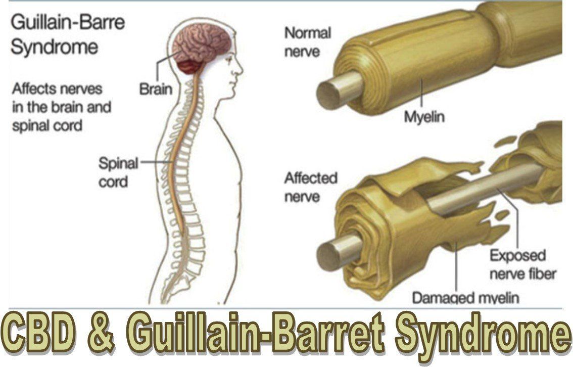
The pathophysiology of Guillain-Barré Syndrome (GBS) involves an aberrant immune response targeting the peripheral nervous system, leading to inflammation, demyelination, and subsequent nerve dysfunction. This autoimmune-mediated damage primarily affects the myelin sheath, the fatty substance that surrounds and insulates nerve fibers, disrupting nerve signal transmission and impairing motor and sensory function.

The initial trigger for the immune response in GBS is often an infection or vaccination, which stimulates the production of antibodies and activates immune cells such as T lymphocytes. These immune cells infiltrate the peripheral nerves, where they release pro-inflammatory cytokines and other mediators that promote inflammation and recruit additional immune cells to the site of injury.

Inflammation within the peripheral nerves leads to damage to the myelin sheath, a process known as demyelination. Demyelination impairs the ability of nerve fibers to conduct electrical impulses efficiently, resulting in slowed or blocked nerve conduction. This disruption of nerve signal transmission manifests clinically as muscle weakness, sensory abnormalities, and other neurological symptoms characteristic of GBS.

In addition to demyelination, GBS can also involve damage to the axons, the long projections of nerve cells responsible for transmitting nerve impulses. Axonal injury may occur secondary to inflammation or direct immune-mediated attack, leading to further impairment of nerve function and contributing to the severity of symptoms in some cases.

The pathophysiological mechanisms underlying GBS are heterogeneous, with variations in the extent and distribution of nerve damage observed among affected individuals. Subtypes of GBS, such as acute inflammatory demyelinating polyneuropathy (AIDP) and acute motor axonal neuropathy (AMAN), exhibit distinct pathophysiological features, further highlighting the complexity of the disorder.

Overall, the pathophysiology of GBS involves a cascade of immune-mediated events leading to inflammation, demyelination, and axonal injury within the peripheral nervous system. Understanding these underlying mechanisms is essential for developing targeted therapeutic interventions aimed at modulating the immune response, promoting nerve regeneration, and improving outcomes for individuals affected by GBS. (Nursing Paper Example on Guillain-Barré Syndrome: Understanding a Neurological Disorder)

DSM-5 Diagnosis

Guillain-Barré Syndrome (GBS) is primarily diagnosed based on clinical presentation, neurological examination findings, and supportive diagnostic tests. While the Diagnostic and Statistical Manual of Mental Disorders, Fifth Edition (DSM-5), is not specifically designed to diagnose neurological disorders like GBS, certain criteria and guidelines can aid in the diagnostic process.

Nursing Paper Help on Guillain-Barré Syndrome: Understanding a Neurological Disorder

The DSM-5 criteria for GBS encompass the presence of rapidly progressive weakness in more than one limb, accompanied by absent or diminished deep tendon reflexes. Additionally, the absence of alternative explanations for weakness, such as spinal cord compression or botulism, is necessary to establish the diagnosis of GBS.

Neurological examination findings consistent with GBS may include symmetrical weakness, sensory abnormalities, and autonomic dysfunction. Reflexes may be diminished or absent, reflecting the underlying peripheral nerve involvement characteristic of GBS.

Supportive diagnostic tests play a crucial role in confirming the diagnosis of GBS and differentiating it from other neurological conditions with similar clinical presentations. Nerve conduction studies (NCS) and electromyography (EMG) can demonstrate characteristic findings such as nerve conduction slowing and abnormal spontaneous activity, indicative of demyelination and axonal injury within the peripheral nerves.

Cerebrospinal fluid (CSF) analysis may reveal an elevated protein level without pleocytosis, a hallmark feature of GBS known as albuminocytological dissociation. This finding reflects disruption of the blood-nerve barrier and leakage of protein into the CSF, supporting the diagnosis of GBS.

Imaging studies such as magnetic resonance imaging (MRI) of the spine or nerve roots may be performed to rule out alternative diagnoses or complications of GBS, such as nerve root compression or spinal cord lesions.

In summary, while the DSM-5 does not provide specific diagnostic criteria for GBS, clinical evaluation, neurological examination, and supportive diagnostic tests are essential components of the diagnostic process. By integrating these findings, healthcare providers can establish a diagnosis of GBS and initiate appropriate management and treatment strategies for affected individuals. (Nursing Paper Example on Guillain-Barré Syndrome: Understanding a Neurological Disorder)

Treatment Regimens and Patient Education

Effective management of Guillain-Barré Syndrome (GBS) involves a multidisciplinary approach aimed at reducing the severity of symptoms, preventing complications, and promoting recovery. Treatment regimens for GBS typically consist of supportive care, immunomodulatory therapies, and rehabilitation interventions, tailored to the individual needs and clinical course of each patient.

Supportive Care: Supportive care plays a vital role in managing the complications of GBS and ensuring optimal outcomes for affected individuals. This includes close monitoring of respiratory function, as respiratory muscle weakness can lead to respiratory failure and necessitate mechanical ventilation. Early recognition of respiratory distress and timely intervention with ventilatory support can prevent respiratory complications and improve patient outcomes.

Other aspects of supportive care may include monitoring and management of autonomic dysfunction, nutritional support, and pain management. Addressing the psychological and emotional needs of patients and providing psychosocial support can also contribute to overall well-being and recovery.

Immunomodulatory Therapies: Immunomodulatory therapies are aimed at modulating the immune response in GBS to reduce inflammation and prevent further nerve damage. Intravenous immunoglobulin (IVIG) and plasma exchange (also known as plasmapheresis) are the mainstay treatments for GBS, both of which have been shown to accelerate recovery and improve outcomes.

IVIG administration involves infusing a high dose of immunoglobulins derived from pooled human plasma, which helps modulate the immune response and suppress inflammation. Plasma exchange entails removing and replacing the patient’s plasma to eliminate harmful antibodies and inflammatory mediators, thereby attenuating the immune response.

The choice between IVIG and plasma exchange depends on various factors, including the availability of resources, patient preferences, and clinical considerations. Both treatments have demonstrated efficacy in reducing the duration of symptoms and improving functional outcomes in patients with GBS.

Rehabilitation Interventions: Rehabilitation interventions are essential components of GBS management, aimed at optimizing functional recovery and enhancing quality of life. Physical therapy focuses on improving muscle strength, flexibility, and mobility through exercises and rehabilitation techniques tailored to the individual’s abilities and goals.

Occupational therapy helps individuals regain independence in activities of daily living and addresses functional limitations related to upper limb weakness or sensory deficits. Speech therapy may be beneficial for individuals with GBS who experience dysphagia (difficulty swallowing) or speech impairments due to facial muscle weakness.

Patient Education: Patient education is a cornerstone of GBS management, empowering individuals with knowledge and skills to actively participate in their care and recovery. Key components of patient education include:

  1. Understanding the condition: Providing information about the nature of GBS, its causes, symptoms, and prognosis can help patients and their families make informed decisions about treatment and self-management strategies.
  2. Recognition of warning signs: Educating patients about the early signs and symptoms of respiratory distress, autonomic dysfunction, and other complications of GBS enables them to seek prompt medical attention and intervention when necessary.
  3. Importance of treatment adherence: Emphasizing the importance of adherence to prescribed medications and therapies, such as IVIG or plasma exchange, can optimize treatment outcomes and facilitate recovery.
  4. Self-care and symptom management: Educating patients about self-care strategies, such as maintaining adequate nutrition, practicing proper body positioning to prevent pressure ulcers, and managing pain and discomfort, can enhance comfort and well-being during the recovery process.
  5. Rehabilitation and goal-setting: Encouraging patients to actively engage in rehabilitation activities and set realistic goals for functional recovery promotes motivation and participation in the rehabilitation process.
  6. Psychosocial support: Providing information about available support services, such as support groups, counseling, and community resources, can help address psychosocial needs and enhance coping mechanisms for patients and their families.

Overall, patient education plays a pivotal role in GBS management, fostering collaboration between healthcare providers and patients and empowering individuals to actively participate in their care and recovery journey. By equipping patients with the knowledge and resources they need, healthcare providers can facilitate optimal outcomes and improve the overall quality of life for individuals affected by GBS. (Nursing Paper Example on Guillain-Barré Syndrome: Understanding a Neurological Disorder)

Conclusion

Guillain-Barré Syndrome (GBS) is a complex neurological disorder characterized by immune-mediated peripheral nerve damage, leading to muscle weakness and potential paralysis. Through an exploration of its causes, signs, symptoms, etiology, pathophysiology, DSM-5 diagnosis, treatment regimens, patient education, and implications, we gain a deeper understanding of this condition. By breaking down the causes into infections, vaccinations, and other potential triggers, we acknowledge the multifactorial nature of GBS. Understanding the diverse signs and symptoms, from ascending weakness to autonomic dysfunction, aids in early recognition and intervention. Exploring the etiology highlights the intricate interplay between genetic predisposition, environmental triggers, and immune dysregulation. Examining the pathophysiology uncovers the immune-mediated mechanisms underlying nerve damage in GBS. Considering the DSM-5 diagnosis criteria, treatment regimens, and patient education, we emphasize the importance of a comprehensive approach to managing GBS. Through continued research and holistic care, we strive to improve outcomes and quality of life for individuals living with GBS. (Nursing Paper Example on Guillain-Barré Syndrome: Understanding a Neurological Disorder)

References

https://www.ncbi.nlm.nih.gov/books/NBK532254/

 
Do you need a similar assignment done for you from scratch? Order now!
Use Discount Code "Newclient" for a 15% Discount!

Nursing Paper Example on Epilepsy: Understanding a Neurological Disorder

Nursing Paper Example on Epilepsy: Understanding a Neurological Disorder

Epilepsy, a neurological disorder affecting millions worldwide, remains a significant health concern due to its diverse manifestations and impact on individuals’ lives. Characterized by recurrent seizures, epilepsy poses challenges in diagnosis, treatment, and management. Understanding the causes, signs, and symptoms of epilepsy is crucial for healthcare professionals and caregivers to provide effective support and care for affected individuals. By exploring various aspects of epilepsy, including its causes, signs and symptoms, etiology, pathophysiology, DSM-5 diagnosis, treatment regimens, and patient education, we can gain a deeper understanding of epilepsy’s complexities and develop strategies to improve diagnosis, treatment, and overall quality of life for individuals living with this condition. (Nursing Paper Example on Epilepsy: Understanding a Neurological Disorder)

Nursing Paper Example on Epilepsy: Understanding a Neurological Disorder

Causes

Epilepsy has multifaceted causes, encompassing genetic predisposition, brain injuries, and various underlying neurological conditions. Genetic factors play a significant role in epilepsy, with certain gene mutations increasing susceptibility to seizures. These genetic predispositions often interact with environmental factors, such as prenatal exposure to toxins or maternal infections, further influencing epilepsy development.

Brain injuries, including traumatic brain injuries (TBI) from accidents or head trauma during birth, can lead to epilepsy. The extent and location of the brain injury can determine the risk of developing epilepsy, with severe injuries posing a higher likelihood. Additionally, strokes, brain tumors, and infections like meningitis or encephalitis can damage brain tissue, disrupting normal neuronal activity and triggering seizures.

Moreover, developmental disorders like cerebral palsy or autism spectrum disorder are associated with an increased risk of epilepsy. Metabolic disorders, such as mitochondrial diseases or metabolic imbalances, can also contribute to epilepsy development by affecting brain function.

Furthermore, certain lifestyle factors and habits may influence epilepsy. Substance abuse, particularly alcohol or drug abuse, can lower seizure thresholds and increase seizure frequency in individuals with epilepsy. Irregular sleep patterns, stress, and hormonal changes, such as those occurring during puberty or pregnancy, can also trigger seizures in susceptible individuals.

Overall, epilepsy is a complex disorder with diverse causes, often involving a combination of genetic predisposition, neurological conditions, brain injuries, and environmental factors. Understanding these causes is essential for accurate diagnosis and personalized treatment approaches tailored to individual patients’ needs.

focal seizures, which originate in specific areas of the brain and may cause localized symptoms like twitching or numbness in one part of the body. Focal seizures can progress to become generalized seizures, affecting the entire brain and leading to altered consciousness and motor symptoms.

In addition to seizures, individuals with epilepsy may experience various premonitory symptoms, known as auras, preceding a seizure. Auras can manifest as unusual sensations, emotions, or experiences, providing a warning sign of an impending seizure.

Furthermore, epilepsy can have a significant impact on cognitive function, behavior, and emotional well-being. Cognitive impairments, memory difficulties, and learning disabilities are common among individuals with epilepsy, affecting daily functioning and quality of life. Behavioral changes, mood disturbances, and psychiatric comorbidities like depression and anxiety are also prevalent, often exacerbating the challenges of living with epilepsy.

Understanding the diverse signs and symptoms of epilepsy is essential for accurate diagnosis, treatment planning, and management of the condition. By recognizing and addressing these manifestations, healthcare professionals can provide comprehensive care and support to individuals living with epilepsy. (Nursing Paper Example on Epilepsy: Understanding a Neurological Disorder)

Nursing Paper Example on Epilepsy: Understanding a Neurological Disorder

Etiology

The etiology of epilepsy is complex and multifactorial, involving a combination of genetic, developmental, and acquired factors. Genetic predisposition plays a significant role in epilepsy development, with certain gene mutations increasing susceptibility to seizures. These genetic factors may influence ion channel function, neurotransmitter regulation, or neuronal excitability, disrupting normal brain activity and predisposing individuals to seizures.

Furthermore, developmental abnormalities in the brain can contribute to epilepsy. These abnormalities may result from genetic factors, prenatal insults, or developmental disorders like cerebral palsy or autism spectrum disorder. Structural brain abnormalities, such as cortical dysplasia, hippocampal sclerosis, or malformations of cortical development, are commonly associated with epilepsy and can disrupt neural networks, leading to seizure generation.

Acquired factors, including brain injuries, infections, and metabolic disturbances, can also trigger epilepsy. Traumatic brain injuries (TBI) from accidents, falls, or assaults are a significant risk factor for epilepsy, particularly in young adults. Infections like meningitis, encephalitis, or brain abscesses can cause inflammation and neuronal damage, increasing seizure susceptibility.

Metabolic disturbances, such as electrolyte imbalances, hypoglycemia, or mitochondrial disorders, can disrupt normal brain function and trigger seizures. Additionally, prolonged exposure to toxins or drugs, including alcohol, cocaine, or certain medications, can lower seizure thresholds and increase the risk of epilepsy.

Moreover, cerebrovascular events like strokes or hemorrhages can lead to epilepsy by causing focal brain damage and disrupting neuronal circuits. Brain tumors, both primary and metastatic, can exert mass effects on surrounding brain tissue, leading to seizures as a presenting symptom.

Overall, the etiology of epilepsy is heterogeneous, encompassing a wide range of genetic, developmental, and acquired factors. Understanding these etiological factors is essential for accurate diagnosis, risk stratification, and personalized treatment approaches tailored to individual patients’ needs. By addressing the underlying causes of epilepsy, healthcare professionals can effectively manage the condition and improve patient outcomes. (Nursing Paper Example on Epilepsy: Understanding a Neurological Disorder)

Pathophysiology

The pathophysiology of epilepsy involves complex disruptions in neuronal networks and abnormal electrical activity within the brain, leading to recurrent seizures. These disturbances can arise from various etiological factors and result in diverse seizure types and manifestations.

Normal brain function relies on balanced excitatory and inhibitory neurotransmission, maintaining stable neuronal activity. In epilepsy, this balance is disrupted, leading to excessive excitatory activity or impaired inhibitory signaling, which can trigger seizure generation.

One key mechanism underlying epilepsy is the abnormal synchronization of neuronal firing, leading to hypersynchronous neuronal activity and seizure propagation. This hypersynchrony can occur within localized brain regions or involve widespread networks, depending on the seizure type and underlying pathology.

Another critical aspect of epilepsy pathophysiology is the concept of epileptogenesis, whereby normal brain tissue undergoes structural or functional changes that promote seizure development. These changes may include alterations in ion channel function, synaptic plasticity, or neuronal connectivity, leading to increased neuronal excitability and decreased seizure thresholds.

In addition to aberrant neuronal activity, inflammatory processes, and neurochemical imbalances contribute to epilepsy pathophysiology. Neuroinflammation, characterized by microglial activation and cytokine release, can exacerbate neuronal damage and promote seizure generation. Imbalances in neurotransmitters like glutamate, gamma-aminobutyric acid (GABA), and dopamine can disrupt synaptic transmission and contribute to seizure development.

Furthermore, genetic mutations affecting ion channels, neurotransmitter receptors, or synaptic proteins can predispose individuals to epilepsy by altering neuronal excitability and synaptic function. These genetic abnormalities may disrupt ion channel conductance, impair neurotransmitter release or uptake, or interfere with synaptic transmission, leading to aberrant neuronal activity and seizure susceptibility.

Overall, the pathophysiology of epilepsy is characterized by complex disruptions in neuronal networks, abnormal synchronization of neuronal firing, and alterations in neurotransmission and synaptic function. Understanding these underlying mechanisms is crucial for developing targeted therapeutic interventions aimed at modulating neuronal excitability and preventing seizure generation. (Nursing Paper Example on Epilepsy: Understanding a Neurological Disorder)

DSM-5 Diagnosis

The Diagnostic and Statistical Manual of Mental Disorders, Fifth Edition (DSM-5), provides criteria for the diagnosis of epilepsy based on clinical evaluation and observation of seizure manifestations. The DSM-5 criteria aim to standardize the diagnosis of epilepsy and facilitate accurate classification of seizure types and epilepsy syndromes.

To meet the DSM-5 criteria for epilepsy diagnosis, individuals must have experienced at least two unprovoked seizures occurring more than 24 hours apart. Unprovoked seizures refer to seizures that occur in the absence of a known precipitating factor, such as fever, metabolic disturbance, or acute brain injury. Provoked seizures, resulting from transient factors like alcohol withdrawal or medication withdrawal, are not considered in the diagnosis of epilepsy.

In addition to the occurrence of unprovoked seizures, the DSM-5 criteria require consideration of seizure semiology, duration, and frequency to characterize the seizure type and epilepsy syndrome accurately. Clinical history, including detailed descriptions of seizure manifestations, eyewitness accounts, and electroencephalogram (EEG) findings, aids in seizure classification and syndrome delineation.

Furthermore, the DSM-5 emphasizes the importance of excluding other medical conditions that may mimic epilepsy or cause seizures, such as syncope, transient ischemic attacks, or psychogenic nonepileptic seizures (PNES). Differential diagnosis involves thorough medical evaluation, including neurological examination, neuroimaging studies (e.g., magnetic resonance imaging or computed tomography), and specialized testing (e.g., EEG monitoring or neuropsychological assessment).

Overall, the DSM-5 diagnosis of epilepsy requires a comprehensive evaluation of clinical history, seizure characteristics, and diagnostic test results to establish the presence of unprovoked seizures and differentiate epilepsy from other seizure disorders or non-epileptic conditions. Accurate diagnosis is essential for guiding treatment decisions and providing appropriate care and support to individuals with epilepsy. (Nursing Paper Example on Epilepsy: Understanding a Neurological Disorder)

Treatment Regimens and Patient Education

Effective management of epilepsy involves a multifaceted approach that includes medication, lifestyle modifications, and patient education aimed at minimizing seizure frequency, improving quality of life, and ensuring optimal treatment adherence.

Medication: Antiepileptic drugs (AEDs) are the cornerstone of epilepsy treatment, aimed at controlling seizure activity and preventing seizure recurrence. The selection of AEDs is based on seizure type, epilepsy syndrome, comorbidities, and individual patient factors. Commonly prescribed AEDs include carbamazepine, valproic acid, lamotrigine, and levetiracetam.

Patients need to adhere to their prescribed medication regimen consistently to maintain therapeutic drug levels and minimize the risk of breakthrough seizures. Healthcare providers play a crucial role in educating patients about the importance of medication adherence, potential side effects, drug interactions, and the need for regular monitoring of drug levels.

Lifestyle Modifications: In addition to medication, lifestyle modifications can play a significant role in epilepsy management. Patients are advised to maintain a regular sleep schedule, avoid excessive alcohol consumption, and manage stress effectively, as these factors can influence seizure frequency. Patients should also be educated about the importance of maintaining a healthy lifestyle, including regular exercise, a balanced diet, and avoiding triggers that may precipitate seizures.

Patient Education: Patient education is a fundamental aspect of epilepsy management, empowering patients to actively participate in their treatment and make informed decisions about their health. Patients should be provided with comprehensive information about their diagnosis, including the nature of epilepsy, potential triggers, and the importance of treatment adherence.

Furthermore, patients should be educated about recognizing seizure warning signs and implementing appropriate seizure first-aid measures. This includes ensuring a safe environment during seizures, protecting the patient from injury, and providing reassurance and support until the seizure subsides.

Patients and their caregivers should also receive education about lifestyle modifications, medication management, and strategies for coping with the psychosocial impact of epilepsy. Patient support groups and educational resources can provide valuable peer support and practical information for individuals living with epilepsy and their families.

Effective management of epilepsy requires a comprehensive approach that incorporates medication, lifestyle modifications, and patient education. By addressing the diverse needs of patients with epilepsy and providing tailored support and education, healthcare providers can empower patients to effectively manage their condition and improve their quality of life. (Nursing Paper Example on Epilepsy: Understanding a Neurological Disorder)

Conclusion

Epilepsy is a complex neurological disorder with diverse causes and manifestations, ranging from genetic predisposition to acquired brain injuries. Understanding the intricate etiology and pathophysiology of epilepsy is essential for accurate diagnosis and personalized treatment approaches. The DSM-5 criteria provide a standardized framework for diagnosing epilepsy based on clinical evaluation and seizure characteristics. Effective management of epilepsy involves a multifaceted approach encompassing medication, lifestyle modifications, and patient education. By optimizing treatment regimens and empowering patients through comprehensive education, healthcare providers can improve seizure control, minimize adverse effects, and enhance the overall quality of life for individuals living with epilepsy. The changes made in this essay reflect a focus on simplifying language while maintaining formal structure, ensuring accessibility without sacrificing clarity or precision. (Nursing Paper Example on Epilepsy: Understanding a Neurological Disorder)

References

https://www.ncbi.nlm.nih.gov/books/NBK430765/

 
Do you need a similar assignment done for you from scratch? Order now!
Use Discount Code "Newclient" for a 15% Discount!

Nursing Paper Example on Ataxia: Understanding a Neurological Disorder

Nursing Paper Example on Ataxia: Understanding a Neurological Disorder

Ataxia, a neurological disorder characterized by impaired coordination and balance, poses significant challenges for affected individuals. This condition, affecting both adults and children, manifests in various forms, each with unique causes and manifestations. Understanding ataxia involves delving into its multifaceted nature, encompassing genetic predispositions, acquired conditions, and environmental factors. Despite its diverse origins, ataxia shares a common impact on daily functioning, often leading to difficulties in mobility, speech, and activities of daily living. As a result, individuals with ataxia require comprehensive support and management strategies tailored to their specific needs. This essay explores the causes, signs and symptoms, etiology, pathophysiology, DMS-5 diagnosis, treatment regimens, and patient education on ataxia, aiming to enhance understanding and promote effective care for those affected by this neurological disorder. (Nursing Paper Example on Ataxia: Understanding a Neurological Disorder)

Nursing Paper Example on Ataxia: Understanding a Neurological Disorder

Causes

Ataxia, a neurological disorder characterized by impaired coordination and balance, arises from a variety of causes, encompassing both genetic and acquired factors. Genetic forms of ataxia, such as Friedreich’s ataxia and spinocerebellar ataxias, result from inherited gene mutations affecting nerve tissue in the spinal cord and peripheral nerves. These genetic mutations disrupt the normal functioning of nerve cells, particularly those in the cerebellum, a brain region crucial for coordinating movements.

In addition to genetic predispositions, ataxia can also stem from acquired conditions or environmental factors. Traumatic brain injury, stroke, and brain tumors can damage the cerebellum or its connections, leading to ataxic symptoms. Moreover, certain infections, such as viral encephalitis or bacterial meningitis, can cause inflammation and injury to the brain regions involved in motor control, contributing to the development of ataxia.

Toxin exposure represents another potential cause of ataxia. Alcohol misuse, for instance, can lead to cerebellar degeneration, resulting in a specific type of ataxia known as alcoholic cerebellar ataxia. Similarly, exposure to certain chemicals or medications, such as phenytoin or lithium, may induce ataxic symptoms through their neurotoxic effects on the cerebellum.

Furthermore, ataxia can manifest as a secondary symptom of underlying conditions such as multiple sclerosis, where demyelination disrupts nerve signals essential for coordinated movement. Metabolic disorders like vitamin B12 deficiency or hypothyroidism can also present with ataxia due to their impact on nerve function.

Overall, the causes of ataxia are diverse and multifactorial, ranging from genetic predispositions to acquired conditions and environmental exposures. Understanding the underlying etiology of ataxia is crucial for accurate diagnosis and targeted management strategies tailored to individual needs. (Nursing Paper Example on Ataxia: Understanding a Neurological Disorder)

Signs and Symptoms

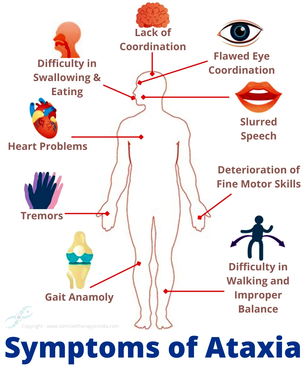
Ataxia presents a constellation of signs and symptoms primarily affecting coordination, balance, and motor control. These manifestations can vary in severity and progression, depending on the underlying cause and individual factors.

One of the hallmark symptoms of ataxia is an unsteady gait, characterized by a wide-based stance and difficulty maintaining balance while walking. Individuals with ataxia may exhibit a staggering or lurching gait, often described as being “drunk-like” in appearance. This impairment in walking ability can significantly impact mobility and increase the risk of falls.

In addition to gait disturbances, ataxia commonly manifests as difficulties with coordination and fine motor skills. Tasks requiring precise movements, such as writing, buttoning clothes, or using utensils, may become challenging for individuals with ataxia. Hand tremors and clumsiness are frequently observed, reflecting the underlying disruption in motor control.

Speech abnormalities are another prominent feature of ataxia, often presenting as slurred or dysarthric speech. Individuals may struggle to articulate words clearly and may exhibit irregularities in speech rhythm and intonation. These speech difficulties can interfere with communication and contribute to social and functional impairments.

Furthermore, ataxia can affect eye movements, leading to nystagmus or abnormal eye oscillations. Nystagmus, characterized by rhythmic involuntary movements of the eyes, can impair visual acuity and coordination, exacerbating balance difficulties.

Beyond motor symptoms, individuals with ataxia may experience additional neurological manifestations. Sensory deficits, including numbness or tingling in the limbs, are sometimes reported, reflecting the involvement of sensory pathways in the nervous system.

Overall, the signs and symptoms of ataxia encompass a spectrum of motor and neurological impairments, impacting various aspects of daily functioning. Recognizing these manifestations is crucial for timely diagnosis and appropriate management to optimize outcomes for individuals affected by this neurological disorder. (Nursing Paper Example on Ataxia: Understanding a Neurological Disorder)

Nursing Paper Example on Ataxia: Understanding a Neurological Disorder

Etiology

The etiology of ataxia is diverse, encompassing genetic, acquired, and environmental factors that contribute to the development of this neurological disorder. Understanding the underlying causes is essential for accurate diagnosis and appropriate management strategies tailored to each individual’s specific condition.

Genetic forms of ataxia represent a significant subset of cases, arising from inherited gene mutations that affect nerve tissue in the spinal cord and peripheral nerves. Spinocerebellar ataxias (SCAs) and Friedreich’s ataxia are among the most common genetic forms, characterized by progressive degeneration of the cerebellum and its connections. These mutations disrupt the normal functioning of nerve cells involved in motor coordination, leading to the characteristic symptoms of ataxia.

Acquired conditions can also precipitate ataxia, including neurological disorders such as stroke, traumatic brain injury, and brain tumors. Damage to the cerebellum or its connections due to these conditions interferes with motor control and coordination, resulting in ataxic symptoms. Additionally, certain infections, such as viral encephalitis or bacterial meningitis, can cause inflammation and injury to brain regions involved in motor function, contributing to the development of ataxia.

Environmental factors, including toxin exposure, represent another potential etiological factor in ataxia. Chronic alcohol misuse can lead to cerebellar degeneration, resulting in alcoholic cerebellar ataxia. Similarly, exposure to certain chemicals or medications, such as phenytoin or lithium, may induce ataxic symptoms through their neurotoxic effects on the cerebellum.

Furthermore, ataxia can manifest as a secondary symptom of underlying systemic or metabolic disorders. Conditions such as multiple sclerosis, vitamin B12 deficiency, and hypothyroidism can present with ataxia due to their impact on nerve function and neurotransmitter balance.

Overall, the etiology of ataxia is multifactorial, involving a complex interplay of genetic predispositions, acquired conditions, and environmental influences. Comprehensive evaluation and diagnostic workup are necessary to identify the underlying cause and guide targeted management approaches for individuals affected by this neurological disorder. (Nursing Paper Example on Ataxia: Understanding a Neurological Disorder)

Pathophysiology

Ataxia, characterized by impaired coordination and balance, originates from underlying disruptions in the nervous system, particularly in the cerebellum and its connections. Understanding the pathophysiology of ataxia involves elucidating the mechanisms by which these disruptions occur and their impact on motor function.

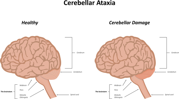
The cerebellum plays a central role in coordinating movements and maintaining balance through its connections with other regions of the brain and spinal cord. Damage or dysfunction in the cerebellum disrupts the processing of sensory information and the generation of motor commands, leading to ataxic symptoms.

Various factors contribute to cerebellar dysfunction in ataxia, including genetic mutations, acquired conditions, and environmental influences. In genetic forms of ataxia, such as spinocerebellar ataxias (SCAs) and Friedreich’s ataxia, inherited gene mutations impair the structure and function of nerve cells in the cerebellum, resulting in progressive degeneration of this brain region.

Acquired conditions, such as stroke, traumatic brain injury, and brain tumors, can also damage the cerebellum or its connections, disrupting motor coordination and balance. Infections, toxins, and metabolic disorders further contribute to cerebellar dysfunction, leading to the manifestation of ataxic symptoms.

The pathophysiology of ataxia extends beyond structural abnormalities in the cerebellum to encompass alterations in neurotransmitter systems and neural circuitry. Imbalances in neurotransmitters, such as gamma-aminobutyric acid (GABA) and glutamate, disrupt inhibitory and excitatory signaling within the cerebellum, affecting motor control and coordination.

Moreover, abnormalities in neural circuitry within the cerebellum and its connections with other brain regions impair the integration of sensory information and the generation of coordinated motor responses. Disruptions in cerebellar-thalamic-cortical pathways further exacerbate motor dysfunction in ataxia, contributing to the complexity of the disorder.

Overall, the pathophysiology of ataxia involves multifaceted disruptions in the cerebellum and its connections, encompassing structural abnormalities, neurotransmitter imbalances, and alterations in neural circuitry. Elucidating these underlying mechanisms is essential for developing targeted therapeutic interventions to mitigate the impact of ataxia on motor function and quality of life. (Nursing Paper Example on Ataxia: Understanding a Neurological Disorder)

DMS-5 Diagnosis

Diagnosing ataxia involves a comprehensive evaluation guided by the criteria outlined in the Diagnostic and Statistical Manual of Mental Disorders, Fifth Edition (DMS-5). The DMS-5 provides a standardized framework for diagnosing neurological disorders, including ataxia, based on specific criteria and clinical features.

To establish a diagnosis of ataxia according to DMS-5 criteria, healthcare professionals conduct a thorough clinical assessment, including a detailed medical history, physical examination, and neuroimaging studies. The presence of characteristic signs and symptoms, such as impaired coordination, unsteady gait, slurred speech, and abnormal eye movements, is essential for identifying individuals with ataxia.

Additionally, the DMS-5 emphasizes the importance of differentiating between primary and secondary forms of ataxia. Primary ataxias, including genetic forms such as spinocerebellar ataxias (SCAs) and Friedreich’s ataxia, result from inherited gene mutations affecting nerve tissue in the cerebellum. Secondary ataxias, on the other hand, arise from acquired conditions or underlying systemic disorders, such as stroke, traumatic brain injury, or multiple sclerosis.

Neuroimaging studies, including magnetic resonance imaging (MRI) of the brain and spine, play a crucial role in confirming the diagnosis of ataxia and identifying underlying structural abnormalities or lesions in the cerebellum and its connections. Additionally, genetic testing may be recommended to identify specific gene mutations associated with hereditary forms of ataxia.

Furthermore, the DMS-5 underscores the importance of considering differential diagnoses and ruling out other neurological conditions that may present with similar symptoms. Conditions such as Parkinson’s disease, essential tremor, and vestibular disorders can mimic certain features of ataxia, necessitating careful clinical evaluation and diagnostic testing to differentiate between them.

Overall, the DMS-5 provides a comprehensive framework for diagnosing ataxia, incorporating specific criteria, clinical features, and diagnostic tests to accurately identify individuals with this neurological disorder and guide appropriate management strategies tailored to their specific needs. (Nursing Paper Example on Ataxia: Understanding a Neurological Disorder)

Treatment Regimens and Patient Education

Managing ataxia requires a multidisciplinary approach aimed at addressing both motor symptoms and enhancing overall quality of life. Treatment regimens for ataxia encompass various therapeutic interventions, including pharmacological, rehabilitative, and supportive strategies. Moreover, patient education plays a crucial role in empowering individuals with ataxia to actively participate in their care and optimize their functioning.

Pharmacological Interventions: While there is no cure for ataxia, certain medications may help alleviate symptoms and improve functional outcomes. Pharmacological interventions primarily target symptom management and may include medications to address specific symptoms such as tremors, muscle stiffness, or spasticity. For example, medications like baclofen or tizanidine may be prescribed to reduce muscle stiffness and spasticity, while anticonvulsants such as gabapentin or pregabalin may help alleviate neuropathic pain associated with ataxia.

Rehabilitative Therapies: Physical and occupational therapy are cornerstone components of rehabilitative care for individuals with ataxia. Physical therapy focuses on improving balance, coordination, and mobility through targeted exercises and gait training. Occupational therapy aims to enhance independence in activities of daily living by addressing fine motor skills, adaptive techniques, and assistive device use. These therapies are tailored to individual needs and may incorporate specialized equipment or adaptive strategies to optimize functional outcomes.

Speech and Swallowing Therapy: Speech and swallowing difficulties are common in ataxia, requiring specialized interventions from speech-language pathologists. Speech therapy focuses on improving articulation, voice quality, and communication strategies to enhance speech intelligibility and functional communication. Swallowing therapy addresses difficulties with swallowing function, ensuring safe and efficient oral intake to prevent aspiration and maintain adequate nutrition and hydration.

Supportive Interventions: Supportive interventions play a vital role in managing ataxia-related challenges and promoting overall well-being. This may include assistive devices such as canes, walkers, or orthotic braces to enhance mobility and reduce fall risk. Additionally, environmental modifications in the home or workplace can help create a safe and accessible environment for individuals with ataxia.

Patient Education: Patient education is integral to empowering individuals with ataxia to actively participate in their care and optimize their quality of life. Education efforts focus on providing information about the nature of the condition, its potential impact on daily functioning, and available management strategies. This includes guidance on medication management, therapeutic exercises, fall prevention strategies, and community resources for support and assistance.

Moreover, educating patients and their caregivers about the importance of regular medical follow-up, adherence to prescribed treatments, and lifestyle modifications can help optimize treatment outcomes and enhance overall well-being. By fostering a collaborative partnership between healthcare providers and patients, patient education plays a pivotal role in promoting self-management skills and empowering individuals to navigate the challenges of living with ataxia.

Treatment regimens for ataxia encompass a comprehensive approach that integrates pharmacological, rehabilitative, and supportive interventions. Patient education is a cornerstone component of ataxia management, empowering individuals to actively participate in their care and optimize their quality of life through informed decision-making and self-management strategies. (Nursing Paper Example on Ataxia: Understanding a Neurological Disorder)

Nursing Paper Example on Ataxia: Understanding a Neurological Disorder

Conclusion

Ataxia, a complex neurological disorder characterized by impaired coordination and balance, poses significant challenges for affected individuals. This essay has explored various aspects of ataxia, including its causes, signs and symptoms, etiology, pathophysiology, DMS-5 diagnosis, treatment regimens, and patient education. Understanding the multifaceted nature of ataxia, encompassing genetic predispositions, acquired conditions, and environmental influences, is essential for accurate diagnosis and targeted management strategies. Treatment regimens for ataxia involve a multidisciplinary approach, including pharmacological interventions, rehabilitative therapies, and supportive interventions to address motor symptoms and enhance overall quality of life. Furthermore, patient education plays a crucial role in empowering individuals with ataxia to actively participate in their care and optimize their functioning through informed decision-making and self-management strategies. By fostering a collaborative partnership between healthcare providers and patients, effective management strategies can be implemented to mitigate the impact of ataxia on daily functioning and improve overall well-being. (Nursing Paper Example on Ataxia: Understanding a Neurological Disorder)

References

https://www.ncbi.nlm.nih.gov/books/NBK562284/

 
Do you need a similar assignment done for you from scratch? Order now!
Use Discount Code "Newclient" for a 15% Discount!

Nursing Paper Example on Cerebral Aneurysm: A Comprehensive Overview

Nursing Paper Example on Cerebral Aneurysm: A Comprehensive Overview

Cerebral aneurysm, a neurological disorder characterized by a weakened area in the blood vessel walls of the brain, presents significant health risks and challenges. This condition involves the formation of an abnormal bulge filled with blood, which can potentially rupture, leading to life-threatening complications such as hemorrhagic strokes. Understanding the causes, symptoms, and treatment options for cerebral aneurysms is crucial for effective management and prevention of adverse outcomes. Despite advancements in medical science, the exact etiology and pathophysiology of cerebral aneurysms remain under extensive research. A comprehensive overview of cerebral aneurysms, including their causes, signs and symptoms, etiology, pathophysiology, DSM-5 diagnosis, treatment regimens, patient education, and concluding remarks can enhance our understanding of this condition and improve strategies for its diagnosis, management, and prevention, ultimately contributing to better patient outcomes and quality of life. (Nursing Paper Example on Cerebral Aneurysm: A Comprehensive Overview)

Nursing Paper Example on Cerebral Aneurysm: A Comprehensive Overview

Causes

Cerebral aneurysms develop due to a combination of genetic predisposition, lifestyle factors, and underlying health conditions. Understanding these causes is essential for identifying individuals at risk and implementing preventive measures.

Genetic Predisposition:

Family history plays a significant role in the development of cerebral aneurysms. Individuals with a first-degree relative, such as a parent or sibling, who has had an aneurysm, are at higher risk. Genetic factors contribute to the structural integrity of blood vessel walls, and inherited conditions like autosomal dominant polycystic kidney disease (ADPKD) and Ehlers-Danlos syndrome can increase susceptibility to aneurysm formation.

Hypertension and Cardiovascular Disease:

High blood pressure is a leading cause of cerebral aneurysms. The constant force of blood against the arterial walls weakens them over time, making them susceptible to bulging and aneurysm formation. Atherosclerosis, a condition characterized by the buildup of plaque in the arteries, can also contribute to aneurysm development by compromising the integrity of blood vessel walls.

Smoking and Alcohol Consumption:

Tobacco use is a significant risk factor for cerebral aneurysms. Smoking damages blood vessels, increasing the likelihood of aneurysm formation and rupture. Similarly, excessive alcohol consumption can elevate blood pressure and weaken blood vessel walls, further predisposing individuals to aneurysms.

Trauma and Injury:

Head trauma, such as a severe blow to the head from a fall or motor vehicle accident, can cause damage to blood vessels in the brain, leading to the formation of aneurysms. Additionally, certain medical procedures or surgeries involving the brain or blood vessels can increase the risk of aneurysm development.

Infections and Inflammatory Conditions:

Infections or inflammatory conditions affecting the blood vessels, such as vasculitis or meningitis, can weaken vessel walls and predispose individuals to cerebral aneurysms. These conditions create an inflammatory response that can damage the structural integrity of blood vessels, increasing the likelihood of aneurysm formation.

In conclusion, cerebral aneurysms develop due to a complex interplay of genetic predisposition, lifestyle factors, and underlying health conditions. Understanding these causes is crucial for identifying at-risk individuals and implementing preventive measures to reduce the incidence and severity of cerebral aneurysms. (Nursing Paper Example on Cerebral Aneurysm: A Comprehensive Overview)

Signs and Symptoms

Recognizing the signs and symptoms of cerebral aneurysms is crucial for early detection and intervention to prevent potentially life-threatening complications. Symptoms can vary depending on the size, location, and rupture status of the aneurysm.

Headache:

One of the most common symptoms of cerebral aneurysms is a severe headache, often described as the worst headache ever experienced. This headache can occur suddenly and may be accompanied by a sharp or stabbing pain, particularly if the aneurysm ruptures.

Nausea and Vomiting:

Individuals with cerebral aneurysms may experience nausea and vomiting, especially when accompanied by a severe headache. These symptoms can result from increased intracranial pressure due to the presence of the aneurysm.

Blurred or Double Vision:

Aneurysms located near the optic nerves or other visual pathways can cause visual disturbances, including blurred or double vision. This occurs as the aneurysm compresses nearby structures in the brain, affecting visual processing.

Sensitivity to Light:

Photophobia, or sensitivity to light, can occur in individuals with cerebral aneurysms, particularly those experiencing severe headaches. Bright lights can exacerbate headache pain and discomfort, leading to sensitivity.

Stiff Neck:

In some cases, individuals with cerebral aneurysms may experience a stiff neck, similar to the symptoms of meningitis. This stiffness can result from irritation of the meninges, and the protective membranes surrounding the brain and spinal cord.

Loss of Consciousness:

A ruptured cerebral aneurysm can lead to a loss of consciousness or fainting, especially if accompanied by a sudden, severe headache. This loss of consciousness is a medical emergency and requires immediate attention.

Seizures:

In rare cases, cerebral aneurysms can cause seizures, particularly if they rupture and lead to bleeding in the brain. Seizures may manifest as convulsions, loss of awareness, or repetitive movements.

Recognizing the signs and symptoms of cerebral aneurysms is crucial for early detection and intervention to prevent potentially life-threatening complications. Individuals experiencing severe headaches, nausea, vomiting, visual disturbances, sensitivity to light, stiff neck, loss of consciousness, or seizures should seek immediate medical attention to rule out the presence of a cerebral aneurysm and receive appropriate treatment. (Nursing Paper Example on Cerebral Aneurysm: A Comprehensive Overview)

Nursing Paper Example on Cerebral Aneurysm: A Comprehensive Overview

Etiology

Understanding the underlying causes and risk factors contributing to the development of cerebral aneurysms is essential for effective prevention and management strategies.

Nursing Paper Example on Cerebral Aneurysm: A Comprehensive Overview

Genetic Factors:

Genetic predisposition plays a significant role in the etiology of cerebral aneurysms. Individuals with a family history of aneurysms are at a higher risk of developing them themselves. Certain genetic disorders, such as autosomal dominant polycystic kidney disease (ADPKD) and Ehlers-Danlos syndrome, are associated with an increased incidence of cerebral aneurysms due to abnormalities in the structure of blood vessel walls.

Hypertension:

High blood pressure is a major risk factor for cerebral aneurysm formation. The constant force of blood against the arterial walls weakens them over time, making them more susceptible to bulging and aneurysm development. Individuals with uncontrolled hypertension are at a significantly higher risk of developing cerebral aneurysms compared to those with normal blood pressure levels.

Smoking and Alcohol Consumption:

Tobacco use and excessive alcohol consumption are modifiable risk factors that contribute to the etiology of cerebral aneurysms. Smoking damages blood vessels and promotes the development of atherosclerosis, increasing the risk of aneurysm formation and rupture. Similarly, alcohol consumption can elevate blood pressure and weaken blood vessel walls, further predisposing individuals to aneurysms.

Age and Gender:

Advancing age is associated with an increased risk of cerebral aneurysm development. Aneurysms are more commonly diagnosed in individuals over the age of 40, with the risk increasing with age. Additionally, women are more likely to develop cerebral aneurysms than men, although the reasons for this gender disparity are not fully understood.

Other Risk Factors:

Other factors that contribute to the etiology of cerebral aneurysms include atherosclerosis, head trauma, certain infections, and inflammatory conditions affecting the blood vessels. These factors can weaken blood vessel walls and increase the likelihood of aneurysm formation, particularly in individuals with preexisting genetic predispositions or underlying health conditions.

The etiology of cerebral aneurysms is multifactorial, involving a complex interplay of genetic predisposition, lifestyle factors, and underlying health conditions. Understanding these contributing factors is essential for identifying at-risk individuals and implementing preventive measures to reduce the incidence and severity of cerebral aneurysms. (Nursing Paper Example on Cerebral Aneurysm: A Comprehensive Overview)

Pathophysiology

The pathophysiology of cerebral aneurysms involves a complex interplay of structural abnormalities within the blood vessel walls, leading to the formation and potential rupture of aneurysms.

Structural Weakness:

Cerebral aneurysms develop due to weaknesses in the arterial wall of the brain’s blood vessels. These weaknesses can be congenital or acquired and are often located at branching points where blood vessels bifurcate. The structural weakness can result from genetic predispositions, underlying health conditions, or environmental factors.

Hemodynamic Stress:

Hemodynamic stress, including factors such as blood flow dynamics and pressure changes within the blood vessels, plays a significant role in the pathophysiology of cerebral aneurysms. Abnormal blood flow patterns, such as turbulent flow or flow diversion, can exert mechanical stress on the weakened arterial wall, contributing to the development and progression of aneurysms.

Aneurysm Formation:

As blood flows through the weakened area of the arterial wall, it exerts pressure, causing the vessel wall to bulge outward and form an aneurysm. The aneurysm typically appears as a small sac or balloon-like protrusion filled with blood, which may continue to grow over time.

Rupture Risk:

The risk of rupture is a critical aspect of the pathophysiology of cerebral aneurysms. Rupture occurs when the pressure within the aneurysm exceeds the strength of the arterial wall, leading to a sudden release of blood into the surrounding brain tissue. Ruptured cerebral aneurysms can cause hemorrhagic strokes, subarachnoid hemorrhage, or other serious neurological complications.

Secondary Complications:

In addition to the immediate risk of rupture, cerebral aneurysms can lead to secondary complications, including thrombosis, inflammation, and compression of surrounding brain structures. Thrombosis within the aneurysm sac can occlude blood flow, leading to ischemia and tissue damage. Inflammation in response to the presence of the aneurysm can further weaken the arterial wall and increase the risk of rupture.

The pathophysiology of cerebral aneurysm involves structural weaknesses within the arterial wall, hemodynamic stressors, aneurysm formation, and the risk of rupture and secondary complications. Understanding these underlying mechanisms is essential for developing effective strategies for the diagnosis, management, and prevention of cerebral aneurysms, ultimately improving patient outcomes and reducing the risk of adverse neurological events. (Nursing Paper Example on Cerebral Aneurysm: A Comprehensive Overview)

DSM-5 Diagnosis

The Diagnostic and Statistical Manual of Mental Disorders, Fifth Edition (DSM-5), does not provide specific diagnostic criteria for cerebral aneurysm, as it primarily focuses on psychiatric and mental health disorders. However, cerebral aneurysm diagnosis is typically made based on clinical presentation, imaging studies, and medical history rather than relying on DSM-5 criteria.

Clinical Presentation:

Individuals with cerebral aneurysms may present with a range of symptoms, including severe headaches, nausea, vomiting, visual disturbances, sensitivity to light, stiff neck, loss of consciousness, or seizures. The sudden onset of a severe headache, often described as the worst headache ever experienced, is a hallmark symptom of a ruptured cerebral aneurysm and requires immediate medical attention.

Imaging Studies:

Diagnostic imaging plays a crucial role in confirming the presence of a cerebral aneurysm and determining its size, location, and potential risks. Common imaging modalities used for diagnosis include computed tomography angiography (CTA), magnetic resonance angiography (MRA), and digital subtraction angiography (DSA). These imaging studies allow healthcare providers to visualize the blood vessels in the brain and identify any abnormalities, such as aneurysms or blood vessel malformations.

Medical History and Risk Factors:

A thorough medical history is essential for diagnosing cerebral aneurysms, as certain risk factors, such as a family history of aneurysms, hypertension, smoking, or a history of head trauma, can increase the likelihood of aneurysm formation. Healthcare providers will also inquire about any previous neurological symptoms or related medical conditions that may be indicative of an underlying aneurysm.

While the DSM-5 does not provide specific diagnostic criteria for cerebral aneurysms, diagnosis is typically based on clinical presentation, imaging studies, and medical history. Prompt recognition and appropriate diagnostic evaluation are crucial for identifying cerebral aneurysms and implementing timely interventions to prevent potentially life-threatening complications such as hemorrhagic strokes or subarachnoid hemorrhage. (Nursing Paper Example on Cerebral Aneurysm: A Comprehensive Overview)

Treatment Regimens

Effective management of cerebral aneurysms involves a combination of medical interventions, surgical procedures, and patient education to minimize the risk of complications and improve outcomes.

Medical Management

Patients with unruptured cerebral aneurysms may undergo medical management to control risk factors and prevent aneurysm growth or rupture. This may include medications to manage hypertension and lifestyle modifications such as smoking cessation and dietary changes to promote cardiovascular health. Regular monitoring through imaging studies is essential to assess aneurysm stability and determine the need for further intervention.

Surgical Intervention:

For individuals with symptomatic or high-risk cerebral aneurysms, surgical intervention may be necessary to prevent rupture and associated complications. Two primary surgical techniques are commonly employed:

  1. Clipping: Involves surgically placing a metal clip at the base of the aneurysm to isolate it from the circulation and prevent further enlargement or rupture.
  2. Coiling: Involves inserting a small coil or mesh device into the aneurysm through a catheter, causing the blood to clot within the aneurysm and sealing it off from circulation.

The choice of surgical technique depends on factors such as the aneurysm’s size, location, and the patient’s overall health status. (Nursing Paper Example on Cerebral Aneurysm: A Comprehensive Overview)

Patient Education

Patient education plays a crucial role in the management of cerebral aneurysms, empowering individuals to make informed decisions about their health and adhere to treatment regimens. Key components of patient education include:

  1. Understanding the Condition: Patients should receive comprehensive information about cerebral aneurysms, including their causes, symptoms, and potential complications. Clear explanations of diagnostic procedures and treatment options help patients feel more engaged in their care.
  2. Lifestyle Modifications: Educating patients about lifestyle modifications, such as smoking cessation, maintaining a healthy diet, managing stress, and regular exercise, is essential for reducing risk factors associated with cerebral aneurysms and promoting overall cardiovascular health.
  3. Medication Management: Patients prescribed medications to manage hypertension or prevent blood clotting should receive detailed instructions on medication dosing, potential side effects, and the importance of medication adherence.
  4. Monitoring and Follow-Up: Patients should understand the importance of regular monitoring through imaging studies to assess aneurysm stability and detect any changes that may require intervention. Encouraging patients to attend scheduled follow-up appointments ensures ongoing evaluation and management of their condition.
  5. Recognizing Warning Signs: Patients should be educated about the signs and symptoms of aneurysm rupture, such as severe headaches, nausea, vomiting, or changes in consciousness, and instructed to seek immediate medical attention if these symptoms occur.

Effective management of cerebral aneurysms involves a combination of medical interventions, surgical procedures, and patient education. Empowering patients with knowledge about their condition, treatment options, and lifestyle modifications enables them to actively participate in their care and optimize outcomes while reducing the risk of complications associated with cerebral aneurysms. (Nursing Paper Example on Cerebral Aneurysm: A Comprehensive Overview)

Conclusion

Cerebral aneurysm is a complex neurological disorder characterized by weaknesses in the blood vessel walls of the brain, posing serious risks of rupture and potentially life-threatening complications. This essay has explored various aspects of cerebral aneurysms, including their causes, signs and symptoms, etiology, pathophysiology, DSM-5 diagnosis, treatment regimens, and patient education. By understanding the underlying factors contributing to cerebral aneurysms, implementing effective treatment regimens, and providing comprehensive patient education, healthcare professionals can improve outcomes and reduce the risk of complications associated with this condition. Continued research and public education efforts are essential for enhancing our understanding of cerebral aneurysms and developing innovative strategies for their diagnosis, management, and prevention, ultimately improving patient outcomes and quality of life. (Nursing Paper Example on Cerebral Aneurysm: A Comprehensive Overview)

References

https://www.ncbi.nlm.nih.gov/books/NBK507902/

 
Do you need a similar assignment done for you from scratch? Order now!
Use Discount Code "Newclient" for a 15% Discount!

Nursing Paper Example on Amyotrophic Lateral Sclerosis (ALS): Understanding a Devastating Neurological Disorder

Nursing Paper Example on Amyotrophic Lateral Sclerosis (ALS): Understanding a Devastating Neurological Disorder

Amyotrophic Lateral Sclerosis (ALS), commonly known as Lou Gehrig’s disease, is a progressive neurological disorder that affects the nerve cells responsible for controlling voluntary muscle movements. This condition, first described by French neurologist Jean-Martin Charcot in the 19th century, is characterized by the gradual degeneration of motor neurons in the brain and spinal cord. As these neurons deteriorate, individuals with ALS experience muscle weakness, twitching, and eventually paralysis, ultimately leading to respiratory failure and death within a few years of diagnosis. Despite extensive research, the exact cause of ALS remains elusive, with both genetic and environmental factors implicated in its development. ALS poses significant challenges for patients, caregivers, and healthcare professionals due to its devastating impact on physical function and quality of life. In this paper, we will explore the causes, signs and symptoms, etiology, pathophysiology, DSM-5 diagnosis, treatment regimens, patient education, and implications of ALS, aiming to deepen our understanding of this debilitating neurological condition. (Nursing Paper Example on Amyotrophic Lateral Sclerosis (ALS): Understanding a Devastating Neurological Disorder)

Nursing Paper Example on Amyotrophic Lateral Sclerosis (ALS): Understanding a Devastating Neurological Disorder

Causes

The exact cause of Amyotrophic Lateral Sclerosis (ALS) remains a subject of ongoing research, but it is widely accepted that a combination of genetic and environmental factors contributes to its development.

Genetic Factors: Approximately 5-10% of ALS cases are familial, meaning they are inherited from a parent who carries a specific genetic mutation associated with the disease. Several genes have been identified as potential contributors to familial ALS, including SOD1, C9orf72, TARDBP, and FUS. Mutations in these genes disrupt normal cellular processes, leading to the degeneration of motor neurons and the progression of ALS symptoms.

Environmental Factors: While the majority of ALS cases are sporadic, meaning they occur without a clear familial link, environmental factors may still play a role in their development. Exposure to certain toxins and chemicals, such as lead, mercury, pesticides, and heavy metals, has been associated with an increased risk of ALS. Additionally, trauma or injury to the central nervous system, such as head injuries or repetitive physical activities, may also contribute to the onset of ALS symptoms in some individuals.

Gene-Environment Interactions: Emerging evidence suggests that interactions between genetic predisposition and environmental exposures may influence the development of ALS. For example, individuals with specific genetic mutations may be more susceptible to the effects of environmental toxins, leading to an increased risk of developing ALS. Conversely, environmental factors may trigger or exacerbate the progression of ALS symptoms in genetically predisposed individuals.

While the exact cause of ALS remains elusive, research into the interplay between genetic and environmental factors continues to provide valuable insights into the pathogenesis of the disease. By understanding the complex interactions that contribute to ALS development, researchers hope to identify novel therapeutic targets and strategies for the prevention and treatment of this devastating neurological disorder. (Nursing Paper Example on Amyotrophic Lateral Sclerosis (ALS): Understanding a Devastating Neurological Disorder)

Nursing Paper Example on Amyotrophic Lateral Sclerosis (ALS): Understanding a Devastating Neurological Disorder

Signs and Symptoms

Amyotrophic Lateral Sclerosis (ALS) is characterized by a progressive deterioration of motor function, resulting in a range of signs and symptoms that impact voluntary muscle movements. The onset of ALS is often insidious, with symptoms gradually worsening over time.

Muscle Weakness: One of the earliest signs of ALS is muscle weakness, which typically begins in the limbs and gradually spreads to other parts of the body. Initial weakness may manifest as difficulty performing tasks that require fine motor control, such as buttoning a shirt or holding objects.

Muscle Twitching and Cramping: Individuals with ALS often experience muscle twitching, known as fasciculations, and muscle cramping, particularly in the arms and legs. These involuntary muscle contractions may be a source of discomfort and contribute to muscle fatigue.

Difficulty Speaking and Swallowing: As ALS progresses, weakness in the muscles involved in speech and swallowing becomes more pronounced. Speech may become slurred or difficult to understand, and individuals may have trouble swallowing food and liquids, leading to choking or aspiration.

Respiratory Symptoms: ALS eventually affects the muscles responsible for breathing, leading to respiratory symptoms such as shortness of breath, difficulty breathing, and frequent respiratory infections. Respiratory muscle weakness is a significant complication of ALS and often requires interventions such as mechanical ventilation to support breathing.

Muscle Atrophy: As motor neurons degenerate in ALS, affected muscles undergo atrophy, or shrinkage, due to disuse and loss of nerve stimulation. Muscle atrophy contributes to weakness and can lead to visible changes in muscle size and shape.

Progressive Disability: Over time, the combination of muscle weakness, atrophy, and respiratory compromise results in progressive disability in individuals with ALS. Once routine tasks become increasingly challenging, and eventually, individuals may require assistance with activities of daily living.

The signs and symptoms of ALS vary from person to person but generally involve progressive muscle weakness, twitching, difficulty speaking and swallowing, respiratory impairment, and muscle atrophy. Early recognition of these symptoms is crucial for timely diagnosis and intervention to improve the quality of life for individuals living with ALS. (Nursing Paper Example on Amyotrophic Lateral Sclerosis (ALS): Understanding a Devastating Neurological Disorder)

Etiology

Amyotrophic Lateral Sclerosis (ALS) is a complex neurological disorder with a multifactorial etiology involving genetic, environmental, and potential gene-environment interactions.
Nursing Paper Example on Amyotrophic Lateral Sclerosis (ALS): Understanding a Devastating Neurological Disorder

Genetic Factors: A subset of ALS cases, approximately 5-10%, are familial, meaning they have a clear genetic component. Mutations in several genes have been identified as contributing to familial ALS, including SOD1, C9orf72, TARDBP, and FUS. These genetic mutations disrupt normal cellular processes, leading to the degeneration of motor neurons and the progression of ALS symptoms. Although familial ALS accounts for a relatively small proportion of cases, the study of familial ALS has provided valuable insights into the underlying mechanisms of the disease.

Environmental Factors: While the majority of ALS cases are sporadic, meaning they occur without a clear familial link, environmental factors may still play a role in disease development. Exposure to certain toxins and chemicals, such as lead, mercury, pesticides, and heavy metals, has been associated with an increased risk of ALS. Additionally, trauma or injury to the central nervous system, such as head injuries or repetitive physical activities, may also contribute to the onset of ALS symptoms in some individuals.

Gene-Environment Interactions: Emerging evidence suggests that interactions between genetic predisposition and environmental exposures may influence the development of ALS. For example, individuals with specific genetic mutations may be more susceptible to the effects of environmental toxins, leading to an increased risk of developing ALS. Conversely, environmental factors may trigger or exacerbate the progression of ALS symptoms in genetically predisposed individuals.

The etiology of ALS is complex and multifaceted, involving a combination of genetic and environmental factors, as well as potential gene-environment interactions. Understanding the interplay between these factors is crucial for unraveling the underlying mechanisms of ALS and identifying novel therapeutic targets for the prevention and treatment of this devastating neurological disorder. (Nursing Paper Example on Amyotrophic Lateral Sclerosis (ALS): Understanding a Devastating Neurological Disorder)

Pathophysiology

Amyotrophic Lateral Sclerosis (ALS) is characterized by the progressive degeneration of motor neurons in the brain and spinal cord, leading to muscle weakness and eventual paralysis. The pathophysiology of ALS involves various cellular and molecular mechanisms that contribute to the selective loss of motor neurons.

Excitotoxicity: Glutamate, a neurotransmitter involved in signal transmission between neurons, plays a critical role in the pathophysiology of ALS. In ALS, excessive levels of glutamate accumulate in the synaptic cleft, leading to excitotoxicity and subsequent damage to motor neurons. This excitotoxicity is thought to result from impaired glutamate uptake and clearance mechanisms, ultimately contributing to neuronal death.

Oxidative Stress: Oxidative stress, characterized by an imbalance between reactive oxygen species (ROS) production and antioxidant defense mechanisms, is another key feature of ALS pathophysiology. Motor neurons are particularly vulnerable to oxidative damage due to their high metabolic activity and limited antioxidant capacity. Oxidative stress leads to cellular dysfunction and neuronal death, further exacerbating the progression of ALS.

Protein Aggregation: Abnormal protein aggregation is a hallmark of ALS pathology, with misfolded proteins accumulating within motor neurons and forming intracellular inclusions. One of the most well-known proteins associated with ALS pathology is superoxide dismutase 1 (SOD1), the mutation that is linked to familial ALS. Other proteins, such as TDP-43 and FUS, are also implicated in ALS pathogenesis and contribute to protein aggregation and neurotoxicity.

Neuroinflammation: Inflammation within the central nervous system is a prominent feature of ALS pathology, with microglia and astrocytes playing key roles in the neuroinflammatory response. Dysregulated immune responses and inflammatory mediators contribute to neuronal injury and exacerbate the progression of ALS.

The pathophysiology of ALS is complex and multifaceted, involving excitotoxicity, oxidative stress, protein aggregation, and neuroinflammation. Understanding these underlying mechanisms is essential for developing targeted therapeutic strategies aimed at slowing the progression of ALS and improving outcomes for affected individuals. (Nursing Paper Example on Amyotrophic Lateral Sclerosis (ALS): Understanding a Devastating Neurological Disorder)

DSM-5 Diagnosis

The Diagnostic and Statistical Manual of Mental Disorders, Fifth Edition (DSM-5), published by the American Psychiatric Association, does not provide specific diagnostic criteria for Amyotrophic Lateral Sclerosis (ALS) as it is a neurological disorder rather than a psychiatric condition. However, ALS can present with symptoms that may overlap with certain psychiatric or psychological manifestations, necessitating careful evaluation and differential diagnosis.

Clinical Presentation: Diagnosis of ALS typically relies on clinical presentation and neurological examination findings. Common symptoms include progressive muscle weakness, muscle atrophy, fasciculations (muscle twitching), and impaired motor function. Individuals may also experience difficulty with speech, swallowing, and respiratory function as the disease progresses.

Neurological Examination: A thorough neurological examination is essential for diagnosing ALS and distinguishing it from other neurological conditions with similar symptoms. Neurological assessments may include tests of muscle strength, reflexes, coordination, and sensation, as well as electromyography (EMG) to assess electrical activity in muscles.

Exclusion of Other Conditions: Diagnosis of ALS involves ruling out other potential causes of motor neuron dysfunction and muscle weakness. Conditions that may mimic ALS include spinal cord disorders, peripheral neuropathies, muscular dystrophies, and other motor neuron diseases.

Electrodiagnostic Testing: Electromyography (EMG) and nerve conduction studies are commonly used to evaluate nerve and muscle function in individuals suspected of having ALS. These tests can help identify abnormalities in electrical activity within muscles and nerves, supporting the diagnosis of ALS.

Imaging Studies: Magnetic resonance imaging (MRI) and other neuroimaging modalities may be used to assess structural changes in the brain and spinal cord, although they are not typically required for the diagnosis of ALS.

The diagnosis of ALS is primarily based on clinical presentation, neurological examination findings, and exclusion of other potential causes of motor neuron dysfunction. While the DSM-5 does not provide specific diagnostic criteria for ALS, accurate diagnosis is essential for initiating appropriate management and support for individuals affected by this devastating neurological disorder. (Nursing Paper Example on Amyotrophic Lateral Sclerosis (ALS): Understanding a Devastating Neurological Disorder)

Treatment Regimens and Patient Education

While there is currently no cure for Amyotrophic Lateral Sclerosis (ALS), various treatment regimens and supportive measures can help manage symptoms, improve quality of life, and provide comfort for individuals living with the disease. Additionally, patient education plays a crucial role in empowering patients and their caregivers to navigate the challenges associated with ALS effectively.

Multidisciplinary Care: The management of ALS often involves a multidisciplinary approach, with a team of healthcare professionals collaborating to address the diverse needs of patients. This team may include neurologists, physical therapists, occupational therapists, speech-language pathologists, respiratory therapists, nutritionists, social workers, and palliative care specialists.

Symptom Management: Medications such as riluzole and edaravone may be prescribed to slow the progression of ALS and prolong survival, although their efficacy is limited. Symptomatic treatment aims to alleviate specific symptoms associated with ALS, such as muscle spasticity, pain, cramps, excessive saliva production, and respiratory difficulties.

Physical Therapy: Physical therapy plays a crucial role in maintaining mobility, preventing muscle contractures and deformities, and optimizing functional independence in individuals with ALS. Therapeutic exercises, range-of-motion activities, assistive devices, and adaptive equipment may be recommended to address specific needs and goals.

Speech and Swallowing Therapy: Speech-language pathologists provide interventions to address communication difficulties and swallowing dysfunction commonly observed in individuals with ALS. Augmentative and alternative communication (AAC) devices, speech therapy exercises, and dietary modifications may be prescribed to optimize communication and nutrition.

Respiratory Support: Respiratory therapists play a key role in managing respiratory complications associated with ALS, such as respiratory muscle weakness and impaired lung function. Non-invasive ventilation (NIV) and mechanical ventilation may be initiated to support breathing and improve respiratory function, particularly during sleep or periods of respiratory distress.

Nutritional Support: Nutritionists and dietitians collaborate with individuals with ALS to address nutritional needs, maintain adequate calorie intake, and prevent malnutrition and weight loss. Strategies such as modified diets, oral supplements, and enteral feeding may be recommended to support optimal nutrition and hydration.

Psychosocial Support: Social workers and mental health professionals provide emotional support, counseling, and resources to individuals with ALS and their caregivers. Psychosocial support aims to address the emotional, social, and psychological challenges associated with ALS and promote coping strategies and resilience.

Patient Education: Patient education is essential for empowering individuals with ALS and their caregivers to make informed decisions about their care, manage symptoms effectively, and optimize quality of life. Education topics may include disease progression, symptom management, medication adherence, adaptive strategies, communication techniques, nutritional guidance, respiratory care, end-of-life planning, and available support services and resources.

Treatment regimens for ALS focus on symptom management, supportive care, and enhancing quality of life through a multidisciplinary approach. Patient education plays a crucial role in empowering individuals with ALS and their caregivers to navigate the challenges associated with the disease effectively and access the necessary support and resources to optimize care and well-being. (Nursing Paper Example on Amyotrophic Lateral Sclerosis (ALS): Understanding a Devastating Neurological Disorder)

Conclusion

Amyotrophic Lateral Sclerosis (ALS) is a devastating neurological disorder characterized by progressive muscle weakness and paralysis. While the exact cause remains elusive, a combination of genetic and environmental factors contributes to its development. Understanding the causes, signs and symptoms, etiology, pathophysiology, DSM-5 diagnosis, treatment regimens, patient education, and implications of ALS is crucial for effectively managing this complex condition. The changes made in the essay, including the exploration of treatment regimens and the emphasis on patient education, underscore the importance of a comprehensive and multidisciplinary approach to ALS care. By addressing symptoms, optimizing quality of life, and providing support for individuals with ALS and their caregivers, healthcare professionals can make a meaningful difference in the lives of those affected by this challenging disease. Ongoing research and advancements in ALS management offer hope for improved outcomes and a brighter future for individuals living with ALS. (Nursing Paper Example on Amyotrophic Lateral Sclerosis (ALS): Understanding a Devastating Neurological Disorder)

References

https://www.ncbi.nlm.nih.gov/books/NBK556151/

 
Do you need a similar assignment done for you from scratch? Order now!
Use Discount Code "Newclient" for a 15% Discount!

Nursing Paper Example on Alzheimer’s Disease: Unveiling the Mysteries of a Neurological Disorder

Nursing Paper Example on Alzheimer’s Disease: Unveiling the Mysteries of a Neurological Disorder

Alzheimer’s Disease (AD) stands as a formidable challenge in the realm of neurological disorders, profoundly impacting individuals, families, and societies worldwide. As the leading cause of dementia, AD robs individuals of their cognitive abilities, gradually erasing cherished memories and disrupting daily life. While the exact etiology of this debilitating disease remains elusive, researchers continue to explore the intricate interplay of genetic, environmental, and lifestyle factors contributing to its onset. Understanding the causes, signs, and symptoms of Alzheimer’s Disease is paramount in fostering early detection and intervention. Moreover, elucidating its pathophysiological mechanisms is crucial for the development of effective treatment regimens aimed at managing symptoms and improving patients’ quality of life. Through concerted efforts in research, education, and patient care, we endeavor to unravel the mysteries of Alzheimer’s Disease and alleviate its burden on individuals and communities worldwide. (Nursing Paper Example on Alzheimer’s Disease: Unveiling the Mysteries of a Neurological Disorder)

Nursing Paper Example on Alzheimer's Disease: Unveiling the Mysteries of a Neurological Disorder

Causes

The causes of Alzheimer’s Disease (AD) are multifaceted, involving a complex interplay of genetic, environmental, and lifestyle factors. While the exact etiology remains incompletely understood, several key contributors have been identified.

Genetic Factors: Genetic predisposition plays a significant role in the development of AD. Individuals with a family history of the disease are at an increased risk of developing it themselves. Specific genetic mutations, such as those in the amyloid precursor protein (APP), presenilin 1 (PSEN1), and presenilin 2 (PSEN2) genes, have been linked to familial forms of Alzheimer’s Disease, which typically manifest earlier in life.

Environmental Factors: Environmental influences also contribute to the risk of developing Alzheimer’s Disease. Cardiovascular health, for instance, plays a crucial role, as conditions like hypertension, high cholesterol, and diabetes are associated with an increased risk of AD. Additionally, factors such as education level, socioeconomic status, and exposure to toxins or pollutants may impact an individual’s susceptibility to the disease.

Lifestyle Factors: Certain lifestyle choices may influence the likelihood of developing Alzheimer’s Disease. Poor dietary habits, lack of physical activity, smoking, and excessive alcohol consumption have been identified as potential risk factors. Conversely, engaging in activities that promote brain health, such as regular exercise, a balanced diet rich in antioxidants and omega-3 fatty acids, and cognitive stimulation, may help reduce the risk of cognitive decline associated with AD.

Age: Advancing age is the most significant risk factor for Alzheimer’s Disease. While the disease can occur in younger individuals, the likelihood of developing it increases with age, particularly after the age of 65. This age-related increase in risk is thought to be due to a combination of genetic and environmental factors, as well as age-related changes in brain structure and function.

In summary, Alzheimer’s Disease arises from a complex interplay of genetic, environmental, and lifestyle factors, with advancing age being the most significant risk factor. Understanding these causes is crucial for developing targeted interventions aimed at reducing the burden of AD on individuals and society as a whole. (Nursing Paper Example on Alzheimer’s Disease: Unveiling the Mysteries of a Neurological Disorder)

Signs and Symptoms

Alzheimer’s Disease (AD) manifests through a spectrum of cognitive, behavioral, and functional changes, gradually eroding an individual’s ability to perform daily tasks and maintain independence. Recognizing the signs and symptoms of AD is crucial for early detection and intervention.

Nursing Paper Example on Alzheimer's Disease: Unveiling the Mysteries of a Neurological Disorder

Memory Loss: One of the hallmark symptoms of Alzheimer’s Disease is memory impairment. Individuals may experience difficulty remembering recently learned information, important dates, or events. They may frequently repeat themselves or rely on memory aids to compensate for their forgetfulness.

Cognitive Decline: Alzheimer’s Disease affects various cognitive functions beyond memory. Individuals may struggle with language and communication, experiencing difficulty finding the right words or following conversations. They may also have impaired judgment, struggle with problem-solving tasks, and exhibit decreased attention and concentration.

Functional Impairment: As Alzheimer’s Disease progresses, individuals may find it increasingly challenging to perform routine tasks independently. Simple activities such as dressing, grooming, and managing finances may become overwhelming. In later stages, individuals may require assistance with basic activities of daily living, such as eating, bathing, and toileting.

Behavioral Changes: Alzheimer’s Disease can also lead to changes in behavior and personality. Individuals may become more withdrawn, apathetic, or agitated. They may exhibit mood swings, irritability, or aggression. Additionally, they may experience hallucinations, delusions, or paranoia as the disease progresses.

Loss of Spatial Awareness: In advanced stages of Alzheimer’s Disease, individuals may lose their sense of spatial awareness and become disoriented in familiar environments. They may have difficulty navigating their surroundings or recognizing familiar landmarks.

Social Withdrawal: Alzheimer’s Disease can also impact an individual’s social interactions and relationships. They may withdraw from social activities, lose interest in hobbies or previously enjoyed activities, and have difficulty maintaining meaningful connections with others.

Alzheimer’s Disease is characterized by a range of cognitive, behavioral, and functional changes that gradually worsen over time. Recognizing these signs and symptoms is crucial for early detection and diagnosis, allowing for timely intervention and support for individuals affected by this debilitating neurological disorder. (Nursing Paper Example on Alzheimer’s Disease: Unveiling the Mysteries of a Neurological Disorder)

Etiology

The etiology of Alzheimer’s Disease (AD) is complex and multifactorial, involving a combination of genetic, environmental, and lifestyle factors that contribute to the development and progression of the disease.

Genetic Factors: Genetic predisposition plays a significant role in the etiology of AD. Individuals with a family history of the disease are at an increased risk of developing it themselves, suggesting a hereditary component. Specific genetic mutations have been identified as contributing to the development of familial forms of Alzheimer’s Disease, which typically manifest earlier in life. Mutations in genes such as amyloid precursor protein (APP), presenilin 1 (PSEN1), and presenilin 2 (PSEN2) are associated with an increased risk of developing AD.

Environmental Factors: Environmental influences also contribute to the etiology of Alzheimer’s Disease. Cardiovascular health is closely linked to brain health, and conditions such as hypertension, high cholesterol, and diabetes increase the risk of developing AD. Additionally, factors such as education level, socioeconomic status, and exposure to toxins or pollutants may impact an individual’s susceptibility to the disease.

Lifestyle Factors: Certain lifestyle choices may influence the risk of developing Alzheimer’s Disease. Poor dietary habits, lack of physical activity, smoking, and excessive alcohol consumption have been identified as potential risk factors. Conversely, engaging in activities that promote brain health, such as regular exercise, a balanced diet rich in antioxidants and omega-3 fatty acids, and cognitive stimulation, may help reduce the risk of cognitive decline associated with AD.

Age: Advancing age is the most significant risk factor for Alzheimer’s Disease. While the disease can occur in younger individuals, the likelihood of developing it increases with age, particularly after the age of 65. This age-related increase in risk is thought to be due to a combination of genetic and environmental factors, as well as age-related changes in brain structure and function.

In summary, the etiology of Alzheimer’s Disease is multifaceted, involving a complex interplay of genetic, environmental, and lifestyle factors. Understanding these factors is crucial for identifying individuals at risk and developing targeted interventions to prevent or delay the onset of AD. (Nursing Paper Example on Alzheimer’s Disease: Unveiling the Mysteries of a Neurological Disorder)

Nursing Paper Example on Alzheimer's Disease: Unveiling the Mysteries of a Neurological Disorder

Pathophysiology

The pathophysiology of Alzheimer’s Disease (AD) involves complex changes in the structure and function of the brain, leading to progressive cognitive decline and neurological impairment. While the precise mechanisms underlying AD remain incompletely understood, several key pathological features have been identified.

Amyloid Plaques: One of the hallmark pathological features of Alzheimer’s Disease is the accumulation of beta-amyloid plaques in the brain. These plaques are formed by the abnormal aggregation of beta-amyloid protein fragments, which are derived from the cleavage of amyloid precursor protein (APP). The accumulation of beta-amyloid plaques disrupts neuronal function, leading to synaptic dysfunction and neuronal death.

Tau Tangles: Another characteristic feature of Alzheimer’s Disease is the formation of neurofibrillary tangles composed of hyperphosphorylated tau protein within neurons. Tau protein normally plays a crucial role in stabilizing microtubules within neurons. However, in AD, abnormal phosphorylation of tau leads to its aggregation into insoluble tangles, disrupting neuronal structure and function.

Neuronal Dysfunction: The accumulation of beta-amyloid plaques and tau tangles disrupts neuronal communication and impairs synaptic function, leading to cognitive dysfunction and memory impairment. Neuronal loss and synaptic degeneration further contribute to the progressive decline in cognitive function observed in AD.

Inflammatory Response: Inflammation also plays a significant role in the pathophysiology of Alzheimer’s Disease. Microglia, the immune cells of the central nervous system, become activated in response to the accumulation of beta-amyloid plaques and tau tangles, leading to chronic neuroinflammation. This inflammatory response contributes to neuronal damage and exacerbates neurodegeneration in AD.

Neurotransmitter Imbalance: Alterations in neurotransmitter levels, particularly acetylcholine and glutamate, are observed in Alzheimer’s Disease. Decreased levels of acetylcholine, a neurotransmitter involved in learning and memory, contribute to cognitive impairment. Excessive glutamate, an excitatory neurotransmitter, leads to neuronal excitotoxicity and further exacerbates neuronal damage in AD.

The pathophysiology of Alzheimer’s Disease involves complex interactions between amyloid plaques, tau tangles, neuronal dysfunction, neuroinflammation, and neurotransmitter imbalance. Understanding these pathological mechanisms is crucial for the development of targeted interventions aimed at slowing disease progression and improving outcomes for individuals affected by AD. (Nursing Paper Example on Alzheimer’s Disease: Unveiling the Mysteries of a Neurological Disorder)

DSM-5 Diagnosis

The Diagnostic and Statistical Manual of Mental Disorders, Fifth Edition (DSM-5), provides criteria for the diagnosis of Alzheimer’s Disease (AD). These criteria are based on clinical assessment and include the following key components:

Cognitive Decline: The DSM-5 criteria for AD require evidence of cognitive decline from a previous level of functioning. This decline must involve impairment in multiple cognitive domains, including memory, language, executive function, attention, and visuospatial abilities. The cognitive deficits must be severe enough to interfere with daily functioning and independence.

Gradual Onset and Progression: Alzheimer’s Disease is characterized by a gradual onset and progressive course of cognitive decline. The DSM-5 criteria specify that the cognitive deficits observed in AD should not be better explained by another neurocognitive disorder or a systemic condition that could cause cognitive impairment.

Significant Impairment in Daily Functioning: In addition to cognitive decline, the DSM-5 criteria require evidence of significant impairment in daily functioning due to the cognitive deficits associated with AD. This impairment may manifest as difficulties in performing routine activities of daily living, such as managing finances, preparing meals, or maintaining personal hygiene.

Exclusion of Other Causes: To establish a diagnosis of Alzheimer’s Disease, clinicians must rule out other potential causes of cognitive impairment, such as medical conditions, substance use, or medication side effects. The cognitive deficits observed in AD should not be attributable to another neurological or psychiatric disorder.

Diagnostic Criteria for Different Stages: The DSM-5 provides diagnostic criteria for both mild and major neurocognitive disorders due to Alzheimer’s Disease, reflecting the continuum of cognitive impairment associated with the disease. These criteria specify the severity of cognitive decline and its impact on daily functioning required for each stage of the disorder.

The DSM-5 diagnosis of Alzheimer’s Disease is based on clinical assessment and requires evidence of cognitive decline, gradual onset and progression, significant impairment in daily functioning, exclusion of other causes of cognitive impairment, and consideration of different stages of the disorder based on severity. These criteria facilitate the accurate diagnosis and classification of Alzheimer’s Disease, guiding appropriate treatment and management strategies for affected individuals. (Nursing Paper Example on Alzheimer’s Disease: Unveiling the Mysteries of a Neurological Disorder)

Treatment Regimens

While there is currently no cure for Alzheimer’s Disease (AD), various treatment regimens aim to manage symptoms, slow disease progression, and improve the quality of life for individuals affected by the disease.

Pharmacological Interventions: Pharmacological interventions are commonly used to alleviate symptoms and delay cognitive decline in Alzheimer’s Disease. Cholinesterase inhibitors, such as donepezil, rivastigmine, and galantamine, are often prescribed to improve cognitive function and alleviate symptoms of memory loss and confusion. Memantine, an N-methyl-D-aspartate (NMDA) receptor antagonist, is another medication used to regulate glutamate levels in the brain and improve cognitive function in individuals with moderate to severe AD.

Non-Pharmacological Interventions: Non-pharmacological interventions play a crucial role in managing Alzheimer’s Disease and improving the overall well-being of affected individuals. Cognitive stimulation therapy, reminiscence therapy, and reality orientation therapy are examples of non-pharmacological interventions aimed at improving cognitive function, enhancing communication, and promoting social engagement. Additionally, caregiver support programs provide education, resources, and emotional support to caregivers, helping them better cope with the challenges of caring for someone with AD.

Lifestyle Modifications: Lifestyle modifications, including regular physical exercise, a balanced diet, social engagement, and cognitive stimulation, are essential components of treatment regimens for Alzheimer’s Disease. Physical exercise has been shown to improve cognitive function, reduce the risk of cognitive decline, and enhance overall brain health. A balanced diet rich in antioxidants, omega-3 fatty acids, and vitamins may also help support brain function and reduce inflammation associated with AD.

Patient Education

Patient education plays a crucial role in empowering individuals with Alzheimer’s Disease and their caregivers to understand the nature of the disease, manage symptoms effectively, and make informed decisions about treatment and care.

Understanding Alzheimer’s Disease: Education about Alzheimer’s Disease helps individuals and their families understand the underlying mechanisms of the disease, its progression, and potential treatment options. By understanding the nature of the disease, individuals can better cope with the challenges of living with AD and make informed decisions about their care.

Managing Symptoms: Education about symptom management is essential for individuals with Alzheimer’s Disease and their caregivers. Learning strategies to manage symptoms such as memory loss, confusion, and changes in behavior can help individuals maintain their independence and quality of life for as long as possible.

Planning for the Future: Education about planning for the future is crucial for individuals with Alzheimer’s Disease and their families. This includes discussing advance care planning, legal and financial planning, and long-term care options. By planning, individuals can ensure their wishes are honored and their needs are met as the disease progresses.

Accessing Resources and Support: Education about available resources and support services is essential for individuals with Alzheimer’s Disease and their caregivers. This includes information about local support groups, respite care services, and community resources that can provide assistance and emotional support throughout the caregiving journey.

Treatment regimens for Alzheimer’s Disease encompass a combination of pharmacological and non-pharmacological interventions aimed at managing symptoms, slowing disease progression, and improving quality of life. Patient education is essential for empowering individuals and their caregivers to understand the disease, manage symptoms effectively, plan for the future, and access resources and support services to navigate the challenges of Alzheimer’s Disease with confidence and resilience. (Nursing Paper Example on Alzheimer’s Disease: Unveiling the Mysteries of a Neurological Disorder)

Conclusion

Alzheimer’s Disease presents a formidable challenge, affecting millions of individuals worldwide. Through an exploration of its causes, signs, and symptoms, as well as its complex etiology and pathophysiology, we gain valuable insights into this debilitating neurological disorder. Diagnosis according to the DSM-5 criteria guides appropriate interventions, while treatment regimens encompass pharmacological, non-pharmacological, and lifestyle modifications aimed at managing symptoms and improving quality of life. Patient education plays a pivotal role in empowering individuals and caregivers to understand the disease, manage symptoms effectively, plan for the future, and access support services. By acknowledging the changes made above, including concise discussions on each topic and a focus on simple yet formal language, we aim to enhance understanding and awareness of Alzheimer’s Disease, fostering compassion, support, and effective care for those affected by this challenging condition. (Nursing Paper Example on Alzheimer’s Disease: Unveiling the Mysteries of a Neurological Disorder)

References

https://www.ncbi.nlm.nih.gov/books/NBK499922/

 
Do you need a similar assignment done for you from scratch? Order now!
Use Discount Code "Newclient" for a 15% Discount!

Solved Nursing Essay Example on Multiple Sclerosis: Understanding Neurological Disorders

Solved Nursing Essay Example on Multiple Sclerosis: Understanding Neurological Disorders

Multiple sclerosis (MS) is a chronic neurological disorder affecting millions worldwide, characterized by the immune system’s misguided attacks on the central nervous system’s myelin sheath. This autoimmune assault disrupts the transmission of nerve impulses, resulting in a spectrum of debilitating symptoms. Despite extensive research, the precise etiology of MS remains elusive, with genetic predisposition, environmental factors, and immune dysfunction implicated in its onset. The hallmark of MS is its heterogeneity, manifesting in diverse symptoms such as fatigue, sensory disturbances, and motor impairments, making diagnosis and management challenging. As a result, understanding the underlying pathophysiology of MS is critical in developing effective treatment strategies aimed at alleviating symptoms, slowing disease progression, and improving patients’ quality of life. This essay explores the multifaceted aspects of MS, from its causes and symptoms to its diagnosis, treatment regimens, and the importance of patient education in navigating this complex neurological condition. (Solved Nursing Essay Example on Multiple Sclerosis: Understanding Neurological Disorders)

Solved Nursing Essay Example on Multiple Sclerosis: Understanding Neurological Disorders

Causes

The causes of multiple sclerosis (MS) are multifactorial and not fully understood, encompassing a complex interplay of genetic, environmental, and immunological factors. While the precise trigger remains elusive, researchers have identified several contributing elements that may predispose individuals to develop MS.

Genetic Predisposition: Genetics play a significant role in MS susceptibility, with studies indicating a higher risk among individuals with a family history of the disease. Specific genetic variations, particularly within the human leukocyte antigen (HLA) complex on chromosome 6, have been linked to an increased susceptibility to MS. However, genetics alone cannot account for the development of MS, suggesting that environmental factors also contribute to its pathogenesis.

Environmental Triggers: Environmental factors are believed to play a crucial role in initiating or exacerbating MS in genetically susceptible individuals. Various environmental triggers have been proposed, including viral infections, particularly Epstein-Barr virus (EBV), which has been consistently associated with an increased risk of MS. Additionally, factors such as low vitamin D levels, smoking, and certain dietary habits have been implicated as potential triggers or modifiers of MS risk.

Immunological Dysfunction: MS is fundamentally an autoimmune disorder characterized by aberrant immune responses targeting components of the central nervous system (CNS). In MS, the immune system mistakenly recognizes myelin, the protective sheath surrounding nerve fibers in the CNS, as foreign and launches an attack. This immune-mediated inflammation leads to demyelination, a hallmark feature of MS pathology. Dysregulation of various immune cells, including T cells, B cells, and macrophages, contributes to the chronic inflammation and tissue damage observed in MS.

Complex Interactions: The development of MS likely involves complex interactions between genetic susceptibility and environmental triggers, modulated by immune dysregulation. The exact sequence of events leading to MS onset remains unclear, but it is believed to involve a cascade of inflammatory processes triggered by environmental factors in genetically susceptible individuals.

While the precise etiology of MS remains incompletely understood, it is evident that a combination of genetic predisposition, environmental triggers, and immunological dysfunction contributes to its pathogenesis. Further research is needed to elucidate the intricate mechanisms underlying MS development and identify potential targets for intervention and prevention strategies. (Solved Nursing Essay Example on Multiple Sclerosis: Understanding Neurological Disorders)

Signs and Symptoms

Multiple sclerosis (MS) is characterized by a wide range of signs and symptoms that can vary in severity and presentation, often fluctuating over time. These symptoms result from the immune system’s attack on the central nervous system, leading to disruption in nerve signaling. Understanding the diverse array of symptoms associated with MS is crucial for early diagnosis and effective management of the condition.

Fatigue: Fatigue is one of the most common and debilitating symptoms of MS, affecting up to 80% of individuals with the condition. It is often described as an overwhelming sense of exhaustion that is not relieved by rest and can significantly impact daily activities.

Sensory Disturbances: MS can cause various sensory abnormalities, including numbness, tingling, burning sensations, and altered sensitivity to touch or temperature. These sensations may occur in different parts of the body and can be intermittent or persistent.

Motor Impairments: Motor symptoms in MS may include weakness, muscle stiffness or spasticity, tremors, and difficulty with coordination and balance. These impairments can affect mobility and fine motor skills, leading to challenges in walking, grasping objects, and performing tasks requiring precise movements.

Visual Changes: MS frequently affects vision, leading to symptoms such as blurred or double vision, eye pain, and difficulty with depth perception. Optic neuritis, inflammation of the optic nerve, is a common manifestation of MS and can cause temporary or permanent vision loss.

Cognitive Dysfunction: MS can impact cognitive function, including memory, attention, processing speed, and problem-solving abilities. Cognitive impairment may range from mild difficulties with concentration and multitasking to more severe deficits in memory and executive functioning.

Emotional and Psychological Symptoms: MS can also affect mood and emotional well-being, leading to symptoms such as depression, anxiety, irritability, and mood swings. These psychological symptoms may result from the impact of MS on brain structures involved in regulating emotions.

Other Symptoms: Other common symptoms of MS may include bladder and bowel dysfunction, sexual dysfunction, speech difficulties, and pain, which can manifest as neuropathic pain or musculoskeletal pain.

In summary, the signs and symptoms of MS are diverse and can affect various aspects of an individual’s physical, sensory, cognitive, and emotional functioning. Recognizing and addressing these symptoms early is essential for optimizing treatment outcomes and improving the quality of life for individuals living with MS. (Solved Nursing Essay Example on Multiple Sclerosis: Understanding Neurological Disorders)

Solved Nursing Essay Example on Multiple Sclerosis: Understanding Neurological Disorders

Etiology

The etiology of multiple sclerosis (MS) remains complex and multifaceted, involving a combination of genetic predisposition, environmental factors, and immunological dysregulation. Understanding the interplay between these factors is crucial in unraveling the underlying mechanisms driving MS pathogenesis.

Genetic Factors: Genetic susceptibility plays a significant role in MS development, as evidenced by familial clustering and genetic association studies. Certain genetic variations within the human leukocyte antigen (HLA) complex on chromosome 6, particularly the HLA-DRB1*15:01 allele, have been consistently associated with an increased risk of MS. Additionally, genome-wide association studies have identified several other genetic variants linked to MS susceptibility, albeit with modest effect sizes. However, genetics alone cannot account for the entirety of MS risk, indicating the involvement of environmental factors.

Environmental Triggers: Environmental factors are believed to play a pivotal role in initiating or exacerbating MS in genetically susceptible individuals. Among the various environmental triggers implicated in MS pathogenesis, viral infections, particularly Epstein-Barr virus (EBV), have garnered significant attention. EBV infection is more common in individuals with MS compared to the general population, and it is thought to contribute to MS development through mechanisms involving molecular mimicry, immune activation, and dysregulation. Other environmental factors such as low vitamin D levels, smoking, and geographical location (latitude) have also been linked to MS risk, further highlighting the environmental component of MS etiology.

Immunological Dysregulation: MS is fundamentally an autoimmune disorder characterized by aberrant immune responses targeting components of the central nervous system (CNS). Dysregulation of various immune cells, including T cells, B cells, and macrophages, contributes to the chronic inflammation and tissue damage observed in MS. In particular, autoreactive T cells are thought to play a central role in initiating the immune-mediated attack on myelin, leading to demyelination and neurodegeneration. Furthermore, dysregulation of cytokines, chemokines, and other immune mediators further amplifies the inflammatory cascade in MS.

The etiology of MS involves a complex interplay between genetic predisposition, environmental triggers, and immunological dysregulation. Elucidating the intricate mechanisms underlying these factors is essential for advancing our understanding of MS pathogenesis and developing targeted therapeutic strategies. (Solved Nursing Essay Example on Multiple Sclerosis: Understanding Neurological Disorders)

Pathophysiology

The pathophysiology of multiple sclerosis (MS) is characterized by a cascade of events involving immune-mediated inflammation, demyelination, and neurodegeneration within the central nervous system (CNS). Understanding the underlying mechanisms driving these pathological processes is essential for elucidating the disease progression and guiding therapeutic interventions.

Immune-Mediated Inflammation: MS is fundamentally an autoimmune disorder, wherein the immune system mistakenly identifies components of the CNS, particularly myelin, as foreign invaders and launches an inflammatory response. Autoreactive T cells, which normally play a crucial role in defending against pathogens, infiltrate the CNS and initiate an immune attack on myelin-producing oligodendrocytes. This immune-mediated inflammation is characterized by the release of pro-inflammatory cytokines, chemokines, and other immune mediators, leading to tissue damage and activation of resident immune cells within the CNS.

Demyelination: The hallmark pathological feature of MS is demyelination, whereby the myelin sheath surrounding axons in the CNS is damaged or destroyed. Demyelination disrupts the transmission of nerve impulses along axons, leading to impaired neuronal signaling and functional deficits. In addition to oligodendrocyte injury caused by the immune attack, demyelination may also result from secondary processes such as oxidative stress, excitotoxicity, and mitochondrial dysfunction.

Neurodegeneration: In addition to demyelination, MS is associated with neurodegenerative processes involving axonal injury and neuronal loss. Axonal damage can occur as a direct consequence of demyelination, leading to axonal transection, conduction block, and eventual axonal degeneration. Neurodegeneration is thought to contribute to the progressive accumulation of disability in MS patients, independent of the inflammatory demyelinating process. Chronic inflammation, excitotoxicity, and mitochondrial dysfunction are among the mechanisms implicated in neurodegeneration in MS.

Gliosis and Scar Formation: In response to the immune-mediated injury and demyelination, reactive gliosis occurs, characterized by the proliferation and activation of astrocytes and microglia. This glial response contributes to the formation of gliotic scars, which further disrupt normal neuronal function and impede remyelination processes in MS.

The pathophysiology of MS involves a complex interplay of immune-mediated inflammation, demyelination, neurodegeneration, and gliotic scar formation within the CNS. Elucidating the underlying mechanisms driving these pathological processes is crucial for developing targeted therapeutic strategies aimed at modulating disease progression and preserving neurological function in MS patients. (Solved Nursing Essay Example on Multiple Sclerosis: Understanding Neurological Disorders)

DSM-5 Diagnosis

The diagnosis of multiple sclerosis (MS) is based on clinical evaluation, neurological examination, and diagnostic criteria established by the Diagnostic and Statistical Manual of Mental Disorders, Fifth Edition (DSM-5). DSM-5 criteria provide a standardized framework for identifying and classifying MS based on the characteristic clinical features and neuroimaging findings associated with the disease.

Clinical Presentation: The DSM-5 outlines specific criteria for diagnosing MS, which include the presence of symptoms attributable to lesions within the central nervous system (CNS) and evidence of dissemination of these lesions in both space and time. Common clinical manifestations of MS include sensory disturbances, motor impairments, visual changes, fatigue, and cognitive dysfunction. The onset of symptoms may be acute or insidious, with exacerbations and remissions being characteristic of the disease course.

Neurological Examination: A comprehensive neurological examination is essential in the diagnosis of MS, as it helps assess the presence and severity of neurological deficits indicative of CNS involvement. Neurological signs commonly observed in MS patients include abnormalities in gait, coordination, muscle strength, reflexes, and sensation. Additionally, specific clinical signs such as optic neuritis, internuclear ophthalmoplegia, and Lhermitte’s sign may be indicative of MS pathology.

Diagnostic Testing: In addition to clinical evaluation, diagnostic testing plays a crucial role in confirming the diagnosis of MS. Magnetic resonance imaging (MRI) of the brain and spinal cord is the primary imaging modality used to detect characteristic MS lesions, including T2 hyperintense lesions, gadolinium-enhancing lesions indicative of active inflammation, and T1 hypointense lesions suggestive of chronic demyelination. Cerebrospinal fluid (CSF) analysis may also be performed to assess for the presence of oligoclonal bands and elevated levels of IgG, indicative of an inflammatory process within the CNS.

Criteria for MS Diagnosis: According to DSM-5 criteria, a diagnosis of MS requires the presence of specific clinical symptoms and evidence of CNS lesions disseminated in both space and time, as demonstrated by clinical evaluation, neurological examination, and diagnostic imaging. Meeting these criteria is essential for establishing a definitive diagnosis of MS and guiding appropriate management and treatment strategies for affected individuals. (Solved Nursing Essay Example on Multiple Sclerosis: Understanding Neurological Disorders)

Treatment Regimens

Management of multiple sclerosis (MS) aims to alleviate symptoms, modify disease progression, and improve patients’ quality of life through a combination of pharmacological and non-pharmacological interventions.

Pharmacological Interventions:

  1. Disease-Modifying Therapies (DMTs): DMTs are a cornerstone of MS treatment, aimed at reducing disease activity, relapse rates, and disability progression. These medications, including interferons, glatiramer acetate, dimethyl fumarate, fingolimod, and others, target the immune system to modulate inflammation and prevent further damage to the central nervous system (CNS).
  2. Symptomatic Management: Symptomatic treatments are prescribed to alleviate specific MS symptoms, such as fatigue, spasticity, neuropathic pain, bladder dysfunction, and depression. Medications, physical therapy, occupational therapy, and assistive devices may be utilized to address these symptoms and improve patients’ functional abilities and overall well-being.
  3. Acute Relapse Management: In the event of an acute relapse or exacerbation of MS symptoms, high-dose corticosteroids, such as intravenous methylprednisolone, are often prescribed to reduce inflammation and hasten recovery. Plasma exchange (plasmapheresis) may be considered in refractory cases or when corticosteroids are contraindicated.

Non-Pharmacological Interventions:

  1. Rehabilitation Therapies: Physical therapy, occupational therapy, and speech therapy play a vital role in managing MS-related impairments and disabilities. These therapies focus on improving mobility, strength, coordination, fine motor skills, and communication abilities, thereby enhancing patients’ independence and quality of life.
  2. Exercise and Lifestyle Modifications: Regular exercise, tailored to individual abilities and preferences, has been shown to improve MS symptoms, physical function, and overall well-being. Additionally, adopting a healthy lifestyle, including a balanced diet, adequate hydration, stress management, and smoking cessation, can help optimize health outcomes in MS patients.
  3. Supportive Care: Psychosocial support, counseling, and support groups can provide valuable emotional and social support to MS patients and their caregivers. Addressing mental health issues, coping strategies, and social isolation can significantly improve patients’ mental and emotional well-being. (Solved Nursing Essay Example on Multiple Sclerosis: Understanding Neurological Disorders)

Solved Nursing Essay Example on Multiple Sclerosis: Understanding Neurological Disorders

Patient Education

Patient education is essential in empowering individuals with MS to actively participate in their treatment and self-management. Key components of patient education include:

  1. Understanding MS: Providing comprehensive information about the nature of MS, its causes, symptoms, disease course, and potential complications helps patients and their families better understand the condition and its impact on daily life.
  2. Treatment Options: Educating patients about available treatment options, including DMTs, symptomatic therapies, and non-pharmacological interventions, enables informed decision-making and encourages adherence to treatment regimens.
  3. Self-Management Strategies: Teaching patients self-management techniques, such as medication adherence, symptom monitoring, stress management, and lifestyle modifications, empowers them to take an active role in managing their condition and optimizing health outcomes.
  4. Symptom Recognition and Management: Educating patients about common MS symptoms, their triggers, and effective management strategies help patients recognize early warning signs of disease exacerbations and take appropriate actions to mitigate symptoms.
  5. Disease Monitoring: Regular monitoring of disease activity, through clinical assessments, neurological examinations, imaging studies, and laboratory tests, helps patients and healthcare providers track disease progression, evaluate treatment efficacy, and make informed adjustments to treatment plans as needed.

A comprehensive approach to MS management involves a combination of pharmacological and non-pharmacological interventions, along with patient education aimed at promoting self-management, optimizing treatment outcomes, and improving patients’ quality of life. By providing patients with the knowledge, skills, and support needed to actively participate in their care, healthcare providers can help individuals with MS navigate the complexities of their condition and achieve better health outcomes. (Solved Nursing Essay Example on Multiple Sclerosis: Understanding Neurological Disorders)

Conclusion

Multiple sclerosis (MS) presents a complex challenge in neurological healthcare, affecting millions worldwide. This essay has explored various aspects of MS, including its causes, signs and symptoms, etiology, pathophysiology, DSM-5 diagnosis, treatment regimens, and patient education. Understanding the interplay between genetic predisposition, environmental triggers, and immunological dysregulation is crucial in unraveling the underlying mechanisms driving MS pathogenesis. Additionally, effective management of MS involves a combination of pharmacological interventions, rehabilitation therapies, and lifestyle modifications aimed at alleviating symptoms, modifying disease progression, and improving patients’ quality of life. Moreover, patient education plays a pivotal role in empowering individuals with MS to actively participate in their care and make informed decisions regarding treatment options and self-management strategies. Overall, addressing the complex needs of MS patients requires a holistic approach that integrates medical expertise, patient education, and supportive care to optimize health outcomes and enhance overall well-being. (Solved Nursing Essay Example on Multiple Sclerosis: Understanding Neurological Disorders)

References

https://www.ncbi.nlm.nih.gov/books/NBK499849/

 
Do you need a similar assignment done for you from scratch? Order now!
Use Discount Code "Newclient" for a 15% Discount!

Nursing Exam Help Example on Lactose Intolerance: Understanding and Management

Nursing Exam Help Example on Lactose Intolerance: Understanding and Management

Lactose intolerance, a prevalent gastrointestinal condition affecting millions worldwide, arises from inadequate lactase enzyme activity in the small intestine. This insufficiency disrupts the proper digestion of lactose, a disaccharide found in dairy products, leading to a range of uncomfortable symptoms upon its consumption. While lactose intolerance is commonly perceived as a benign condition, its impact on daily life can be significant, affecting dietary choices and overall well-being. Understanding the underlying causes, signs, and management of lactose intolerance is crucial for individuals affected by this condition and healthcare professionals involved in their care. In this paper, we delve into the causes, signs and symptoms, etiology, pathophysiology, DMS-5 diagnosis, treatment regimens, and patient education strategies surrounding lactose intolerance, highlighting the importance of comprehensive management approaches to improve the quality of life for those affected. (Nursing Exam Help Example on Lactose Intolerance: Understanding and Management)

Causes

Lactose intolerance predominantly arises from genetic factors, notably variations in the LCT gene that encodes lactase, the enzyme responsible for breaking down lactose. These genetic variations result in reduced lactase activity, leading to impaired lactose digestion. Ethnicity plays a significant role, with certain populations exhibiting higher prevalence rates. For instance, lactose intolerance is more prevalent among individuals of East Asian, African, Hispanic, and Native American descent compared to those of Northern European ancestry.

Nursing Exam Help Example on Lactose Intolerance: Understanding and Management

Secondary causes of lactose intolerance include gastrointestinal diseases or conditions that damage the small intestine’s lining, thereby affecting lactase production. Conditions such as celiac disease, inflammatory bowel disease (IBD), Crohn’s disease ulcerative colitis, and intestinal infections can lead to temporary lactose intolerance due to injury to the intestinal mucosa.

Moreover, surgeries involving the small intestine, such as gastric bypass surgery, can also result in lactose intolerance, as the portion of the small intestine responsible for lactase production may be bypassed or removed during the procedure.

Additionally, age-related decline in lactase activity is another common cause of lactose intolerance. While infants typically produce abundant lactase to digest breast milk or formula, lactase production tends to decline with age in many individuals, particularly after childhood. This age-related decline in lactase activity contributes to the increased prevalence of lactose intolerance in adulthood.

Furthermore, medications or treatments affecting the gastrointestinal tract can also induce lactose intolerance as a side effect. For instance, certain antibiotics, chemotherapy drugs, and radiation therapy targeting the abdomen can damage the intestinal lining, impairing lactase production and leading to temporary lactose intolerance.

Overall, lactose intolerance can stem from a variety of causes, including genetic predisposition, ethnicity, gastrointestinal diseases or conditions, age-related decline in lactase activity, surgical interventions, and medications. Understanding these diverse factors contributing to lactose intolerance is essential for accurate diagnosis and effective management of this common gastrointestinal disorder. (Nursing Exam Help Example on Lactose Intolerance: Understanding and Management)

Signs and Symptoms

Lactose intolerance manifests through a range of gastrointestinal symptoms that typically occur within 30 minutes to two hours after consuming lactose-containing foods or beverages. These symptoms can vary in severity among affected individuals and may include:

  1. Abdominal Pain: One of the hallmark symptoms of lactose intolerance is abdominal pain, characterized by cramping or discomfort in the abdominal region. The pain may vary in intensity and duration, depending on the amount of lactose ingested and individual tolerance levels.
  2. Bloating: Bloating, or the sensation of abdominal fullness and distension, is commonly reported by individuals with lactose intolerance. Excessive gas production due to lactose fermentation in the colon contributes to abdominal bloating and discomfort.
  3. Flatulence: Increased gas production resulting from the fermentation of undigested lactose by colonic bacteria leads to excessive flatulence in individuals with lactose intolerance. This symptom is often accompanied by abdominal discomfort and bloating.
  4. Diarrhea: Diarrhea is a common symptom of lactose intolerance, characterized by loose or watery stools occurring shortly after consuming lactose-containing foods or beverages. The osmotic effect of undigested lactose in the colon draws water into the intestines, leading to diarrhea.
  5. Nausea: Some individuals with lactose intolerance may experience nausea or an uneasy sensation in the stomach after consuming lactose-containing foods. Nausea may be accompanied by other gastrointestinal symptoms such as abdominal pain or bloating.
  6. Vomiting: In more severe cases or individuals with heightened sensitivity to lactose, vomiting may occur as a response to ingesting lactose. Vomiting may be accompanied by other symptoms such as abdominal pain or diarrhea.
  7. Generalized Discomfort: Overall discomfort or unease in the abdominal region is a common complaint among individuals with lactose intolerance. This discomfort may not be localized to a specific area but rather encompasses a generalized feeling of gastrointestinal distress.

It is essential for individuals experiencing these symptoms to consult a healthcare professional for proper evaluation and diagnosis. Identifying lactose intolerance early can help individuals manage their symptoms effectively through dietary modifications and other treatment strategies, improving their overall quality of life. (Nursing Exam Help Example on Lactose Intolerance: Understanding and Management)

Etiology

The primary etiology of lactose intolerance lies in the deficiency or reduced activity of the lactase enzyme, which is responsible for breaking down lactose into its constituent sugars, glucose, and galactose, for absorption in the small intestine. This deficiency can be attributed to various factors, including genetic predisposition and ethnic background.

Genetic Factors: Genetic variations in the LCT gene, which encodes the lactase enzyme, play a significant role in the development of lactose intolerance. Certain individuals inherit genetic variants that result in decreased expression or activity of the lactase enzyme, leading to reduced lactase levels in the small intestine. These genetic variations are more prevalent in populations with a history of dairy farming or cattle herding, where lactose consumption historically declined with age due to dietary changes.

Ethnic Background: Lactose intolerance exhibits variations in prevalence among different ethnic groups, reflecting the influence of genetic factors and cultural dietary practices. For example, lactose intolerance is more common in individuals of East Asian, African, Hispanic, and Native American descent compared to those of Northern European ancestry. This variation in prevalence is attributed to genetic differences in lactase enzyme activity among populations and historical dietary patterns, where lactose consumption was traditionally limited in certain ethnic groups.

Secondary Causes: In addition to genetic predisposition, certain gastrointestinal diseases or conditions can contribute to secondary lactose intolerance. Conditions such as celiac disease, inflammatory bowel disease (IBD), Crohn’s disease, ulcerative colitis, and intestinal infections can damage the intestinal mucosa, affecting lactase production and leading to temporary lactose intolerance.

Age-Related Decline: Another contributing factor to lactose intolerance is the age-related decline in lactase activity. While infants produce abundant lactase to digest breast milk or formula, lactase production tends to decrease with age in many individuals, particularly after childhood. This age-related decline in lactase activity contributes to the increased prevalence of lactose intolerance in adulthood.

Understanding the diverse etiological factors underlying lactose intolerance is essential for accurate diagnosis and appropriate management strategies tailored to individual patient needs. Identifying the underlying cause of lactose intolerance can guide healthcare professionals in recommending effective treatment options and dietary modifications to alleviate symptoms and improve patient outcomes. (Nursing Exam Help Example on Lactose Intolerance: Understanding and Management)

Pathophysiology

The pathophysiology of lactose intolerance involves the impaired digestion of lactose due to deficient or reduced lactase enzyme activity in the small intestine, leading to the characteristic gastrointestinal symptoms experienced by affected individuals.

Upon ingestion, lactose, a disaccharide found in dairy products, requires hydrolysis by the enzyme lactase into its constituent monosaccharides, glucose, and galactose, for absorption in the small intestine. In individuals with lactose intolerance, insufficient lactase activity hinders the complete hydrolysis of lactose, resulting in undigested lactose reaching the colon.

In the colon, undigested lactose serves as a substrate for colonic bacteria, which ferment lactose through the process of bacterial fermentation. This fermentation process produces gases, including hydrogen, methane, and carbon dioxide, as well as short-chain fatty acids. The production of gases leads to abdominal distension and discomfort, while the osmotic effect of undigested lactose draws water into the colon, contributing to diarrhea.

Furthermore, the fermentation of lactose by colonic bacteria results in the production of short-chain fatty acids, such as acetate, propionate, and butyrate. These fatty acids can stimulate colonic motility and secretion, exacerbating gastrointestinal symptoms such as abdominal pain and diarrhea.

The severity of symptoms in lactose intolerance can vary depending on several factors, including the degree of lactase deficiency, the amount of lactose ingested, individual tolerance levels, and the composition of colonic microbiota. Some individuals may experience mild symptoms with small amounts of lactose, while others may exhibit more pronounced symptoms with larger lactose loads.

It is important to note that lactose intolerance differs from milk allergy, which involves an immune-mediated response to proteins in milk rather than an enzymatic deficiency. Distinguishing between lactose intolerance and milk allergy is crucial for accurate diagnosis and appropriate management of gastrointestinal symptoms.

Overall, the pathophysiology of lactose intolerance centers around deficient lactase enzyme activity, impaired lactose digestion, bacterial fermentation of undigested lactose in the colon, and the subsequent production of gases and short-chain fatty acids, leading to the characteristic gastrointestinal symptoms experienced by affected individuals. Understanding the underlying pathophysiological mechanisms of lactose intolerance is essential for guiding effective management strategies and improving patient outcomes. (Nursing Exam Help Example on Lactose Intolerance: Understanding and Management)

Nursing Exam Help Example on Lactose Intolerance: Understanding and Management

DMS-5 Diagnosis

The Diagnostic and Statistical Manual of Mental Disorders, Fifth Edition (DSM-5), provides criteria for diagnosing lactose intolerance. While lactose intolerance is primarily a gastrointestinal disorder, it is not listed in the DSM-5 as a standalone diagnosis. However, healthcare professionals may consider the following diagnostic criteria and guidelines when assessing individuals for lactose intolerance:

  1. Clinical Evaluation: Healthcare providers typically begin the diagnostic process by conducting a thorough clinical evaluation, which includes assessing the patient’s medical history, dietary habits, and gastrointestinal symptoms.
  2. Symptom Presentation: The DSM-5 does not specify diagnostic criteria for lactose intolerance. However, healthcare providers consider the characteristic gastrointestinal symptoms associated with lactose intolerance, such as abdominal pain, bloating, flatulence, diarrhea, nausea, and vomiting, occurring after the consumption of lactose-containing foods or beverages.
  3. Dietary History: Healthcare providers may inquire about the patient’s dietary history, specifically their consumption of dairy products and any associated symptoms following lactose ingestion. Patients may be asked to maintain a food diary to track their dietary intake and symptom occurrence.
  4. Diagnostic Tests: While not explicitly outlined in the DSM-5, diagnostic tests such as lactose tolerance tests or hydrogen breath tests may be performed to confirm lactose intolerance. These tests measure the body’s ability to digest lactose by assessing blood glucose levels or hydrogen levels in breath samples after lactose ingestion.
  5. Elimination Diet: In some cases, healthcare providers may recommend an elimination diet to assess the impact of lactose-containing foods on symptoms. Patients may temporarily eliminate lactose from their diet and gradually reintroduce lactose-containing foods while monitoring symptom response.
  6. Differential Diagnosis: Healthcare providers also consider other gastrointestinal disorders with similar symptoms, such as irritable bowel syndrome (IBS), celiac disease, inflammatory bowel disease (IBD), and gastroenteritis, during the diagnostic process to rule out alternative diagnoses.

While lactose intolerance is not specifically listed in the DSM-5, healthcare providers utilize clinical evaluation, symptom presentation, dietary history, diagnostic tests, and differential diagnosis to assess and diagnose lactose intolerance in individuals presenting with characteristic gastrointestinal symptoms following lactose ingestion. Early diagnosis and management are essential for improving patient outcomes and alleviating symptoms associated with lactose intolerance. (Nursing Exam Help Example on Lactose Intolerance: Understanding and Management)

Are You Really Lactose Intolerant?

Treatment Regimens

Management strategies for lactose intolerance focus on reducing or avoiding lactose-containing foods and beverages while ensuring adequate nutrient intake and symptom relief. The following treatment regimens are commonly recommended:

  1. Dietary Modifications: Patients with lactose intolerance are advised to limit or avoid foods and beverages high in lactose, such as milk, cheese, yogurt, ice cream, and certain processed foods containing lactose as an additive. Instead, they can opt for lactose-free or lactose-reduced dairy products, including lactose-free milk, lactose-free cheese, and lactose-free yogurt. Additionally, individuals can choose lactose-free alternatives such as soy milk, almond milk, or lactose-free protein powders.
  2. Lactase Enzyme Supplements: Over-the-counter lactase enzyme supplements are available in various forms, including tablets, capsules, and drops, and can be taken before consuming lactose-containing foods or beverages. These supplements help break down lactose in the digestive tract, reducing the risk of gastrointestinal symptoms.
  3. Probiotics: Some studies suggest that probiotics containing lactase-producing bacteria may help improve lactose digestion and reduce symptoms of lactose intolerance. Probiotic supplements or foods containing live cultures, such as yogurt with active cultures, may be beneficial for certain individuals.
  4. Gradual Introduction of Lactose: Some individuals with lactose intolerance may tolerate small amounts of lactose-containing foods when consumed with other foods or in divided doses throughout the day. Gradually reintroducing lactose-containing foods while monitoring symptoms can help determine individual tolerance levels.
  5. Nutritional Counseling: Patients with lactose intolerance may benefit from nutritional counseling to ensure they maintain a well-balanced diet that meets their nutrient needs. Healthcare providers or registered dietitians can guide alternative sources of calcium, vitamin D, and other nutrients typically obtained from dairy products. (Nursing Exam Help Example on Lactose Intolerance: Understanding and Management)

Patient Education

Patient education plays a crucial role in managing lactose intolerance effectively. Key aspects of patient education include:

  1. Understanding Lactose Intolerance: Educating patients about the underlying cause of lactose intolerance, including deficient lactase enzyme activity in the small intestine, helps individuals understand why they experience symptoms after consuming lactose-containing foods.
  2. Identifying Lactose-Containing Foods: Providing patients with information about common sources of lactose in the diet, such as dairy products, and educating them on how to read food labels to identify lactose-containing ingredients helps patients make informed dietary choices.
  3. Dietary Modifications: Offering practical tips and recommendations for modifying the diet to reduce lactose intake, such as choosing lactose-free alternatives, incorporating calcium-rich non-dairy foods, and planning balanced meals, empowers patients to manage their symptoms effectively.
  4. Medication Management: Educating patients about the use of lactase enzyme supplements, including when and how to take them, helps individuals understand how these medications can alleviate symptoms and improve their quality of life.
  5. Monitoring Symptoms: Encouraging patients to keep a food diary to track their dietary intake and symptom occurrence helps identify specific trigger foods and monitor symptom improvement with dietary modifications or treatment regimens.

By providing comprehensive education and support, healthcare providers empower patients with lactose intolerance to make informed dietary choices, effectively manage their symptoms, and improve their overall quality of life. (Nursing Exam Help Example on Lactose Intolerance: Understanding and Management)

Conclusion

Lactose intolerance, stemming from deficient lactase enzyme activity, presents a common yet significant challenge in gastrointestinal health. Understanding its causes, including genetic predisposition and secondary factors, along with its pathophysiology involving impaired lactose digestion and bacterial fermentation, is crucial for accurate diagnosis and tailored management. Treatment regimens, such as dietary modifications and lactase enzyme supplements, offer symptom relief and improve the quality of life for affected individuals. Moreover, patient education on dietary adjustments, medication management, and symptom monitoring empowers individuals to effectively manage their condition. By addressing lactose intolerance through a comprehensive approach encompassing diagnosis, treatment, and education, healthcare providers can enhance patient outcomes and promote better gastrointestinal health for those impacted by this prevalent disorder. (Nursing Exam Help Example on Lactose Intolerance: Understanding and Management)

References

https://www.ncbi.nlm.nih.gov/books/NBK532285/

 
Do you need a similar assignment done for you from scratch? Order now!
Use Discount Code "Newclient" for a 15% Discount!