Nursing Paper Example on Taeniasis

Nursing Paper Example on Taeniasis

Taeniasis is a parasitic infection caused by adult tapeworms in the genus Taenia, particularly Taenia saginata (beef tapeworm), Taenia solium (pork tapeworm), and Taenia asiatica. This infection is transmitted through the consumption of undercooked or raw meat containing tapeworm larvae. Common in areas with poor sanitation, taeniasis can lead to both minor and serious health issues, especially if the larvae infect human tissues, leading to a more severe form of the disease known as cysticercosis.


Nursing Paper Example on Taeniasis

Causes and Transmission

Causative Agent and Life Cycle

Taeniasis is primarily caused by three Taenia species:

  • Taenia saginata (beef tapeworm)
  • Taenia solium (pork tapeworm)
  • Taenia asiatica (often associated with pigs but less common worldwide)

The tapeworm’s life cycle involves two hosts: humans, who harbor the adult tapeworms, and intermediate hosts (cattle for T. saginata and pigs for T. solium and T. asiatica) in which the larval cysts develop. Humans acquire the infection by eating undercooked or raw meat containing viable cysticerci (tapeworm larvae). Once ingested, cysticerci develop into adult tapeworms in the small intestine, where they attach to the intestinal wall and absorb nutrients from the host (Centers for Disease Control and Prevention [CDC], 2022).

(Nursing Paper Example on Taeniasis)

Human Transmission

Humans serve as the definitive hosts for Taenia tapeworms. Infection occurs through:

  1. Ingesting Contaminated Meat: Eating infected beef or pork that is undercooked or raw is the primary mode of transmission.
  2. Poor Hygiene and Sanitation: In regions with limited sanitation, eggs from human feces can contaminate soil or water, leading to infection in livestock, which continues the transmission cycle.

Signs and Symptoms

Taeniasis is often asymptomatic but can present with mild gastrointestinal symptoms such as:

  • Abdominal Pain: A common symptom due to irritation from the tapeworm in the intestines.
  • Nausea: Occasional nausea and discomfort may occur.
  • Loss of Appetite or Increased Hunger: Due to nutrient absorption by the tapeworm.
  • Weight Loss: A potential outcome in cases with high parasite burden.

Some infected individuals may also notice the passage of tapeworm segments (proglottids) in their stool, which can be alarming and prompt medical consultation (World Health Organization [WHO], 2022).

(Nursing Paper Example on Taeniasis)


Diagnosis

Diagnosis of taeniasis involves clinical assessment and laboratory testing:

  1. Stool Examination: Microscopic examination of stool samples can reveal eggs or proglottids, aiding in the identification of the specific Taenia species.
  2. Antigen Detection: Enzyme-linked immunosorbent assays (ELISA) can detect antigens associated with T. solium.
  3. Polymerase Chain Reaction (PCR): Molecular tests like PCR provide more specific results by detecting parasite DNA in stool samples, though they may be less accessible in resource-limited settings (Garcia et al., 2021).

In some cases, imaging techniques like CT or MRI scans are employed if cysticercosis, particularly neurocysticercosis, is suspected.


Treatment

The treatment of taeniasis generally involves antiparasitic medications:

  1. Praziquantel: A commonly prescribed drug effective in eliminating adult tapeworms. Dosage varies based on infection severity.
  2. Niclosamide: An alternative drug that is effective and has few side effects.

Both medications are effective in curing taeniasis, though follow-up stool examinations are advised to ensure the complete clearance of the parasite (CDC, 2022).

For cysticercosis, especially neurocysticercosis, treatment is more complex and may require:

  • Anticonvulsants: To manage seizures if the central nervous system is involved.
  • Anti-inflammatory Agents: To control inflammation during cyst breakdown.
  • Surgery: In cases where cysts cause significant damage or obstructive symptoms, surgical intervention may be necessary.

Complications

While taeniasis itself often remains asymptomatic or causes mild symptoms, complications can arise with T. solium due to the risk of cysticercosis. Complications include:

  1. Neurocysticercosis: If tapeworm eggs are ingested, they can migrate to the brain, forming cysts and causing neurological issues, including seizures, headaches, and potentially life-threatening conditions.
  2. Intestinal Blockage: A high parasite load can lead to bowel obstruction, though this is rare.
  3. Nutritional Deficiencies: In cases with heavy parasite loads, the tapeworm competes for nutrients, potentially causing deficiencies, particularly in malnourished individuals.

These complications are most common in areas with inadequate healthcare access and limited sanitation, particularly in regions of Latin America, Africa, and Asia where cysticercosis poses a significant public health concern (WHO, 2022).


Prevention

Preventing taeniasis involves a combination of food safety practices, personal hygiene, and public health initiatives:

  1. Proper Cooking of Meat: Cooking beef and pork to safe internal temperatures (at least 63°C/145°F for whole cuts and 71°C/160°F for ground meat) kills tapeworm larvae.
  2. Improved Sanitation: Proper disposal of human waste reduces environmental contamination and the risk of transmission to livestock.
  3. Health Education: Public health campaigns focused on hygiene, food safety, and awareness about taeniasis and cysticercosis are essential in endemic regions.
  4. Meat Inspection: Regular inspection of livestock can identify infected animals before they enter the food supply, reducing infection risk.

Vaccination efforts for pigs and cattle are also under research, aiming to reduce transmission rates and infection prevalence in both livestock and humans (Flisser et al., 2023).

(Nursing Paper Example on Taeniasis)


Conclusion

Taeniasis is a parasitic disease with significant public health implications, particularly in regions with poor sanitation and limited access to healthcare. While taeniasis alone may cause minimal symptoms, T. solium infection poses a higher risk due to cysticercosis, which can result in severe neurological complications. Diagnosis is based on stool examination and molecular testing, and treatment generally involves antiparasitic medications. Preventive measures, such as proper meat cooking, sanitation improvement, and public health education, are crucial in controlling taeniasis and reducing its associated complications.


References

Centers for Disease Control and Prevention (CDC). (2022). Taeniasis: Epidemiology and diagnosis. https://www.cdc.gov/parasites/taeniasis/index.html

Flisser, A., Sarti, E., & Lightowlers, M. W. (2023). Taenia solium control and elimination programs: A systematic review of experiences, challenges, and new insights. Current Infectious Disease Reports, 25(4), 135-148. https://www.springer.com/taenia-solium-review

Garcia, H. H., Gonzalez, A. E., & Evans, C. A. W. (2021). Taenia solium cysticercosis and taeniasis. The Lancet Infectious Diseases, 21(12), 1491-1503. https://www.thelancet.com/journals

 
Do you need a similar assignment done for you from scratch? Order now!
Use Discount Code "Newclient" for a 15% Discount!

Nursing Paper Example on Swine Influenza

Nursing Paper Example on Swine Influenza

Swine influenza, also known as swine flu, is a respiratory disease in pigs caused by type A influenza viruses. While primarily affecting swine, certain strains can infect humans, leading to outbreaks and, in some cases, pandemics.


Nursing Paper Example on Swine Influenza

Causes and Transmission

Causative Agent

Swine influenza is caused by influenza A viruses, primarily subtypes H1N1, H3N2, and H1N2. These viruses commonly circulate among pigs and occasionally jump species to infect humans. The 2009 H1N1 influenza pandemic was linked to a novel H1N1 virus with genetic elements from swine, avian, and human influenza viruses (World Health Organization [WHO], 2023).

Transmission to Humans

Swine influenza primarily spreads among pigs through respiratory droplets, contaminated surfaces, and direct contact. Transmission from pigs to humans can occur through close contact, particularly in settings like pig farms and livestock markets. Once in humans, the virus can spread from person to person through respiratory droplets when an infected person coughs or sneezes, similar to seasonal flu (Centers for Disease Control and Prevention [CDC], 2022).


Signs and Symptoms

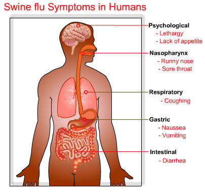
The symptoms of swine influenza in humans are similar to those of seasonal flu and may include:

  1. Fever: Often the first noticeable symptom.
  2. Cough: Dry or productive, with sore throat.
  3. Body Aches: Muscle pain, fatigue, and general weakness.
  4. Headache: Often accompanied by chills and shivering.
  5. Respiratory Symptoms: Shortness of breath, particularly in severe cases.
  6. Gastrointestinal Symptoms: Some patients experience nausea, vomiting, and diarrhea.

These symptoms usually appear 1-4 days after exposure and range from mild to severe, depending on the individual’s overall health and immune response (CDC, 2022).


Diagnosis

Diagnosis of swine influenza involves clinical evaluation and laboratory testing. Key diagnostic methods include:

  1. Rapid Influenza Diagnostic Tests (RIDTs): Provide preliminary results by detecting viral antigens but have limited sensitivity.
  2. Reverse Transcriptase-Polymerase Chain Reaction (RT-PCR): The gold standard test for swine flu, providing highly accurate results by detecting viral RNA.
  3. Viral Culture: Involves growing the virus in a laboratory setting for detailed analysis, though it is more time-consuming.
  4. Serological Tests: Assess immune response by detecting antibodies, especially helpful for retrospective diagnosis (Matsumoto et al., 2021).

RT-PCR is the preferred diagnostic tool due to its accuracy, especially in detecting various influenza A subtypes, including those associated with swine influenza.

(Nursing Paper Example on Swine Influenza)


Treatment

Treatment for swine influenza primarily focuses on antiviral medications and supportive care:

  1. Antiviral Medications: Oseltamivir (Tamiflu) and zanamivir (Relenza) are the main antiviral drugs used to treat influenza, including swine influenza. Early administration, preferably within 48 hours of symptom onset, improves efficacy.
  2. Symptom Management: Rest, hydration, and over-the-counter pain relievers can help manage symptoms such as fever and body aches.
  3. Hospitalization: Severe cases may require hospitalization for respiratory support or additional medical care.

Antiviral resistance is a growing concern, so healthcare providers should prescribe antivirals based on current resistance patterns and guidelines (WHO, 2023).


Complications

In most cases, swine influenza is self-limiting, but certain populations are at higher risk for complications:

  1. Respiratory Complications: Viral pneumonia, acute respiratory distress syndrome, and secondary bacterial infections.
  2. Cardiovascular Complications: Myocarditis and pericarditis, particularly in elderly individuals and those with underlying heart disease.
  3. Neurological Complications: Rare cases of encephalitis, seizures, and other neurological symptoms, especially in children.

High-risk groups for complications include young children, elderly adults, pregnant individuals, and people with chronic health conditions like asthma, diabetes, or heart disease (CDC, 2022).


Prevention

Preventing swine influenza requires a combination of personal hygiene, vaccination, and public health interventions:

  1. Vaccination: Seasonal influenza vaccines protect against common human influenza strains, although they do not typically cover all swine flu variants. In high-risk settings, such as pig farms, vaccination of pigs may help reduce transmission.
  2. Personal Protective Measures: Handwashing, mask-wearing, and avoiding contact with infected pigs can reduce the risk of transmission.
  3. Public Health Monitoring: Surveillance of pig populations and human cases is essential to detect outbreaks early and mitigate spread.
  4. Isolation of Infected Individuals: Infected individuals should self-isolate to prevent further transmission, particularly in healthcare settings or densely populated areas (CDC, 2022).

Effective prevention requires coordination between public health agencies, healthcare providers, and agricultural sectors to monitor and control outbreaks in both swine and human populations.


Conclusion

Swine influenza is a zoonotic disease caused by influenza A viruses that can jump from pigs to humans, posing significant public health risks. While symptoms in humans are often similar to seasonal flu, the disease can lead to severe complications, especially in vulnerable populations. Diagnosis relies on clinical symptoms and laboratory testing, with RT-PCR as the most reliable method. Treatment involves antiviral drugs, supportive care, and symptom management. Prevention includes vaccination, personal hygiene, and surveillance. Public awareness and early intervention are essential for controlling swine influenza and preventing pandemics.


References

Centers for Disease Control and Prevention (CDC). (2022). Swine influenza: Causes, symptoms, and prevention. https://www.cdc.gov/flu/swineflu

Matsumoto, K., Oka, T., Suzuki, K., & Kinoshita, H. (2021). Influenza virus and public health implications: Diagnosis, treatment, and control. Japanese Journal of Infectious Diseases, 74(2), 112-123. https://www.jstage.jst.go.jp/article/yoken/74/2/74_112/_pdf

World Health Organization (WHO). (2023). Influenza at the human-animal interface: Zoonotic influenza viruses. https://www.who.int/news-room/fact-sheets

 
Do you need a similar assignment done for you from scratch? Order now!
Use Discount Code "Newclient" for a 15% Discount!

Nursing Paper Example on Syphilis

Nursing Paper Example on Syphilis

Syphilis is a sexually transmitted infection (STI) caused by the bacterium Treponema pallidum. This condition, known for its multi-stage progression and diverse symptoms, has significant implications for public health. If untreated, syphilis can advance through several stages, leading to severe health complications.


Nursing Paper Example on Syphilis

Causes and Transmission

Causative Agent

Syphilis is caused by the spirochete bacterium Treponema pallidum. This spiral-shaped bacterium is highly mobile and able to penetrate mucous membranes, allowing it to infect the body easily.

Transmission

The primary mode of transmission is through sexual contact, including vaginal, anal, and oral sex. It can also be transmitted vertically from mother to fetus during pregnancy, leading to congenital syphilis. Transmission occurs when T. pallidum enters the body through microscopic abrasions or mucous membranes (CDC, 2021).


Stages of Syphilis and Associated Symptoms

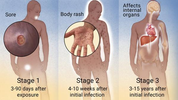
Syphilis progresses in four stages, each with distinct symptoms and characteristics:

1. Primary Syphilis

Primary syphilis is marked by the appearance of a single sore, known as a chancre, at the infection site. This sore is typically firm, round, and painless, appearing within 10-90 days post-exposure. Chancres can occur on the genitals, rectum, or mouth and heal without treatment after 3-6 weeks. Even if the sore heals, syphilis will progress if untreated.

2. Secondary Syphilis

Secondary syphilis can develop weeks to months after the initial infection, marked by a skin rash and mucous membrane lesions. The rash often appears as rough, reddish-brown spots on the palms and soles. Other symptoms include fever, sore throat, muscle aches, swollen lymph nodes, and fatigue. These symptoms may resolve independently, but the infection persists without treatment (CDC, 2021).

3. Latent Syphilis

In the latent stage, syphilis becomes asymptomatic, and no clinical symptoms are present. However, the infection remains active in the body. The latent stage is further divided into early (within the first year) and late (after the first year) phases. Some individuals may remain in this stage without progressing, while others may develop tertiary syphilis.

4. Tertiary Syphilis

Tertiary syphilis can occur years or decades after the initial infection if untreated. It causes severe damage to multiple organs, including the heart, brain, liver, bones, and nerves. Symptoms vary based on affected organs, leading to life-threatening complications such as cardiovascular damage, neurosyphilis, and gummatous lesions (micro-granulomatous lesions).


Diagnosis

Clinical Examination

A healthcare provider may perform a physical exam and assess patient history to identify signs of syphilis.

Laboratory Tests

  1. Serological Testing: Blood tests are the primary diagnostic tool for syphilis.
    • Nontreponemal Tests: Rapid plasma reagin (RPR) and Venereal Disease Research Laboratory (VDRL) tests detect antibodies produced in response to T. pallidum.
    • Treponemal Tests: Fluorescent treponemal antibody absorption (FTA-ABS) and treponemal pallidum particle agglutination assay (TP-PA) detect antibodies specific to T. pallidum and confirm diagnosis.
  2. Direct Detection Methods: Dark-field microscopy or polymerase chain reaction (PCR) tests can detect T. pallidum in samples from lesions, primarily used in primary syphilis.
  3. Neurosyphilis Testing: Cerebrospinal fluid (CSF) analysis is used if neurosyphilis is suspected based on neurological symptoms (Workowski & Bachmann, 2021).

Treatment

Penicillin remains the most effective treatment for all stages of syphilis. The treatment varies based on the infection stage:

  1. Primary, Secondary, and Early Latent Syphilis: A single intramuscular injection of Benzathine penicillin G is recommended.
  2. Late Latent and Tertiary Syphilis: Treatment requires three doses of Benzathine penicillin G administered weekly.
  3. Neurosyphilis: Intravenous (IV) penicillin G is required for individuals with neurosyphilis due to its need to cross the blood-brain barrier.

For penicillin-allergic individuals, doxycycline or tetracycline may be used as alternatives, although penicillin is preferred. Post-treatment, patients should be monitored with follow-up serological tests to ensure treatment efficacy (CDC, 2021).


Complications

Untreated syphilis can result in severe complications:

  1. Cardiovascular Syphilis: Damage to the heart and blood vessels, potentially leading to aneurysms and aortitis.
  2. Neurosyphilis: Syphilis affecting the nervous system can cause cognitive impairments, vision problems, and paralysis.
  3. Gummatous Syphilis: Development of soft, tumor-like growths, called gummas, that damage bones, skin, and soft tissues.
  4. Congenital Syphilis: Infants born to untreated mothers may suffer from congenital syphilis, resulting in severe birth defects, organ damage, and even infant mortality (French et al., 2020).

Prevention

Preventing syphilis primarily involves safe sexual practices and regular STI screening:

  1. Barrier Protection: Consistent condom use can significantly reduce the risk of transmission.
  2. Regular Testing: Regular syphilis testing is crucial, especially for sexually active individuals and high-risk populations.
  3. Prompt Treatment: Treating syphilis promptly prevents its spread to partners and reduces the risk of long-term complications.
  4. Prenatal Screening: Pregnant individuals should be screened for syphilis early to prevent congenital syphilis in infants.

Public health campaigns and education about syphilis prevention can further reduce its prevalence and transmission (French et al., 2020).


Conclusion

Syphilis is a complex STI caused by Treponema pallidum, progressing through four stages if left untreated. Recognizing and treating syphilis in its early stages prevents its progression to more severe forms that affect multiple organs. Diagnosis relies on clinical examination and laboratory tests, with penicillin remaining the primary treatment. Preventing syphilis requires safe sexual practices, regular screenings, and timely treatment, particularly in high-risk groups. Public awareness and education are critical to controlling the spread of this infection.


References

Centers for Disease Control and Prevention (CDC). (2021). Syphilis – CDC fact sheet (detailed). https://www.cdc.gov/std/syphilis/stdfact-syphilis-detailed.htm

French, P., Gomberg, M., Janier, M., Schmidt, B., & van Voorst Vader, P. (2020). Syphilis: Clinical features, diagnosis, and treatment. Clinical Microbiology and Infection, 26(4), 425-431. https://www.clinicalmicrobiologyandinfection.com/article/S1198-743X(19)30698-4/fulltext

Workowski, K. A., & Bachmann, L. H. (2021). Sexually transmitted infections treatment guidelines, 2021. Morbidity and Mortality Weekly Report, 70(4), 1-190. https://www.cdc.gov/std/treatment-guidelines/syphilis.htm

 
Do you need a similar assignment done for you from scratch? Order now!
Use Discount Code "Newclient" for a 15% Discount!

Nursing Paper Example on Synovitis

Nursing Paper Example on Synovitis

Synovitis is an inflammatory condition affecting the synovial membrane, which lines joint cavities. The inflammation leads to pain, swelling, and stiffness in the affected joints. It commonly occurs in association with various forms of arthritis but can also result from injury, infection, or other diseases. A thorough understanding of synovitis includes exploring its causes, symptoms, diagnostic methods, treatment options, complications, and preventive measures.


Nursing Paper Example on Synovitis

Causes and Risk Factors

Primary Causes

Synovitis results from inflammation in the synovial membrane. This inflammation can stem from:

  1. Rheumatoid Arthritis: The most common cause of chronic synovitis, resulting in autoimmunity where the body mistakenly attacks the synovium.
  2. Trauma: Joint injuries can trigger inflammation as the body attempts to repair itself.
  3. Infections: Bacterial or viral infections can lead to septic synovitis, where the joint becomes infected and inflamed.
  4. Gout and Pseudogout: The deposition of uric acid or calcium pyrophosphate crystals within the joints can trigger synovitis.
  5. Other Systemic Diseases: Conditions like lupus and psoriatic arthritis can also contribute to synovial inflammation (Smolen et al., 2018).

(Nursing Paper Example on Synovitis)

Risk Factors

Several factors increase the likelihood of developing synovitis:

  • Age: Synovitis risk increases with age due to the higher likelihood of arthritis.
  • Previous Joint Injuries: Injuries from sports or accidents can predispose individuals to synovitis.
  • Autoimmune Diseases: A history of autoimmune conditions may heighten the risk.
  • Occupational Activities: Jobs involving repetitive joint movement can lead to synovial inflammation over time (McInnes & Schett, 2017).

Signs and Symptoms

Synovitis symptoms vary depending on the severity and underlying cause but typically include:

  • Joint Swelling: Visible swelling occurs due to fluid accumulation in the joint.
  • Pain: Joints feel sore, especially during movement or pressure.
  • Stiffness: Morning stiffness or difficulty moving the joint, which may improve with activity.
  • Warmth and Redness: Inflamed joints often feel warm and may have a reddish hue.
  • Limited Range of Motion: Due to swelling and pain, the joint’s normal movement may become restricted.

These symptoms are commonly seen in joints such as the knees, elbows, wrists, and ankles. Chronic synovitis can eventually cause damage to the joint and lead to joint deformities if left untreated (Filer, 2018).


Pathophysiology

Synovitis involves an immune response in the synovial membrane. In cases of autoimmune disease, the body mistakenly targets synovial cells, leading to inflammation. The inflammatory process is marked by the accumulation of immune cells like macrophages and T cells, which release pro-inflammatory cytokines such as interleukin-1 (IL-1) and tumor necrosis factor-alpha (TNF-α). These cytokines promote further inflammation, leading to swelling, increased blood flow, and joint damage. Over time, persistent inflammation can cause the thickening of the synovial lining, called pannus formation, which further restricts joint mobility and damages cartilage (McInnes & Schett, 2017).


Diagnosis

Clinical Examination

A healthcare provider typically starts by examining the affected joint for swelling, warmth, and limited motion. Patient history, including recent injuries, infections, or pre-existing autoimmune conditions, is also considered.

Imaging Tests

  • Ultrasound: An ultrasound of the joint helps visualize the inflamed synovial membrane and fluid accumulation.
  • Magnetic Resonance Imaging (MRI): MRI can provide a detailed image of the joint structure and is particularly useful in identifying synovitis in deep joints.

Laboratory Tests

  • Blood Tests: Used to detect markers of inflammation, including C-reactive protein (CRP) and erythrocyte sedimentation rate (ESR), and to test for autoimmune markers like rheumatoid factor (RF) and anti-cyclic citrullinated peptide (anti-CCP) antibodies.
  • Joint Aspiration (Arthrocentesis): A sample of synovial fluid is collected and analyzed for signs of infection, crystals, or inflammatory cells (Filer, 2018).

(Nursing Paper Example on Synovitis)


Treatment

Synovitis treatment focuses on reducing inflammation, alleviating pain, and addressing the underlying cause:

  1. Medication:
    • Non-Steroidal Anti-Inflammatory Drugs (NSAIDs): Commonly used to reduce inflammation and relieve pain.
    • Corticosteroids: Can be injected directly into the joint for rapid inflammation reduction.
    • Disease-Modifying Antirheumatic Drugs (DMARDs): Used for chronic autoimmune-related synovitis, these drugs, including methotrexate, help slow disease progression.
    • Biologic Agents: For rheumatoid arthritis-related synovitis, biologics like TNF inhibitors may be used to target specific immune pathways (Smolen et al., 2018).
  2. Physical Therapy:
    • Exercises to maintain joint flexibility and strength can help patients maintain function and prevent stiffness.
  3. Surgery:
    • In severe cases, surgical options like synovectomy (removal of the inflamed synovium) may be considered to relieve pain and prevent further joint damage.
  4. Lifestyle Modifications:
    • Rest and Joint Protection: Reducing stress on affected joints can help manage pain and inflammation.
    • Weight Management: For weight-bearing joints, weight loss can reduce joint strain and inflammation.

Timely diagnosis and treatment are essential to prevent the progression of joint damage in synovitis (McInnes & Schett, 2017).


Complications

Untreated or recurrent synovitis can lead to significant complications:

  1. Chronic Joint Damage: Persistent inflammation can cause cartilage and bone damage, resulting in reduced joint mobility.
  2. Joint Deformities: Severe cases may lead to structural changes in the joint, impairing function.
  3. Infection: Synovitis caused by infections, if left untreated, can spread and result in septic arthritis, a serious and potentially life-threatening condition (Filer, 2018).

Prevention

Preventing synovitis involves reducing risk factors and addressing underlying conditions:

  • Prompt Treatment of Joint Injuries: Reduces the risk of post-traumatic synovitis.
  • Manage Autoimmune Disorders: Early diagnosis and management of autoimmune diseases can help prevent inflammation in the joints.
  • Infection Control: Maintaining proper hygiene, treating infections promptly, and avoiding exposure to pathogens can reduce the risk of infectious synovitis.
  • Protective Equipment: Individuals with high-impact jobs or those involved in sports should use joint protection to minimize trauma (Smolen et al., 2018).

Conclusion

Synovitis, a condition characterized by synovial membrane inflammation, often coexists with other joint disorders and diseases. From autoimmune origins to traumatic injury, multiple factors can initiate and exacerbate synovitis. Prompt diagnosis through physical exams, imaging, and laboratory tests can significantly improve treatment outcomes. Treatment approaches focus on reducing inflammation, managing symptoms, and addressing underlying causes. Preventing synovitis requires both managing primary risk factors and adopting protective measures, particularly for those prone to joint injuries or autoimmune conditions.


References

Filer, A. (2018). The role of synovitis in the pathophysiology of rheumatoid arthritis. Arthritis Research & Therapy, 20(1), 1-7. https://arthritis-research.biomedcentral.com/articles/10.1186/s13075-018-1533-z

McInnes, I. B., & Schett, G. (2017). Pathogenetic insights from the synovium in autoimmunity and inflammation. Current Opinion in Rheumatology, 29(2), 125-130. https://journals.lww.com/co-rheumatology/Abstract/2017/03000/Pathogenetic_insights_from_the_synovium_in.4.aspx

Smolen, J. S., Aletaha, D., & McInnes, I. B. (2018). Rheumatoid arthritis. The Lancet, 388(10055), 2023-2038. https://www.thelancet.com/journals/lancet/article/PIIS0140-6736(18)31791-5/fulltext

 
Do you need a similar assignment done for you from scratch? Order now!
Use Discount Code "Newclient" for a 15% Discount!

Nursing Paper Example on German measles

Nursing Paper Example on German Measles

Rubella, also known as German measles, is a contagious viral infection known for its distinctive red rash. Caused by the rubella virus, it poses significant health risks, especially in pregnant women due to its potential to cause congenital rubella syndrome (CRS) in the fetus. Understanding rubella’s causes, transmission, signs and symptoms, diagnosis, treatment, complications, and prevention is crucial in managing this disease and preventing outbreaks.


Nursing Paper Example on German Measles

Causes and Transmission

Causes

Rubella is caused by the rubella virus, a member of the Togaviridae family and Rubivirus genus. The virus primarily infects the upper respiratory tract and is transmitted via airborne droplets or direct contact with respiratory secretions.

Transmission

Rubella is highly contagious and spreads through:

  1. Airborne Transmission: When an infected person coughs or sneezes, they release respiratory droplets containing the virus.
  2. Direct Contact with Infected Secretions: The virus can spread through direct contact with secretions from the nose or throat.
  3. Vertical Transmission: From mother to fetus during pregnancy, particularly dangerous as it can lead to CRS.

People with rubella are typically contagious for a week before and about a week after the appearance of the rash. Individuals can contract rubella by inhaling viral particles or by contact with infected surfaces (CDC, 2022).


Signs and Symptoms

The symptoms of rubella generally appear 2-3 weeks after exposure and are often mild. They include:

  • Rash: A red or pink rash that starts on the face and spreads downward.
  • Low-Grade Fever: Commonly under 102°F (38.9°C).
  • Swollen Lymph Nodes: Especially behind the ears and at the back of the head.
  • Other Symptoms: Joint pain, sore throat, and red eyes.

In many cases, rubella infection is mild, particularly in children, and may go unnoticed. However, adults tend to experience more severe symptoms, especially joint pain and arthritis, which is common in women (Rogers & Bitnun, 2021).


Pathophysiology

The rubella virus enters the respiratory tract and spreads to the lymphoid tissue, where it undergoes replication. From there, the virus enters the bloodstream (viremia) and disseminates throughout the body. The immune response includes the development of neutralizing antibodies and cellular immunity, which typically leads to the clearance of the virus within two weeks.

In pregnant women, the virus can cross the placenta, infecting fetal tissues. This can result in CRS, a condition characterized by various severe fetal abnormalities, such as cataracts, hearing impairment, heart defects, and developmental delays. The severity of CRS depends on the gestational period at which the fetus is exposed, with the first trimester posing the highest risk (Patrono et al., 2019).


Diagnosis

Rubella is typically diagnosed based on clinical symptoms, but laboratory confirmation is necessary:

  1. Serology: Blood tests detect rubella-specific IgM antibodies, which usually indicate recent infection.
  2. PCR (Polymerase Chain Reaction): Can detect rubella RNA in respiratory specimens, blood, urine, or cerebrospinal fluid, especially valuable in congenital rubella.
  3. Prenatal Testing: In pregnant women with suspected exposure, amniocentesis can be used to test for rubella virus RNA in amniotic fluid to assess fetal infection.

Proper diagnosis is essential for confirming rubella cases, particularly in preventing further spread in high-risk environments like schools or healthcare settings (Banatvala & Brown, 2019).


Treatment

There is no specific antiviral treatment for rubella. Instead, management focuses on symptom relief and supportive care:

  • Fever and Pain Relief: Use of acetaminophen or ibuprofen to reduce fever and alleviate joint pain.
  • Isolation: Prevents the spread of rubella, particularly in community settings. Infected individuals should avoid public spaces until they are no longer contagious.

Patients, especially pregnant women exposed to rubella, are encouraged to seek medical care to monitor for potential complications and discuss preventive measures (CDC, 2022).


Complications

While rubella is generally mild, it can result in complications, particularly in adults and pregnant women:

  1. Arthritis: Common in adults, particularly women, and can last for weeks to months.
  2. Thrombocytopenia: A decrease in blood platelets, which can lead to bleeding issues.
  3. Encephalitis: A rare but serious complication causing inflammation of the brain.
  4. Congenital Rubella Syndrome (CRS): Occurs when a pregnant woman contracts rubella, especially during the first trimester. CRS can result in severe birth defects, including:
    • Cardiac Defects: Such as patent ductus arteriosus.
    • Ocular Abnormalities: Cataracts and glaucoma.
    • Sensorineural Deafness: One of the most common complications.
    • Neurological Impairments: Including microcephaly and developmental delays.

Due to these potential complications, the prevention of rubella, especially in pregnant women, is essential (Patrono et al., 2019).


Prevention

Vaccination

The rubella vaccine, typically combined with the measles and mumps vaccines (MMR), is highly effective. Key aspects include:

  • MMR Vaccine: Recommended for children, with the first dose administered at 12-15 months and a second dose at 4-6 years of age.
  • MMRV Vaccine: An option for children, which includes protection against varicella (chickenpox).
  • Vaccination of Women of Childbearing Age: Women planning to conceive should confirm immunity to rubella, as the vaccine cannot be given during pregnancy.

Vaccination has led to the near eradication of rubella in many developed countries, though periodic outbreaks can occur, especially in areas with low vaccination coverage (WHO, 2020).

Hygiene Practices and Isolation

For those infected, strict hygiene practices and isolation are essential to prevent transmission. Individuals should stay home from work, school, or daycare for at least a week after the rash appears (CDC, 2022).


Conclusion

Rubella is a preventable viral infection with mild symptoms in most cases but serious consequences when contracted by pregnant women due to the risk of CRS. Vaccination remains the most effective preventive measure, nearly eradicating rubella in many regions with high vaccination rates. Prompt diagnosis, effective symptom management, and public health measures, including vaccination, are essential for controlling rubella and preventing congenital infections.


References

Banatvala, J. E., & Brown, D. W. (2019). Rubella. The Lancet, 363(9415), 1127-1137. https://www.thelancet.com/journals/lancet/article/PIIS0140-6736(04)15897-2/fulltext

Centers for Disease Control and Prevention. (2022). Rubella (German Measles). Centers for Disease Control and Prevention. https://www.cdc.gov/rubella/index.html

Patrono, L. V., Poletti, M., Fedele, G., & Borgia, P. (2019). Congenital Rubella Syndrome: A Systematic Review of Diagnosis, Management, and Complications. Journal of Medical Virology, 91(9), 1651-1657. https://onlinelibrary.wiley.com/doi/full/10.1002/jmv.25571

Rogers, S., & Bitnun, A. (2021). Rubella and Congenital Rubella Syndrome. Canadian Journal of Infectious Diseases and Medical Microbiology, 2021, 1-8. https://www.hindawi.com/journals/cjidmm/2021/6638299

 
Do you need a similar assignment done for you from scratch? Order now!
Use Discount Code "Newclient" for a 15% Discount!

Nursing Paper Example on Streptococcal Infection

Nursing Paper Example on Streptococcal Infection

Streptococcal infection refers to an array of infections caused by the Streptococcus bacteria. These bacteria are divided into several groups, primarily Groups A and B, which are responsible for a range of diseases, from mild throat infections to severe, life-threatening illnesses. Understanding the causes, types, signs and symptoms, pathophysiology, diagnosis, and treatment options for streptococcal infections is critical for effective disease management and prevention.


Nursing Paper Example on Streptococcal Infection

Causes and Types

Causes of Streptococcal Infection

Streptococcal infections are caused by different species of the Streptococcus bacteria. The two most common groups causing infections are:

  • Group A Streptococcus (GAS): Primarily causes throat infections, skin infections, and more invasive diseases like toxic shock syndrome and necrotizing fasciitis.
  • Group B Streptococcus (GBS): Often found in the gastrointestinal and genital tracts, GBS can cause infections in newborns, pregnant women, and immunocompromised individuals (CDC, 2022).

Types of Streptococcal Infections

  1. Strep Throat: Caused by GAS, this infection affects the throat and tonsils, resulting in sore throat, pain while swallowing, and fever.
  2. Scarlet Fever: Occurs when GAS releases toxins, leading to a red, sandpaper-like rash, sore throat, and high fever.
  3. Impetigo: A skin infection caused by GAS, leading to red sores on the skin, especially around the mouth and nose.
  4. Invasive Streptococcal Diseases: Conditions like necrotizing fasciitis and streptococcal toxic shock syndrome, caused by GAS invading deeper tissues or the bloodstream, can be life-threatening.
  5. Group B Strep Infection: Particularly dangerous in newborns, GBS can lead to sepsis, pneumonia, and meningitis (Hockenberry & Wilson, 2020).

(Nursing Paper Example on Streptococcal Infection)


Pathophysiology

The pathophysiology of streptococcal infections depends on the specific strain and infection type. Streptococcus pyogenes (GAS) and Streptococcus agalactiae (GBS) have different mechanisms for infecting the host.

  • Group A Streptococcus: These bacteria produce various enzymes and toxins that allow them to adhere to epithelial cells, evade the immune system, and cause tissue destruction. The production of exotoxins, such as streptolysins and superantigens, leads to the intense immune response and tissue damage characteristic of conditions like scarlet fever and toxic shock (Stevens, 2021).
  • Group B Streptococcus: This strain primarily affects newborns due to maternal transmission during childbirth. GBS bacteria can evade the immune system by producing a polysaccharide capsule, allowing them to colonize mucosal surfaces and spread within the host (Schuchat, 2022).

Signs and Symptoms

Streptococcal infections present with a variety of symptoms, depending on the site and severity of the infection:

  • Strep Throat: Sore throat, red and swollen tonsils, fever, and swollen lymph nodes.
  • Scarlet Fever: Red rash, high fever, strawberry-like appearance of the tongue, and sore throat.
  • Impetigo: Red sores that rupture and form a honey-colored crust.
  • Invasive GAS Infections: Severe pain, swelling, and redness in the affected area, fever, and, in cases of toxic shock, low blood pressure and multi-organ failure.
  • Group B Strep in Newborns: Fever, difficulty breathing, lethargy, and irritability.

These symptoms vary significantly based on the type of streptococcal infection and the age or health of the patient (Hockenberry & Wilson, 2020).


Diagnosis

Diagnosing streptococcal infections requires both clinical examination and laboratory tests:

  • Rapid Antigen Detection Test (RADT): Commonly used to diagnose strep throat. This test detects streptococcal antigens and provides quick results, though it may sometimes require confirmation by culture.
  • Throat or Skin Culture: For accurate diagnosis, especially in cases where RADT is inconclusive. Cultures involve growing the bacteria from a sample in a lab to identify the specific streptococcal strain.
  • Blood Tests: For invasive infections, blood cultures can detect the presence of streptococcus in the bloodstream.
  • Polymerase Chain Reaction (PCR): Highly specific and sensitive, PCR tests can detect bacterial DNA in clinical specimens, used mainly for invasive cases (Stevens, 2021).

Treatment

Antibiotics

The primary treatment for streptococcal infections is antibiotics, which are highly effective when administered early:

  • Penicillin and Amoxicillin: These antibiotics are the first-line treatment for non-invasive streptococcal infections, such as strep throat and skin infections.
  • Erythromycin or Clindamycin: Used for patients with penicillin allergies or for more serious infections where GAS is present.
  • Intravenous Antibiotics: Necessary for severe infections like necrotizing fasciitis or streptococcal toxic shock syndrome.

Supportive Care

  • Pain Relief: Nonsteroidal anti-inflammatory drugs like ibuprofen are used to manage pain and fever.
  • Hydration and Rest: Important for aiding the immune system’s response.
  • Surgical Intervention: In invasive cases like necrotizing fasciitis, surgery may be needed to remove infected tissues.

Proper treatment reduces symptoms, prevents complications, and decreases the likelihood of spreading the infection to others (CDC, 2022).

(Nursing Paper Example on Streptococcal Infection)


Complications

Streptococcal infections can result in serious complications, especially if left untreated:

  • Acute Rheumatic Fever: An inflammatory disease affecting the heart, joints, and brain, usually following untreated strep throat or scarlet fever.
  • Post-Streptococcal Glomerulonephritis: A kidney disease resulting from GAS infections.
  • Pneumonia: Particularly dangerous in immunocompromised individuals and newborns with GBS infection.
  • Sepsis and Toxic Shock: Can occur if streptococcal bacteria invade the bloodstream, causing life-threatening conditions.

Preventing these complications relies on timely diagnosis, treatment, and monitoring for signs of progression (Hockenberry & Wilson, 2020).


Prevention

Good Hygiene Practices

  • Handwashing: Reduces the risk of spread, especially after coughing, sneezing, or handling contaminated objects.
  • Covering Mouth and Nose: Reduces the risk of droplet transmission.

Vaccination and Screening

  • Group B Strep Screening: Pregnant women are often screened for GBS during late pregnancy to prevent newborn infections.
  • Potential Vaccine Research: Currently, no vaccine exists for streptococcal infections, though research is ongoing to develop preventive measures for GAS and GBS (Stevens, 2021).

Conclusion

Streptococcal infections are a diverse group of bacterial infections with varying symptoms, from mild to life-threatening. Effective diagnosis relies on rapid tests and cultures, while treatment is primarily antibiotic-based. Early intervention prevents serious complications, including rheumatic fever and toxic shock. Preventive measures, especially in healthcare settings, are crucial for minimizing transmission and protecting vulnerable populations. Further research and potential vaccine development hold promise for reducing the burden of streptococcal infections globally.


References

Centers for Disease Control and Prevention. (2022). Group A Streptococcal (GAS) Disease. Centers for Disease Control and Prevention. https://www.cdc.gov/groupastrep/diseases-public/index.html

Hockenberry, M. J., & Wilson, D. (2020). Wong’s Essentials of Pediatric Nursing. Elsevier. https://www.elsevier.com/books/wongs-essentials-of-pediatric-nursing/hockenberry/978-0-323-60805-4

Schuchat, A. (2022). Group B streptococcus. In Infectious Diseases. Elsevier. https://www.elsevier.com/books/infectious-diseases/cohen/978-0-7020-6285-8

Stevens, D. L. (2021). Invasive Group A Streptococcal Infections. New England Journal of Medicine, 384(12), 1150-1161. https://www.nejm.org

 
Do you need a similar assignment done for you from scratch? Order now!
Use Discount Code "Newclient" for a 15% Discount!

Nursing Paper Example on Strep Throat

Nursing Paper Example on Strep Throat

Strep throat, or streptococcal pharyngitis, is a bacterial infection caused by Streptococcus pyogenes, also known as group A Streptococcus. It primarily affects the throat and tonsils, leading to a sore throat, fever, and other symptoms. While strep throat can affect people of all ages, it is most common in children and adolescents. Left untreated, strep throat may lead to complications, some of which can be severe, such as rheumatic fever and kidney inflammation. This overview addresses the causes, pathophysiology, signs and symptoms, diagnosis, treatment options, and potential complications of strep throat.


Nursing Paper Example on Strep Throat

Causes and Pathophysiology

Primary Cause

Strep throat is caused by the bacterium Streptococcus pyogenes, a gram-positive pathogen that spreads through respiratory droplets. The infection is highly contagious and spreads via close contact, sharing of personal items, or exposure to contaminated surfaces. Once the bacteria enter the body, they can colonize the mucosal tissues in the throat, leading to inflammation and infection (Cohen-Poradosu & Kasper, 2022).

Pathophysiology

When Streptococcus pyogenes enters the throat, it produces various virulence factors that enable it to adhere to and invade epithelial cells. These include M proteins, hyaluronic acid capsules, and exotoxins. The M proteins allow the bacteria to evade the host’s immune response, while the exotoxins cause tissue damage and inflammation, resulting in the characteristic symptoms of strep throat (Shulman et al., 2012).

(Nursing Paper Example on Strep Throat)


Signs and Symptoms

Primary Symptoms

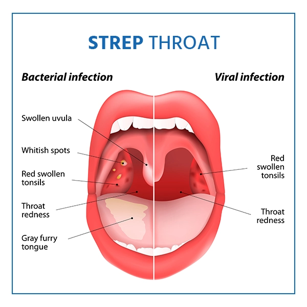
The common symptoms of strep throat include:

  • Severe sore throat
  • Pain while swallowing
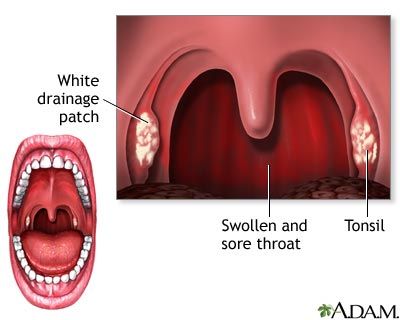
  • Red and swollen tonsils, sometimes with white patches or streaks of pus
  • Fever, usually over 101°F (38.3°C)
  • Swollen lymph nodes, particularly in the neck

Additional Symptoms

Some individuals may also experience symptoms such as headache, abdominal pain, nausea, and vomiting, especially in children. Unlike viral infections, strep throat typically lacks cough, hoarseness, or runny nose, helping differentiate it from other throat infections (Mayo Clinic, 2023).


Diagnosis

Physical Examination and History

Healthcare providers initially assess symptoms and examine the throat, noting redness, swelling, and any white patches. Swollen lymph nodes are another indicator that may prompt further testing.

Rapid Antigen Detection Test (RADT)

The RADT is a quick test used to identify the presence of group A Streptococcus antigens. This test provides results within minutes and has high specificity, but a lower sensitivity. A negative result may require confirmation with a throat culture (Shulman et al., 2012).

Throat Culture

A throat culture remains the gold standard for diagnosing strep throat due to its high accuracy. Although it takes 24-48 hours to get results, it is crucial for confirming strep throat in cases where RADT is negative. Throat cultures are particularly recommended for children and adolescents to prevent complications (Cohen-Poradosu & Kasper, 2022).


Treatment and Management

Antibiotic Therapy

The standard treatment for strep throat is antibiotics, with penicillin and amoxicillin being the most common choices due to their effectiveness and low risk of resistance. For patients allergic to penicillin, alternatives include cephalexin, clindamycin, and azithromycin. Antibiotic therapy generally shortens the duration of symptoms, reduces transmission risk, and prevents complications like rheumatic fever (Shulman et al., 2012).

Symptomatic Relief

In addition to antibiotics, symptomatic treatment includes:

  • Over-the-counter pain relievers like ibuprofen or acetaminophen to reduce fever and relieve pain.
  • Warm saltwater gargles to soothe throat discomfort.
  • Increased fluid intake to stay hydrated and reduce throat irritation.

Rest and Isolation

Rest is advised to help the immune system recover. Patients should remain home from school or work for at least 24 hours after starting antibiotics to prevent spreading the infection (Mayo Clinic, 2023).

(Nursing Paper Example on Strep Throat)


Complications

Acute Complications

Without prompt treatment, strep throat can lead to several acute complications, including:

  • Peritonsillar abscess: A collection of pus near the tonsils that can cause severe pain and may require drainage.
  • Otitis media: Middle ear infection that can develop from the spread of bacteria.

Post-Streptococcal Complications

If strep throat is untreated or inadequately treated, certain immune-mediated complications can occur, including:

  • Rheumatic fever: An inflammatory disease that can affect the heart, joints, skin, and brain. Rheumatic fever is associated with long-term cardiac complications, including rheumatic heart disease.
  • Post-streptococcal glomerulonephritis: Inflammation of the kidney, leading to hematuria, edema, and hypertension.

These complications underscore the importance of early diagnosis and treatment of strep throat (Cohen-Poradosu & Kasper, 2022).


Prevention

Hygiene Practices

Preventive measures include regular hand washing, covering the mouth when coughing or sneezing, and avoiding sharing personal items. In communal settings like schools, proper hygiene and isolation of infected individuals are vital to minimize the spread.

Awareness in Close Contacts

Household members or close contacts of those infected with strep throat should remain vigilant for symptoms. In some cases, antibiotic prophylaxis may be considered for high-risk individuals to prevent transmission.


Conclusion

Strep throat is a common yet potentially serious bacterial infection that requires prompt medical attention. Caused by Streptococcus pyogenes, it primarily affects the throat, leading to symptoms like sore throat, fever, and swollen lymph nodes. Diagnosis involves clinical examination, RADT, and throat culture. Antibiotic therapy is essential for treatment and prevention of complications such as rheumatic fever and kidney inflammation. Good hygiene practices and isolation during the infectious period are critical to preventing the spread of strep throat in communal settings. Early intervention with antibiotics and supportive care is essential to reduce symptom duration and avoid severe outcomes.


References

Cohen-Poradosu, R., & Kasper, D. L. (2022). Streptococcal infections. In Kasper, D. L., et al. (Eds.), Harrison’s Principles of Internal Medicine, 21st Edition. McGraw Hill. https://accessmedicine.mhmedical.com/content.aspx?bookid=2129&sectionid=192014500

Shulman, S. T., Bisno, A. L., Clegg, H. W., Gerber, M. A., Kaplan, E. L., Lee, G., … & Van Beneden, C. (2012). Clinical Practice Guideline for the Diagnosis and Management of Group A Streptococcal Pharyngitis: 2012 Update by the Infectious Diseases Society of America. Clinical Infectious Diseases, 55(10), e86-e102. https://academic.oup.com/cid/article/55/10/e86/321183

 
Do you need a similar assignment done for you from scratch? Order now!
Use Discount Code "Newclient" for a 15% Discount!

Nursing Paper Example on Fever

Nursing Paper Example on Fever

Fever, or pyrexia, is a temporary elevation in body temperature, often as a result of an infection or other medical condition. Fever is a natural physiological response, triggered by the immune system, to help combat infections. Defined as a body temperature above the normal range of 98.6°F (37°C), fever is commonly recognized at 100.4°F (38°C) or higher. While fever is generally beneficial, helping to reduce the growth of pathogens, high or prolonged fever can be harmful, particularly in vulnerable populations such as infants and elderly individuals. This overview addresses the causes, pathophysiology, types, symptoms, diagnosis, and treatment of fever.

Nursing Paper Example on Fever

Causes and Pathophysiology

Common Causes of Fever

Fever is often a symptom of an underlying infection, but it can result from a variety of causes, including:

  1. Infections: Bacterial, viral, fungal, or parasitic infections are primary causes.
  2. Autoimmune Conditions: Diseases like lupus or rheumatoid arthritis can trigger a fever.
  3. Inflammatory Disorders: Fever can accompany inflammatory conditions, including inflammatory bowel disease.
  4. Heat Exhaustion: Prolonged exposure to high temperatures can result in heat-related fever.
  5. Medication Reaction: Some drugs, such as antibiotics, can cause drug-induced fever.
  6. Vaccination: Fever may occur as an immune response post-vaccination, typically mild and short-lived.

Pathophysiology of Fever

The pathophysiology of fever involves the hypothalamus, which regulates body temperature. When pyrogens (fever-inducing agents) enter the body, either from external (e.g., bacteria, viruses) or internal sources (e.g., cytokines from the immune system), they stimulate the hypothalamus to raise the body’s set-point temperature. This elevation leads to thermoregulatory adjustments, including shivering and peripheral vasoconstriction, which produce heat and elevate body temperature (Dinarello & Porat, 2020).


Types of Fever

Fever is generally classified based on its pattern and duration:

  1. Intermittent Fever: Body temperature fluctuates but returns to normal within a 24-hour period, common in diseases like malaria.
  2. Remittent Fever: Fluctuating temperature that does not return to normal but varies throughout the day, often seen in bacterial infections.
  3. Sustained or Continuous Fever: The temperature remains elevated with minimal fluctuation, typically associated with infections like typhoid.
  4. Relapsing Fever: Characterized by recurrent fever episodes, as seen in diseases like Lyme disease and some viral infections (Harrison, 2022).

Symptoms of Fever

Aside from elevated body temperature, symptoms accompanying fever can vary widely:

  • Chills and Shivering: Body responses to generate warmth as the temperature rises.
  • Sweating: Occurs as fever begins to break or resolve.
  • Headache and Muscle Aches: Due to inflammatory responses.
  • Fatigue and Weakness: The immune system’s response can cause energy depletion.
  • Irritability or Confusion: In children and elderly individuals, fever can lead to changes in behavior or cognition.

These symptoms, however, can vary based on the underlying cause and the patient’s age and overall health condition (Mayo Clinic, 2023).


Diagnosis of Fever

Clinical Assessment

The diagnosis of fever starts with a detailed medical history and physical examination. Healthcare providers assess the pattern, duration, and associated symptoms to identify possible causes.

Temperature Measurement

Measuring body temperature accurately is crucial. Common methods include oral, axillary (underarm), rectal, and tympanic (ear) thermometers. Rectal thermometry is considered the gold standard, especially in infants and children, for accurately detecting fever (Dinarello & Porat, 2020).

Laboratory Tests

When the cause of fever is unclear, additional tests may include:

  • Blood Tests: To identify infections or inflammatory markers.
  • Urinalysis: To detect urinary tract infections.
  • Chest X-ray: If a respiratory infection, like pneumonia, is suspected.
  • Cultures: Blood, urine, and other body fluids may be cultured to identify bacterial or fungal infections.

In cases of prolonged or unexplained fever, known as fever of unknown origin (FUO), more extensive testing may be warranted to investigate non-infectious causes (Harrison, 2022).


Treatment and Management

Medication

  • Antipyretics: Drugs like acetaminophen and ibuprofen are common choices to lower fever and relieve associated discomfort.
  • Antibiotics: Used only if a bacterial infection is confirmed or strongly suspected. Antibiotic use is avoided for viral infections, where supportive care is the primary management.
  • Anti-inflammatory Drugs: For fevers due to inflammatory or autoimmune conditions, corticosteroids or nonsteroidal anti-inflammatory drugs may be prescribed.

Supportive Care

  • Hydration: Fever increases fluid loss, making adequate hydration essential.
  • Rest: Contributes to immune function and speeds recovery.
  • Cool Compresses: Can help relieve discomfort and reduce temperature slightly.

Potential Complications

While fever generally supports immune function, it may cause complications in some populations:

  • Febrile Seizures: A complication seen mainly in young children, often harmless but requiring monitoring.
  • Dehydration: Due to increased sweating and respiratory water loss.
  • Delirium and Confusion: Especially in older adults, high fever may lead to temporary cognitive impairments.
  • Organ Stress: High fevers, if sustained, can strain vital organs, particularly in individuals with underlying health conditions.

In these cases, monitoring and immediate medical attention can help prevent further complications (Dinarello & Porat, 2020).


Prevention of Fever

Preventive measures primarily focus on avoiding infections and managing chronic health conditions that may predispose individuals to fever:

  • Hand Hygiene: Regular hand washing to prevent infections.
  • Vaccination: Helps prevent diseases like influenza and pneumonia, common fever causes.
  • Avoiding Contaminated Sources: Particularly food and water sources in endemic regions.

Conclusion

Fever is a common symptom with a range of causes, from infections to immune responses. It plays a critical role in fighting infection, as the body’s elevated temperature can inhibit pathogen growth. Proper diagnosis involves physical examination, laboratory tests, and accurate temperature assessment. Treatment includes antipyretics, supportive care, and, if necessary, specific antimicrobial therapy for infections. Prevention focuses on hygiene, vaccination, and managing predisposing conditions. While fever itself is usually beneficial, monitoring is essential to avoid potential complications, especially in vulnerable populations.


References

Dinarello, C. A., & Porat, R. (2020). Fever in Infectious Diseases. In Mandell, Douglas, and Bennett’s Principles and Practice of Infectious Diseases, 9th Edition. Elsevier. https://www.sciencedirect.com/science/article/pii/B978032348255400039X

Harrison, T. R. (2022). Fever and Hyperthermia. In Harrison’s Principles of Internal Medicine, 21st Edition. McGraw-Hill Education. https://accessmedicine.mhmedical.com/content.aspx?bookid=2129&sectionid=192012433

Mayo Clinic. (2023). Fever: Causes, Diagnosis, and Treatment. Mayo Clinic. https://www.mayoclinic.org

 
Do you need a similar assignment done for you from scratch? Order now!
Use Discount Code "Newclient" for a 15% Discount!

Nursing Paper Example on Strabismus

Nursing Paper Example on Strabismus

Strabismus, commonly referred to as “crossed eyes” or “squint,” is a visual disorder where the eyes are misaligned and point in different directions. This misalignment can lead to various vision issues, as the eyes fail to work together to focus on the same object. Strabismus affects people of all ages but is most common in children. Early diagnosis and treatment are essential to prevent complications like amblyopia or “lazy eye.” This overview will explore the causes, types, pathophysiology, signs and symptoms, diagnostic methods, and treatment options for strabismus.

Nursing Paper Example on Strabismus

Causes and Pathophysiology

Primary Causes

Strabismus results from an imbalance in the eye muscles, an issue with the nerves transmitting information to these muscles, or problems within the brain responsible for coordinating eye movements. The disorder can be congenital, often due to genetics, or acquired through conditions such as stroke, traumatic brain injury, or neurological issues. In children, refractive errors like farsightedness can also lead to strabismus as the eye muscles strain to focus (Holmes & Clarke, 2006).

Pathophysiology

Strabismus arises when the eyes fail to maintain alignment due to weakened or overactive eye muscles. Six muscles control eye movement, allowing for up, down, side, and rotational motions. In healthy eyes, these muscles coordinate for both eyes to focus on a single point. However, with strabismus, one or more muscles may be underactive or overactive, causing the affected eye to deviate. Over time, the brain may ignore input from the misaligned eye to avoid double vision, which can result in amblyopia (Scott & Mashige, 2016).

(Nursing Paper Example on Strabismus)


Types of Strabismus

Esotropia

Esotropia occurs when one or both eyes turn inward. It is the most common type of strabismus in infants and can be constant or intermittent. Esotropia is often related to congenital issues or neurological conditions (Burian, 2015).

Exotropia

Exotropia, or “wall-eye,” is characterized by outward deviation of the eye. It may be present intermittently, often during times of tiredness or when focusing on distant objects. Exotropia is more common in older children and adults than in infants (Holmes & Clarke, 2006).

Hypertropia and Hypotropia

In hypertropia, the eye turns upward, while hypotropia involves downward turning. These types of vertical strabismus are less common but may indicate a neurological disorder or be associated with congenital abnormalities (Scott & Mashige, 2016).


Signs and Symptoms

Individuals with strabismus may experience several symptoms, which vary based on the severity and type of strabismus.

Primary Symptoms

Common symptoms include:

  • Eyes pointing in different directions
  • Double vision (diplopia)
  • Frequent squinting or closing one eye, especially in bright light
  • Head tilting to improve focus

In Children

Children may exhibit symptoms such as misaligned eyes, frequent eye rubbing, or lack of depth perception. Untreated strabismus in children often leads to amblyopia, as the brain suppresses images from the misaligned eye (Burian, 2015).


Diagnosis

Eye Examination

An ophthalmologist or optometrist diagnoses strabismus through a comprehensive eye exam. The Hirschberg test (corneal light reflex test) and cover test are commonly used to evaluate eye alignment and identify the type of strabismus.

Visual Acuity and Refraction Tests

These tests help assess the sharpness of vision and any refractive errors contributing to strabismus. Hyperopia, or farsightedness, is often associated with strabismus in children (Scott & Mashige, 2016).

Neurological Assessment

In cases where strabismus arises suddenly in adults, a neurological exam may be necessary to rule out serious conditions like brain tumors, stroke, or head trauma. Imaging studies, such as MRI or CT scans, can be employed if a neurological cause is suspected.

(Nursing Paper Example on Strabismus)


Treatment and Management

Corrective Lenses

For strabismus associated with refractive errors, prescription glasses or contact lenses can improve focus and alignment. Prism lenses are sometimes used to alter the light entering the eye, helping reduce double vision (Holmes & Clarke, 2006).

Vision Therapy

Vision therapy is often effective for certain types of strabismus, particularly for intermittent or mild forms. It involves exercises to strengthen eye muscles, improve coordination, and enhance depth perception.

Surgical Intervention

Eye muscle surgery is the primary treatment for severe or persistent strabismus. Surgeons adjust the position or length of eye muscles to achieve proper alignment. The procedure is commonly performed on children but may also be effective for adults. While surgery often improves alignment, it may not restore perfect depth perception, and follow-up treatments may be necessary (Burian, 2015).

Botulinum Toxin (Botox) Injections

Botulinum toxin injections may be used as a non-surgical option to temporarily weaken overactive eye muscles, particularly in adults with acquired strabismus. Botox is typically considered when surgery is not an option or as a temporary solution (Scott & Mashige, 2016).


Complications and Prognosis

Untreated strabismus, especially in children, can lead to amblyopia or permanent vision loss in the misaligned eye. Early diagnosis and treatment are essential to prevent these complications. In adults, untreated strabismus may lead to chronic double vision, impacting daily life and quality of life.

While children respond well to treatment, adults may experience limited success with certain interventions due to established neural pathways. However, with appropriate management, strabismus can be effectively controlled, reducing symptoms and improving visual alignment.


Conclusion

Strabismus is a complex eye condition that can significantly affect vision and quality of life if left untreated. With varied causes, including genetics, neurological factors, and refractive errors, it requires a multi-faceted approach for effective management. Treatment options range from corrective lenses and vision therapy to surgery and Botox injections, depending on the type and severity of the condition. Early intervention, especially in children, is key to preventing complications like amblyopia and preserving normal vision. For adults, treatment options remain available, although results may vary.


References

Burian, H. M. (2015). Pediatric Strabismus and Amblyopia. Journal of Pediatric Ophthalmology and Strabismus, 52(1), 23-30. https://journals.healio.com/journal/jpos

Holmes, J. M., & Clarke, M. P. (2006). Amblyopia. The Lancet, 367(9519), 1343-1351. https://www.thelancet.com/journals/lancet/article/PIIS0140-6736(06)68581-3/fulltext

Scott, W. E., & Mashige, K. P. (2016). Clinical management of strabismus in children and adults. Clinical Eye and Vision Care, 9(4), 233-246. https://www.sciencedirect.com/science/article/abs/pii/S0959270916000209

 
Do you need a similar assignment done for you from scratch? Order now!
Use Discount Code "Newclient" for a 15% Discount!

Nursing Paper Example on Stomach Ulcers

Nursing Paper Example on Stomach Ulcers

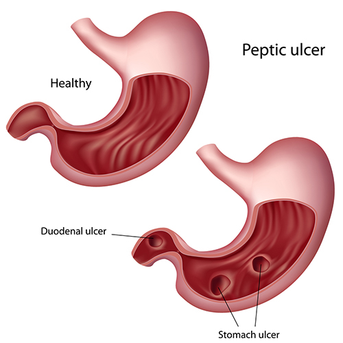
Stomach ulcers, or gastric ulcers, are painful lesions in the stomach lining. They fall under a broader category of peptic ulcers, which also includes duodenal ulcers in the upper part of the small intestine. These ulcers are largely attributed to bacterial infections, particularly from Helicobacter pylori (H. pylori), and to the excessive use of certain medications like nonsteroidal anti-inflammatory drugs (NSAIDs). This paper provides a comprehensive overview of the causes, pathophysiology, signs and symptoms, diagnostic approaches, and treatments for stomach ulcers.

Nursing Paper Example on Stomach Ulcers

Causes and Pathophysiology

Primary Causes

The two primary causes of stomach ulcers are H. pylori infections and NSAID usage. H. pylori, a spiral-shaped bacterium, colonizes the stomach lining and produces enzymes and toxins that damage the mucosal layer, making it more vulnerable to stomach acid. Prolonged NSAID usage inhibits prostaglandin production, which disrupts the protective mucus in the stomach, increasing the risk of ulcer formation (Sung et al., 2009).

Pathophysiology

In a healthy stomach, a thick mucus layer lines the stomach walls, protecting them from hydrochloric acid, which aids in digestion. However, when H. pylori bacteria are present, they weaken the stomach lining through a series of biochemical reactions, including the release of urease that neutralizes stomach acid and creates an alkaline environment favorable to the bacteria. This weakening, combined with the corrosive effect of acid and digestive enzymes, leads to ulcer formation. NSAIDs further exacerbate this by reducing mucus production, leaving the stomach wall unprotected (Malfertheiner et al., 2012).

(Nursing Paper Example on Stomach Ulcers)


Signs and Symptoms

Primary Symptoms

The most common symptom of a stomach ulcer is a burning or gnawing pain in the upper abdomen, which may improve or worsen with food intake. Other symptoms include bloating, heartburn, and nausea.

Severe Symptoms

In advanced cases, ulcers can cause severe complications, such as bleeding, perforation, or obstruction of the stomach. Blood in vomit or stools, unintentional weight loss, and severe abdominal pain are indicative of serious complications requiring immediate medical attention (Laine et al., 2008).


Diagnosis

Clinical Examination

Initial diagnosis is based on the patient’s symptoms and medical history, including any use of NSAIDs or symptoms of infection.

Endoscopic Examination

Endoscopy is the most definitive diagnostic tool for detecting stomach ulcers, allowing direct visualization and biopsy of the stomach lining. The procedure also helps assess the ulcer’s severity and rule out malignancies (Malfertheiner et al., 2012).

Non-Invasive Tests for H. pylori

For identifying H. pylori infections, non-invasive tests like the urea breath test, stool antigen test, and blood antibody test are commonly used. The urea breath test, considered the most accurate, involves ingesting a urea solution. If H. pylori is present, the bacteria break down urea, releasing carbon dioxide that can be detected in the patient’s breath (Sung et al., 2009).


Treatment and Management

Antibiotic Therapy for H. pylori

To eradicate H. pylori, a combination of antibiotics such as amoxicillin, clarithromycin, and metronidazole is prescribed. Known as triple therapy, this regimen is highly effective, especially when combined with proton pump inhibitors to reduce stomach acid and promote healing (Graham & Shiotani, 2008).

Acid-Suppressive Therapy

Proton pump inhibitors (PPIs) and histamine-2 blockers reduce stomach acid production, giving the stomach lining time to heal. PPIs, such as omeprazole, are generally preferred for their potent acid-suppressive effect.

Lifestyle Modifications

Patients are advised to avoid foods that irritate the stomach lining, such as spicy foods, caffeine, and alcohol. Smoking cessation is also critical, as smoking impedes ulcer healing and increases the likelihood of recurrence (Laine et al., 2008).

NSAID Alternatives

For patients with NSAID-induced ulcers, discontinuing or reducing NSAID use is essential. If pain management is necessary, alternative medications like acetaminophen may be recommended, as they are gentler on the stomach lining.

(Nursing Paper Example on Stomach Ulcers)


Prevention

Hygiene Practices

Since H. pylori infection is often acquired through contaminated food or water, maintaining good hygiene practices—such as regular handwashing and consuming clean, safe food—can lower infection risk.

Safe Medication Use

Limiting NSAID use and using protective medications, like PPIs, in conjunction with NSAIDs can help prevent NSAID-induced ulcers. Physicians may also recommend NSAID alternatives when feasible (Sung et al., 2009).


Conclusion

Stomach ulcers, predominantly caused by H. pylori infections and prolonged NSAID use, represent a significant health burden due to their potential complications. While treatable through a combination of antibiotics, acid-suppressive medications, and lifestyle modifications, severe cases may require further medical intervention to manage complications like bleeding or perforation. Preventive measures, including good hygiene and careful NSAID use, are crucial in reducing the prevalence of stomach ulcers. Advancements in diagnosis and treatment continue to improve patient outcomes, offering relief and healing to those affected by this condition.


References

Graham, D. Y., & Shiotani, A. (2008). New concepts of resistance in the treatment of Helicobacter pylori infections. Nature Clinical Practice Gastroenterology & Hepatology, 5(6), 321-331. https://www.nature.com/articles/ncpgasthep1141

Laine, L., Takeuchi, K., & Tarnawski, A. (2008). Gastric mucosal defense and cytoprotection: Bench to bedside. Gastroenterology, 135(1), 41-60. https://www.gastrojournal.org/article/S0016-5085(08)00650-1/fulltext

Malfertheiner, P., Megraud, F., O’Morain, C. A., Gisbert, J. P., Kuipers, E. J., & Axon, A. T. (2012). Management of Helicobacter pylori infection—the Maastricht IV/ Florence consensus report. Gut, 61(5), 646-664. https://gut.bmj.com/content/61/5/646

Sung, J. J., Kuipers, E. J., & El-Serag, H. B. (2009). Systematic review: the global incidence and prevalence of peptic ulcer disease. Alimentary Pharmacology & Therapeutics, 29(9), 938-946. https://onlinelibrary.wiley.com/doi/full/10.1111/j.1365-2036.2009.03960.x

 
Do you need a similar assignment done for you from scratch? Order now!
Use Discount Code "Newclient" for a 15% Discount!