Nursing Paper Example on Stomach Ulcers

Nursing Paper Example on Stomach Ulcers

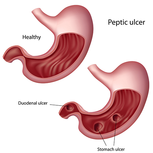
Stomach ulcers, or gastric ulcers, are painful lesions in the stomach lining. They fall under a broader category of peptic ulcers, which also includes duodenal ulcers in the upper part of the small intestine. These ulcers are largely attributed to bacterial infections, particularly from Helicobacter pylori (H. pylori), and to the excessive use of certain medications like nonsteroidal anti-inflammatory drugs (NSAIDs). This paper provides a comprehensive overview of the causes, pathophysiology, signs and symptoms, diagnostic approaches, and treatments for stomach ulcers.
Nursing Paper Example on Stomach Ulcers

Causes and Pathophysiology

Primary Causes

The two primary causes of stomach ulcers are H. pylori infections and NSAID usage. H. pylori, a spiral-shaped bacterium, colonizes the stomach lining and produces enzymes and toxins that damage the mucosal layer, making it more vulnerable to stomach acid. Prolonged NSAID usage inhibits prostaglandin production, which disrupts the protective mucus in the stomach, increasing the risk of ulcer formation (Sung et al., 2009).

Pathophysiology

In a healthy stomach, a thick mucus layer lines the stomach walls, protecting them from hydrochloric acid, which aids in digestion. However, when H. pylori bacteria are present, they weaken the stomach lining through a series of biochemical reactions, including the release of urease that neutralizes stomach acid and creates an alkaline environment favorable to the bacteria. This weakening, combined with the corrosive effect of acid and digestive enzymes, leads to ulcer formation. NSAIDs further exacerbate this by reducing mucus production, leaving the stomach wall unprotected (Malfertheiner et al., 2012).

(Nursing Paper Example on Stomach Ulcers)


Signs and Symptoms

Primary Symptoms

The most common symptom of a stomach ulcer is a burning or gnawing pain in the upper abdomen, which may improve or worsen with food intake. Other symptoms include bloating, heartburn, and nausea.

Severe Symptoms

In advanced cases, ulcers can cause severe complications, such as bleeding, perforation, or obstruction of the stomach. Blood in vomit or stools, unintentional weight loss, and severe abdominal pain are indicative of serious complications requiring immediate medical attention (Laine et al., 2008).


Diagnosis

Clinical Examination

Initial diagnosis is based on the patient’s symptoms and medical history, including any use of NSAIDs or symptoms of infection.

Endoscopic Examination

Endoscopy is the most definitive diagnostic tool for detecting stomach ulcers, allowing direct visualization and biopsy of the stomach lining. The procedure also helps assess the ulcer’s severity and rule out malignancies (Malfertheiner et al., 2012).

Non-Invasive Tests for H. pylori

For identifying H. pylori infections, non-invasive tests like the urea breath test, stool antigen test, and blood antibody test are commonly used. The urea breath test, considered the most accurate, involves ingesting a urea solution. If H. pylori is present, the bacteria break down urea, releasing carbon dioxide that can be detected in the patient’s breath (Sung et al., 2009).


Treatment and Management

Antibiotic Therapy for H. pylori

To eradicate H. pylori, a combination of antibiotics such as amoxicillin, clarithromycin, and metronidazole is prescribed. Known as triple therapy, this regimen is highly effective, especially when combined with proton pump inhibitors to reduce stomach acid and promote healing (Graham & Shiotani, 2008).

Acid-Suppressive Therapy

Proton pump inhibitors (PPIs) and histamine-2 blockers reduce stomach acid production, giving the stomach lining time to heal. PPIs, such as omeprazole, are generally preferred for their potent acid-suppressive effect.

Lifestyle Modifications

Patients are advised to avoid foods that irritate the stomach lining, such as spicy foods, caffeine, and alcohol. Smoking cessation is also critical, as smoking impedes ulcer healing and increases the likelihood of recurrence (Laine et al., 2008).

NSAID Alternatives

For patients with NSAID-induced ulcers, discontinuing or reducing NSAID use is essential. If pain management is necessary, alternative medications like acetaminophen may be recommended, as they are gentler on the stomach lining.

(Nursing Paper Example on Stomach Ulcers)


Prevention

Hygiene Practices

Since H. pylori infection is often acquired through contaminated food or water, maintaining good hygiene practices—such as regular handwashing and consuming clean, safe food—can lower infection risk.

Safe Medication Use

Limiting NSAID use and using protective medications, like PPIs, in conjunction with NSAIDs can help prevent NSAID-induced ulcers. Physicians may also recommend NSAID alternatives when feasible (Sung et al., 2009).


Conclusion

Stomach ulcers, predominantly caused by H. pylori infections and prolonged NSAID use, represent a significant health burden due to their potential complications. While treatable through a combination of antibiotics, acid-suppressive medications, and lifestyle modifications, severe cases may require further medical intervention to manage complications like bleeding or perforation. Preventive measures, including good hygiene and careful NSAID use, are crucial in reducing the prevalence of stomach ulcers. Advancements in diagnosis and treatment continue to improve patient outcomes, offering relief and healing to those affected by this condition.


References

Graham, D. Y., & Shiotani, A. (2008). New concepts of resistance in the treatment of Helicobacter pylori infections. Nature Clinical Practice Gastroenterology & Hepatology, 5(6), 321-331. https://www.nature.com/articles/ncpgasthep1141

Laine, L., Takeuchi, K., & Tarnawski, A. (2008). Gastric mucosal defense and cytoprotection: Bench to bedside. Gastroenterology, 135(1), 41-60. https://www.gastrojournal.org/article/S0016-5085(08)00650-1/fulltext

Malfertheiner, P., Megraud, F., O’Morain, C. A., Gisbert, J. P., Kuipers, E. J., & Axon, A. T. (2012). Management of Helicobacter pylori infection—the Maastricht IV/ Florence consensus report. Gut, 61(5), 646-664. https://gut.bmj.com/content/61/5/646

Sung, J. J., Kuipers, E. J., & El-Serag, H. B. (2009). Systematic review: the global incidence and prevalence of peptic ulcer disease. Alimentary Pharmacology & Therapeutics, 29(9), 938-946. https://onlinelibrary.wiley.com/doi/full/10.1111/j.1365-2036.2009.03960.x

 
Do you need a similar assignment done for you from scratch? Order now!
Use Discount Code "Newclient" for a 15% Discount!

Nursing Paper Example on Viral Gastroenteritis

Nursing Paper Example on Viral Gastroenteritis

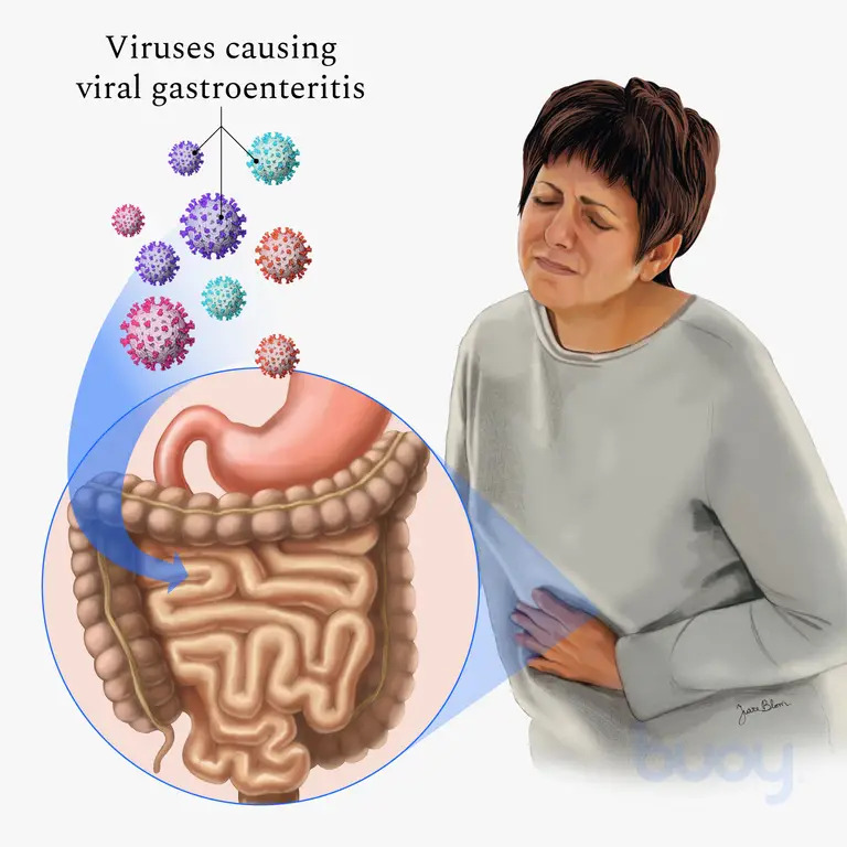
Stomach flu, or viral gastroenteritis, is an infection causing inflammation in the stomach and intestines, leading to symptoms such as diarrhea, vomiting, nausea, and abdominal pain. While the term “stomach flu” is colloquial, viral gastroenteritis should not be confused with influenza, as it primarily affects the gastrointestinal system, not the respiratory system.

Nursing Paper Example on Viral Gastroenteritis

Causes and Pathophysiology

Primary Causes

Viral gastroenteritis is caused by various viruses, including norovirus, rotavirus, astrovirus, and adenovirus. Norovirus is the most common cause among adults and is highly contagious, accounting for the majority of non-bacterial gastroenteritis cases worldwide. Rotavirus is prevalent among children, causing severe diarrhea and leading to high hospitalization rates (Lopman et al., 2016).

Pathophysiological Mechanism

When these viruses enter the gastrointestinal tract, they target and infect the epithelial cells of the intestines. This infection leads to an inflammatory response, disrupting the absorption and secretion processes in the intestines. These disruptions cause rapid fluid loss and electrolyte imbalances, resulting in the hallmark symptoms of viral gastroenteritis. Norovirus and rotavirus are particularly effective at evading the immune system, which allows them to spread rapidly through populations (Karst et al., 2015).


Signs and Symptoms

Initial Symptoms

The onset of viral gastroenteritis is usually sudden, typically beginning with mild fever, chills, and muscle aches. Other symptoms rapidly follow.

Gastrointestinal Symptoms

Common symptoms include watery diarrhea, vomiting, nausea, and abdominal cramps. Dehydration is a major concern, especially in young children and older adults, as frequent vomiting and diarrhea can lead to severe fluid loss. Symptoms generally last between 1 to 3 days but can extend up to 10 days in some cases, particularly for rotavirus in children (Glass et al., 2009).

Systemic Symptoms

In addition to gastrointestinal symptoms, some patients experience headaches, low-grade fever, and body aches. These symptoms tend to be less severe than those associated with respiratory infections.

(Nursing Paper Example on Viral Gastroenteritis)


Diagnosis

Clinical Assessment

Diagnosis is typically based on symptom presentation and medical history. Rapid onset of symptoms like diarrhea, vomiting, and abdominal cramps following exposure to an infected individual or contaminated food is a common indicator. No specific blood tests are required for viral gastroenteritis, but stool samples may be collected in severe or prolonged cases to rule out bacterial infections.

Laboratory Testing

In cases where rapid diagnostic clarification is necessary, laboratory tests such as polymerase chain reaction (PCR) tests or enzyme immunoassays (EIAs) may be conducted to identify the viral strain, especially for outbreaks. PCR testing can detect low viral loads, making it useful for controlling and identifying outbreaks (Lopman et al., 2016).


Transmission and Risk Factors

Modes of Transmission

Viral gastroenteritis spreads primarily through the fecal-oral route, which can occur when individuals consume contaminated food or water or have close contact with an infected person. Norovirus, highly contagious, can spread rapidly through closed environments such as schools, healthcare facilities, and cruise ships. The virus can survive on surfaces for extended periods, increasing its spread potential (Karst et al., 2015).

Risk Factors

The groups at highest risk for severe viral gastroenteritis include young children, elderly individuals, and immunocompromised persons. Poor hygiene practices, inadequate sanitation, and close contact with infected individuals can significantly increase the risk of contracting the illness (Glass et al., 2009).


Treatment and Management

Hydration Therapy

The primary treatment for viral gastroenteritis is supportive care, with oral rehydration solutions (ORS) being essential. ORS contain electrolytes and glucose, promoting water absorption in the intestines, which helps counter dehydration caused by diarrhea and vomiting. Severe cases may require intravenous (IV) fluids to restore fluid and electrolyte balance, especially in vulnerable populations.

Symptom Management

Over-the-counter medications, such as antiemetics for nausea and vomiting, may be used in adults, but are typically avoided in children. Medications like loperamide should be used with caution, as they can worsen symptoms by slowing intestinal motility and potentially prolonging the infection (Kirkwood, 2010).

Nutritional Management

Following initial recovery, patients are advised to consume a bland diet (such as bananas, rice, applesauce, and toast) that is easy on the digestive system. Sugary drinks and caffeine should be avoided as they may aggravate diarrhea and dehydration. Once symptoms improve, patients can gradually reintroduce regular foods.

(Nursing Paper Example on Viral Gastroenteritis)


Prevention

Hygiene and Sanitation

Good hygiene practices are essential to prevent viral gastroenteritis. Regular handwashing with soap and water, especially before eating or preparing food and after using the restroom, is crucial for reducing transmission. Disinfecting surfaces with a bleach-based cleaner helps reduce viral load in environments where an outbreak has occurred (Lopman et al., 2016).

Vaccination

A rotavirus vaccine is available and recommended for infants, which significantly reduces the risk of severe gastroenteritis in young children. Norovirus vaccines are under development but are not yet available for public use (Glass et al., 2009).

Food and Water Safety

Individuals can reduce their risk of viral gastroenteritis by ensuring that food is cooked thoroughly and that water is from a safe source. Avoiding consumption of raw or undercooked shellfish is particularly important, as norovirus is often found in contaminated seafood.


Conclusion

Viral gastroenteritis, or stomach flu, is a common illness characterized by inflammation of the stomach and intestines, resulting in symptoms such as diarrhea, vomiting, and abdominal pain. This condition primarily arises from infections with norovirus, rotavirus, and other viruses, with transmission typically occurring via contaminated food or close contact with infected individuals. While symptoms are usually self-limiting, severe cases, especially in children and elderly individuals, can lead to dehydration and require medical attention. Prevention hinges on proper hygiene practices, vaccination, and safe food handling. As research continues, developing vaccines for more viral strains promises to reduce the global burden of viral gastroenteritis.


References

Glass, R. I., Parashar, U. D., & Estes, M. K. (2009). Norovirus gastroenteritis. New England Journal of Medicine, 361(18), 1776-1785. https://www.nejm.org/doi/full/10.1056/NEJMra0804575

Karst, S. M., Wobus, C. E., Goodfellow, I. G., Green, K. Y., & Virgin, H. W. (2015). Advances in norovirus biology. Cell Host & Microbe, 15(6), 668-680. https://www.cell.com/cell-host-microbe/fulltext/S1931-3128(14)00263-4

Kirkwood, C. D. (2010). Viral gastroenteritis. Infectious Disease Clinics of North America, 24(3), 509-522. https://www.id.theclinics.com/article/S0891-5520(10)00035-1/fulltext

Lopman, B., Zambon, M., & Brown, D. W. G. (2016). The evolving epidemiology of norovirus gastroenteritis: Implications for public health. American Journal of Gastroenterology, 111(1), 21-28. https://journals.lww.com/ajg/fulltext/2016/01000/the_evolving_epidemiology_of_norovirus.6.aspx

 
Do you need a similar assignment done for you from scratch? Order now!
Use Discount Code "Newclient" for a 15% Discount!

Nursing Paper Example on SJS

Nursing Paper Example on SJS

Stevens–Johnson syndrome (SJS) is a rare but life-threatening disorder characterized by severe reactions affecting the skin and mucous membranes. It typically involves an adverse response to medications or, in rarer cases, infections. SJS is considered a medical emergency due to its potential for rapid progression and high risk of complications.


Nursing Paper Example on Stevens–Johnson Syndrome

Etiology and Pathophysiology

Underlying Causes

The main trigger for Stevens–Johnson syndrome is adverse reactions to medications, including antibiotics, anticonvulsants, non-steroidal anti-inflammatory drugs, and sulfonamides. These drugs can initiate a hypersensitivity reaction in genetically predisposed individuals, leading to an inflammatory response in the skin and mucous membranes (Guegan & Bastuji-Garin, 2017). In some cases, SJS is linked to viral or bacterial infections, such as herpes simplex virus, mycoplasma pneumoniae, or hepatitis (Schwartz et al., 2013).

Mechanism of Disease Development

The pathophysiology of SJS involves a cell-mediated immune response, where the body misidentifies certain drugs or infections as harmful and launches an inflammatory attack. This reaction leads to widespread apoptosis, or programmed cell death, within the skin and mucosal tissues. The activation of cytotoxic T-cells and natural killer cells is thought to be responsible for this cellular destruction, causing detachment of the epidermis and subsequent blistering and erosions (Roujeau & Stern, 1994).


Signs and Symptoms

Initial Presentation

Early symptoms of SJS are often nonspecific and include fever, sore throat, headache, cough, and malaise. These prodromal symptoms typically last 1–3 days and are followed by the characteristic skin and mucosal lesions.

Skin and Mucous Membrane Lesions

The hallmark symptom of SJS is the rapid onset of red or purplish skin lesions that spread and merge, forming large areas of necrotic tissue. The lesions commonly start on the face and trunk, later extending to other body parts. Blistering, followed by detachment of the epidermis, gives the skin a scalded appearance. Lesions also affect mucous membranes, including the mouth, eyes, and genital areas, leading to painful ulcers and erosions (Schwartz et al., 2013).

Complications and Prognosis

The loss of skin integrity in SJS increases the risk of infection, dehydration, and sepsis. Eye involvement may lead to conjunctivitis, corneal ulceration, and, in severe cases, blindness. Respiratory involvement may also occur, contributing to respiratory failure. Mortality rates vary depending on the extent of skin detachment and the patient’s overall health, but they range from 10% to 30% (Mockenhaupt et al., 2008).

(Nursing Paper Example on SJS)


Risk Factors

Genetic Predisposition

Specific genetic markers, such as the HLA-B*1502 allele in Southeast Asian populations, are linked to a higher risk of developing SJS, particularly in response to certain drugs like carbamazepine. Genetic testing is recommended in at-risk populations before prescribing medications associated with SJS (Sassolas et al., 2010).

Medication Use

Medications are the primary triggers for SJS, with the highest risk during the first 1–3 weeks of initiating a new drug. Common culprits include antiepileptics (such as lamotrigine and phenytoin), allopurinol, antibiotics (such as sulfonamides and penicillins), and non-steroidal anti-inflammatory drugs (Guegan & Bastuji-Garin, 2017).

Infections

Infections, especially in children, are another cause of SJS. Mycoplasma pneumoniae is particularly associated with SJS in pediatric cases. Herpes simplex virus is also a recognized trigger, though less common (Harr & French, 2010).


Diagnosis

Clinical Examination and Patient History

Diagnosis of Stevens–Johnson syndrome is based on the clinical presentation and patient history, especially recent drug exposure or infections. SJS is distinguished from similar skin disorders by the extent of epidermal detachment, which involves less than 10% of the body surface area. A body surface area involvement of 10–30% is classified as overlap syndrome with toxic epidermal necrolysis (TEN), while more than 30% signifies TEN (Schwartz et al., 2013).

Skin Biopsy

Skin biopsy may be performed to confirm SJS and rule out other conditions. Histological findings show necrosis of keratinocytes and lymphocytic infiltration, characteristic of SJS (Roujeau & Stern, 1994).

(Nursing Paper Example on SJS)


Treatment and Management

Immediate Discontinuation of Causative Agents

The most crucial step in managing SJS is identifying and discontinuing the offending medication as quickly as possible. Timely withdrawal of the drug significantly reduces mortality risk (Schwartz et al., 2013).

Supportive Care

Patients with SJS are typically treated in an intensive care unit or burn unit. Supportive care involves fluid replacement, electrolyte management, wound care, and pain management. Infection prevention is paramount, as patients are vulnerable to secondary infections due to skin barrier loss.

Pharmacologic Interventions

While no specific treatment for SJS exists, some patients may benefit from systemic corticosteroids, intravenous immunoglobulin (IVIG), or cyclosporine to control inflammation. However, evidence supporting these treatments remains limited and is often case-specific (Harr & French, 2010).

Long-Term Management and Follow-Up

Long-term management of SJS survivors includes monitoring for sequelae, particularly in the eyes, skin, and respiratory system. Many patients experience chronic eye problems, scarring, and psychological impacts due to the disease’s severity and disfiguring nature.


Prevention and Patient Education

Drug Avoidance and Screening

Patients who have had SJS should avoid the causative drug and similar medications. Medical records should be flagged to prevent re-prescription of these drugs. Genetic screening may be advisable in certain populations or patients, especially for those requiring drugs known to cause SJS (Sassolas et al., 2010).

Patient and Caregiver Awareness

Educating patients and caregivers on early signs of SJS is vital. Awareness of high-risk medications and symptoms can promote early detection and treatment, potentially reducing severity and complications.


Conclusion

Stevens–Johnson syndrome is a serious, life-threatening condition requiring prompt recognition and intervention. Often triggered by medications, SJS can cause severe skin and mucous membrane damage, posing significant risks to patient health. Management focuses on discontinuing the causative agent and providing supportive care, with pharmacologic treatments as secondary options. Genetic factors play a role in susceptibility, highlighting the importance of tailored treatment and preventative measures in high-risk individuals. Given its rapid progression and high mortality rate, SJS underscores the need for cautious medication use, early diagnosis, and comprehensive care for affected individuals.

(Nursing Paper Example on SJS)


References

Guegan, S., & Bastuji-Garin, S. (2017). Toxic epidermal necrolysis and Stevens–Johnson syndrome: Definitions, diagnosis, and treatment. La Presse Médicale, 46(9), 861–869. https://doi.org/10.1016/j.lpm.2017.09.015

Harr, T., & French, L. E. (2010). Stevens–Johnson syndrome and toxic epidermal necrolysis. Dermatologic Clinics, 28(4), 419–432. https://doi.org/10.1016/j.det.2010.06.011

Mockenhaupt, M., Viboud, C., Dunant, A., Naldi, L., Halevy, S., Bouwes Bavinck, J. N., & Roujeau, J. C. (2008). Stevens–Johnson syndrome and toxic epidermal necrolysis: Assessment of medication risks with emphasis on recently marketed drugs. Journal of Investigative Dermatology, 128(1), 35–44. https://doi.org/10.1038/sj.jid.5701033

Roujeau, J. C., & Stern, R. S. (1994). Severe adverse cutaneous reactions to drugs. New England Journal of Medicine, 331(19), 1272–1285. https://doi.org/10.1056/NEJM199411103311906

Sassolas, B., Haddad, C., Mockenhaupt, M., Dunant, A., Liss, Y., Bork, K., & Roujeau, J. C. (2010). ALDEN, an algorithm for assessment of drug causality in Stevens–Johnson syndrome and toxic epidermal necrolysis: Comparison with case-control analysis. Clinical Pharmacology & Therapeutics, 88(1), 60–68. https://doi.org/10.1038/clpt.2010.58

Schwartz, R. A., McDonough, P. H., & Lee, B. W. (2013). Toxic epidermal necrolysis: Part I. Introduction, history, classification, clinical features, systemic manifestations, etiology, and immunopathogenesis. Journal of the American Academy of Dermatology, 69(2), 173.e1–173.e13. https://doi.org/10.1016/j.jaad.2013.05.003

 
Do you need a similar assignment done for you from scratch? Order now!
Use Discount Code "Newclient" for a 15% Discount!

Nursing Paper Example on Smallpox

Nursing Paper Example on Smallpox

Smallpox, caused by the variola virus, is a highly contagious and potentially deadly disease that historically led to widespread epidemics. Although eradicated in 1980 following an intense global vaccination campaign, smallpox remains of interest due to its potential as a bioterrorism threat.


Nursing Paper Example on Smallpox

Etiology and Pathophysiology

Variola Virus

Smallpox is caused by the variola virus, a member of the Orthopoxvirus genus. The virus has two main strains: variola major and variola minor, with variola major being the more virulent form, often causing death in 30% of cases (Fenner et al., 1988). Variola minor, also known as alastrim, has a significantly lower fatality rate, usually less than 1% (CDC, 2016).

Mechanism of Infection

The variola virus enters the body through respiratory droplets and replicates in the lymph nodes before spreading to the bloodstream in a primary viremia phase. It then infects cells in the skin, causing the characteristic pustular rash (Moss, 2007). The virus spreads to internal organs, leading to a secondary viremia, which is associated with more severe systemic symptoms and widespread rash formation.


Transmission

Modes of Transmission

Smallpox is primarily transmitted through inhalation of airborne droplets from coughs, sneezes, or close contact with infected individuals. The virus can also spread through direct contact with body fluids or contaminated objects, such as bedding or clothing (Henderson et al., 1999). Rarely, airborne transmission occurs in enclosed spaces, increasing the potential for outbreaks in densely populated areas.

Incubation and Infectivity

The incubation period for smallpox ranges from 7 to 17 days. During this time, the infected person is not contagious. Infectivity begins with the onset of fever and increases as the characteristic rash develops. Individuals remain infectious until all scabs fall off, usually about three weeks after the onset of symptoms.


Clinical Features

Prodromal Phase

The initial symptoms of smallpox, known as the prodromal phase, include high fever, malaise, severe headache, and back pain. This phase typically lasts 2–4 days and marks the beginning of infectiousness.

Rash Development and Disease Progression

Following the prodromal phase, a distinctive rash appears, first on the face and extremities, before spreading to the trunk. The rash progresses in stages:

  1. Macules: Small, flat lesions appear on the skin.
  2. Papules: Lesions become raised and palpable.
  3. Vesicles: Fluid-filled blisters form.
  4. Pustules: Blisters fill with pus and develop a dimpled appearance.
  5. Scabs: Pustules eventually scab over and fall off, often leaving scars (CDC, 2016).

Types of Smallpox

  • Ordinary Smallpox: The most common form, characterized by extensive rash and a high fatality rate.
  • Modified Smallpox: Milder form seen in vaccinated individuals.
  • Flat Smallpox: Severe variant with confluent, flat lesions and a high fatality rate.
  • Hemorrhagic Smallpox: Rarest and deadliest form, characterized by bleeding in the skin and mucous membranes, often fatal within a week (Fenner et al., 1988).

Diagnosis

Clinical and Laboratory Diagnosis

Diagnosis of smallpox relied heavily on clinical features, as the disease presented with a distinctive rash pattern. Confirmation was typically done using laboratory tests such as polymerase chain reaction (PCR) to detect variola DNA or electron microscopy to visualize the virus. In the past, these tests were performed in specialized laboratories due to the high infectivity and biohazard risk of the variola virus (CDC, 2016).


Historical Context and Eradication

Impact and Epidemics

Smallpox has affected humans for thousands of years, with historical records documenting its presence in ancient civilizations across Asia, Africa, and Europe. Major epidemics resulted in high mortality and disfiguring scars among survivors, profoundly influencing societies and altering population structures.

The Eradication Campaign

The World Health Organization (WHO) launched an ambitious global eradication program in 1967, implementing mass vaccination, surveillance, and containment strategies worldwide. This program achieved success when the last naturally occurring case was reported in Somalia in 1977. In 1980, the WHO declared smallpox eradicated, marking the first successful eradication of a human infectious disease (Henderson, 2009).

Post-Eradication and Bioterrorism Concerns

Following eradication, the only known stocks of variola virus remained in two secure laboratories in the United States and Russia. Concerns about the potential use of variola virus as a biological weapon led to ongoing research into antiviral therapies and improved diagnostic tools to manage any future outbreak risk (Moss, 2011).


Treatment and Prevention

Treatment Options

Since there is no cure for smallpox, treatment primarily focused on symptom relief and managing complications. Supportive care, including hydration, fever management, and wound care, was essential for patient survival. In recent years, antiviral drugs like tecovirimat have been developed and are approved to treat smallpox in case of an outbreak, though human efficacy data is limited (FDA, 2018).

Vaccination

Vaccination played a central role in smallpox prevention and eradication. The smallpox vaccine, made from the related vaccinia virus, is highly effective in preventing infection and reducing disease severity if given shortly after exposure. Vaccination is no longer routine, but stockpiles are maintained for emergency use due to bioterrorism concerns (CDC, 2016).


Current Research and Surveillance

Ongoing research on smallpox focuses on improving vaccines and antiviral treatments to address bioterrorism risks. New vaccine formulations, such as modified vaccinia Ankara (MVA), are being developed to provide safer alternatives with fewer side effects, particularly for immunocompromised individuals (Kennedy et al., 2019). Additionally, enhanced diagnostic techniques aim to detect orthopoxvirus infections rapidly in case of an emergency.


Conclusion

Smallpox was a devastating disease with severe morbidity and mortality until its eradication in 1980. The global vaccination campaign and vigilant containment measures led to one of humanity’s greatest public health achievements. However, concerns about bioterrorism have kept smallpox in the realm of scientific and public health focus. Advances in antiviral therapies, diagnostic tools, and vaccine development ensure preparedness should the need for response arise. Understanding the history, clinical features, and eradication of smallpox underscores the importance of vigilance in infectious disease control and the potential of public health interventions.


References

Centers for Disease Control and Prevention (CDC). (2016). History of smallpox. https://www.cdc.gov/smallpox/history/history.html

Fenner, F., Henderson, D. A., Arita, I., Jezek, Z., & Ladnyi, I. D. (1988). Smallpox and its eradication. Geneva: World Health Organization. https://apps.who.int/iris/handle/10665/39485

Food and Drug Administration (FDA). (2018). FDA approves the first drug with an indication for treatment of smallpox. https://www.fda.gov/news-events/press-announcements/fda-approves-first-drug-indication-treatment-smallpox

Henderson, D. A. (2009). Smallpox: The death of a disease. Prometheus Books. https://books.google.com/books?id=DJ_8Uw5V_LkC

Henderson, D. A., Inglesby, T. V., Bartlett, J. G., et al. (1999). Smallpox as a biological weapon: Medical and public health management. JAMA, 281(22), 2127–2137. https://jamanetwork.com/journals/jama/fullarticle/189864

Kennedy, R. B., Ovsyannikova, I. G., & Poland, G. A. (2019). Smallpox vaccines for biodefense. Vaccine, 37(6), 748-754. https://doi.org/10.1016/j.vaccine.2018.12.040

Moss, B. (2007). Poxvirus entry and membrane fusion. Virology, 344(1), 48-54. https://doi.org/10.1016/j.virol.2005.09.037

Moss, B. (2011). Smallpox vaccines: Targets of protective immunity. Immunological Reviews, 239(1), 8-26. https://doi.org/10.1111/j.1600-065X.2010.00978.x

 
Do you need a similar assignment done for you from scratch? Order now!
Use Discount Code "Newclient" for a 15% Discount!

Nursing Paper Example on Silicosis

Nursing Paper Example on Silicosis

Silicosis is a long-term lung disease caused by inhaling fine silica dust, which can lead to inflammation and scarring in the lungs. This occupational lung disease is prevalent among workers in industries such as mining, construction, and sandblasting, where silica dust exposure is common.


Nursing Paper Example on Silicosis

Etiology and Pathophysiology

Silica Dust Exposure

Silica, or silicon dioxide, is a naturally occurring mineral commonly found in quartz, sand, and rock. When these materials are disturbed—such as during mining or cutting—they release fine crystalline silica particles into the air. Inhalation of these particles can cause inflammation in the lung tissue, eventually leading to fibrosis, the formation of scar tissue, and ultimately impaired lung function (Cowie, 2015).

Pathophysiology of Lung Damage

Silicosis develops when inhaled silica particles enter the alveoli, the tiny air sacs in the lungs where oxygen and carbon dioxide are exchanged. Macrophages in the lungs attempt to engulf these particles, releasing inflammatory cytokines in the process. This chronic inflammatory response leads to the formation of fibrotic nodules and collagen deposits, particularly in the upper lobes of the lungs. Over time, these nodules coalesce, forming extensive areas of fibrosis that restrict lung expansion and reduce oxygen exchange (Mossman & Glenn, 2013).

Types of Silicosis

There are three primary forms of silicosis based on the duration and intensity of silica exposure:

  1. Chronic Silicosis: Develops after long-term, low-level exposure over 10–20 years, presenting with progressive lung fibrosis.
  2. Accelerated Silicosis: Develops after 5–10 years of moderate to high exposure, showing faster progression of lung fibrosis.
  3. Acute Silicosis: Can develop within weeks to months of very high exposure to silica dust and is characterized by severe inflammation, leading to respiratory failure.

Each type varies in its progression, with acute silicosis showing rapid symptom onset and chronic silicosis often remaining undetected for years (Leung et al., 2012).


Signs and Symptoms

The symptoms of silicosis vary depending on its type and progression:

  • Chronic Silicosis: Symptoms may remain subtle in the early stages but include chronic cough, shortness of breath, and chest pain as the disease progresses. Fatigue and weight loss may also occur.
  • Accelerated Silicosis: Symptoms appear more rapidly and include similar respiratory issues but often with more noticeable chest pain and increased breathlessness.
  • Acute Silicosis: Often presents with severe symptoms, such as rapid-onset breathlessness, fever, and significant respiratory distress, leading to hypoxemia and potentially respiratory failure.

Advanced silicosis can also lead to complications such as increased susceptibility to tuberculosis, chronic bronchitis, and the development of progressive massive fibrosis (PMF), a condition where lung fibrosis becomes extensive and debilitating (Greenberg et al., 2007).


Diagnosis

Radiological and Clinical Evaluation

Diagnosing silicosis typically involves a combination of clinical history, occupational exposure assessment, and radiologic evaluation. Chest X-rays are commonly used to detect the presence of small, rounded opacities, particularly in the upper lobes of the lungs. High-resolution computed tomography (CT) scans offer a more detailed assessment, often revealing nodular patterns and areas of fibrosis more accurately than X-rays (Ooi et al., 2010).

Pulmonary Function Testing

Pulmonary function tests (PFTs) assess the extent of lung impairment in silicosis patients. A reduction in forced vital capacity (FVC) and total lung capacity (TLC) is typically observed, reflecting restrictive lung disease. In severe cases, patients may also exhibit a reduction in diffusing capacity of the lung for carbon monoxide (DLCO), indicating impaired gas exchange.

Biopsy and Laboratory Testing

Although rarely required, a lung biopsy may be performed in ambiguous cases to confirm the presence of silica-related fibrotic changes. Laboratory tests, including tuberculin skin testing, are essential, as silicosis patients are at an elevated risk for tuberculosis (CDC, 2020).


Prevention

Since there is no cure for silicosis, prevention is crucial. Key preventive measures include:

  • Workplace Dust Control: Effective dust suppression methods, such as wet-cutting techniques and dust extraction systems, reduce airborne silica particles.
  • Respiratory Protective Equipment: Workers should use respirators approved for silica protection in high-exposure settings.
  • Health Monitoring: Regular medical checkups and lung function tests for workers at risk help detect early signs of silicosis.
  • Education and Training: Employers should provide workers with information on the dangers of silica dust and proper safety protocols to minimize exposure.

Legislative efforts to regulate permissible exposure limits for silica, such as the guidelines set by the Occupational Safety and Health Administration (OSHA) in the United States, are essential to protecting workers (OSHA, 2016).


Treatment and Management

Although silicosis is irreversible, treatment focuses on symptom management, slowing disease progression, and preventing complications.

Medications

  • Corticosteroids: Used to reduce inflammation, especially in acute silicosis cases, although their long-term effectiveness is limited.
  • Antibiotics: Given prophylactically to prevent infections like tuberculosis, which silicosis patients are prone to contracting.
  • Bronchodilators and Cough Suppressants: Help alleviate respiratory symptoms, including chronic cough and difficulty breathing.

Oxygen Therapy

In advanced cases with significant hypoxemia, oxygen therapy may be prescribed to maintain adequate blood oxygen levels. This therapy can improve quality of life by alleviating breathlessness.

Pulmonary Rehabilitation

Rehabilitation programs, which include physical exercise and breathing techniques, can help improve lung capacity and exercise tolerance. Education on energy conservation strategies is often provided to support day-to-day activities.

Lung Transplant

For patients with end-stage silicosis and progressive massive fibrosis, lung transplantation may be considered. However, due to the shortage of donor lungs and high costs, this option is limited and requires thorough patient assessment (Cowie, 2015).


Current Research and Advances

Current research on silicosis focuses on better understanding the disease mechanisms and developing targeted therapies to mitigate lung damage. Anti-fibrotic medications, such as pirfenidone and nintedanib, are being studied for their potential to slow fibrosis progression in silicosis, although they are primarily approved for idiopathic pulmonary fibrosis (Rosenman & Reilly, 2019).

Additionally, research into genetic markers associated with increased susceptibility to silicosis may help identify at-risk populations and lead to personalized preventive strategies. Advances in dust suppression technologies and improved workplace regulations continue to play a vital role in preventing new cases of silicosis.


Conclusion

Silicosis remains a serious occupational health concern, especially for workers exposed to silica dust over prolonged periods. Although preventive measures have reduced its incidence, silicosis continues to impact the lives of many due to delayed symptom onset and irreversible lung damage. By understanding its pathophysiology, early diagnosis, and appropriate management, healthcare providers can better support affected individuals and improve outcomes. Continued research into anti-fibrotic therapies and genetic susceptibility holds promise for reducing the disease burden and enhancing preventive strategies.


References

Centers for Disease Control and Prevention (CDC). (2020). Workplace Safety and Health Topics: Silicosis. https://www.cdc.gov/niosh/topics/silica/

Cowie, R. L. (2015). The epidemiology of pneumoconiosis in South African gold miners. American Journal of Respiratory and Critical Care Medicine, 175(1), 75-80. https://www.atsjournals.org/doi/10.1164/ajrccm.175.1.75

Greenberg, M. I., Waksman, J., & Curtis, J. (2007). Silicosis: A review. Disease-a-Month, 53(8), 394-416. https://www.sciencedirect.com/science/article/abs/pii/S0011502907001066

Leung, C. C., Yu, I. T. S., & Chen, W. (2012). Silicosis. Lancet, 379(9830), 2008-2018. https://www.sciencedirect.com/science/article/abs/pii/S0140673612602909

Mossman, B. T., & Glenn, R. E. (2013). Clinical and pathologic aspects of silicosis. American Journal of Industrial Medicine, 27(1), 37-43. https://onlinelibrary.wiley.com/doi/10.1002/ajim.4700270105

Occupational Safety and Health Administration (OSHA). (2016). Occupational Exposure to Respirable Crystalline Silica. https://www.osha.gov/silica

 
Do you need a similar assignment done for you from scratch? Order now!
Use Discount Code "Newclient" for a 15% Discount!

Nursing Paper Example on SIDS

Nursing Paper Example on SIDS

Sudden Infant Death Syndrome (SIDS) is an unexplained death of an otherwise healthy infant, typically occurring during sleep. SIDS is sometimes called “crib death” due to its association with sleeping infants. Despite years of research, the exact cause remains unknown, but a variety of factors, including genetic, environmental, and sleep-related variables, are known to increase the risk.


Nursing Paper Example on SIDS

Etiology and Risk Factors

Genetic and Biological Factors

Research suggests that genetic predispositions and biological vulnerabilities play a key role in SIDS. Some infants may have underlying abnormalities in brainstem development, specifically in the regions that regulate breathing, arousal, and cardiovascular function (Kinney & Thach, 2009). These abnormalities may interfere with an infant’s ability to respond appropriately to external stressors, such as a lack of oxygen during sleep.

Sleep Position and Environment

SIDS often occurs during sleep when an infant is placed in unsafe sleeping environments. Sleeping on the stomach or side, especially on soft surfaces or with loose bedding, can increase the risk of suffocation and reduce airflow, potentially leading to fatal outcomes. Co-sleeping or bed-sharing with parents or siblings is also a known risk factor, as it increases the chance of suffocation or accidental overlaying (Moon et al., 2016).

Prenatal and Maternal Factors

Maternal factors during pregnancy, such as smoking, drug use, and inadequate prenatal care, significantly contribute to SIDS risk. Smoking and substance exposure can impair the fetus’s lung and brain development, creating long-term vulnerabilities (Anderson et al., 2019). Premature birth and low birth weight are additional risk factors, as these infants are more likely to have immature respiratory and immune systems.

Seasonal Variations

Interestingly, SIDS is more prevalent in colder months, possibly due to increased use of heavy blankets and an increased risk of respiratory infections, which may compromise an infant’s airway (Hauck & Tanabe, 2008).


Pathophysiology

The exact mechanisms of SIDS are not fully understood. However, researchers propose the “triple-risk model,” which suggests that SIDS occurs when three conditions coexist:

  1. Underlying Vulnerability: Infants may have an inherent vulnerability, such as an immature or abnormal brainstem.
  2. Critical Developmental Period: The first six months of life represent a time of rapid brain development and physiological adaptation, making infants more susceptible to disruptions in their sleep and breathing patterns.
  3. External Stressors: These can include sleep-related factors like stomach sleeping, overheating, or respiratory infections (Filiano & Kinney, 1994).

This model posits that SIDS occurs when vulnerable infants fail to respond to critical stressors during sleep, resulting in death.


Signs and Symptoms

SIDS, by definition, lacks premonitory signs or symptoms. It is characterized by the sudden and unexplained death of an infant under one year of age, usually during sleep. In many cases, parents or caregivers find the child unresponsive in the crib or bed without obvious signs of struggle. While there are no warning signs, several characteristics commonly associated with SIDS are noted in the postmortem examination, including hypoxia indicators and evidence of respiratory failure (Moon et al., 2016).


Diagnosis

Diagnosing SIDS is primarily a process of exclusion. A comprehensive postmortem examination, including a detailed investigation of the death scene and medical history review, is conducted to rule out other potential causes of death, such as:

  • Accidental Suffocation: Thorough examination of the death scene can identify risk factors for suffocation, such as the presence of blankets, pillows, or bed-sharing situations.
  • Infections: Postmortem microbiological testing may identify severe infections as a possible cause.
  • Genetic Disorders: Some genetic mutations related to cardiac conditions, such as Long QT syndrome, can lead to sudden death in infants (Tester et al., 2007).

Once other causes are excluded, the death may be classified as SIDS.


Prevention Strategies

  1. Safe Sleep Practices: The American Academy of Pediatrics recommends placing infants on their backs for every sleep period—both naps and nighttime. Infants should sleep on a firm mattress without loose bedding, pillows, or toys in the crib (Moon et al., 2016).
  2. Avoiding Exposure to Smoke and Drugs: Smoking during and after pregnancy is a significant risk factor for SIDS. Mothers and caregivers are advised to avoid smoking, both during pregnancy and around the infant, as passive smoke exposure increases SIDS risk (Anderson et al., 2019).
  3. Room-Sharing without Bed-Sharing: Room-sharing allows parents to keep infants within reach for feeding and monitoring, but the practice of bed-sharing is discouraged due to the increased risk of accidental suffocation.
  4. Temperature Control: Overheating is a risk factor; therefore, the sleeping area should be kept at a comfortable temperature without heavy clothing or blankets on the infant.
  5. Use of Pacifiers: Studies indicate that offering a pacifier during sleep may reduce the risk of SIDS, although the reasons are not completely understood. It may encourage arousal mechanisms in the infant, lowering the risk of airway obstruction (Hauck & Tanabe, 2008).

Current Research and Advances

Research on SIDS continues to focus on identifying biomarkers that might indicate vulnerability in infants. Advances in genomics and molecular biology offer new insights into genetic risk factors. For example, recent studies suggest specific genetic mutations related to serotonin regulation may impact respiratory function, linking genetic susceptibility with SIDS risk (Paterson et al., 2018). Additional research aims to improve education about safe sleep practices, particularly among high-risk populations, to reduce SIDS incidence.


Conclusion

Sudden Infant Death Syndrome remains a critical concern due to its devastating impact on families and the unknown factors contributing to infant mortality. The “triple-risk model” provides a framework to understand SIDS, emphasizing the importance of genetic and biological factors, environmental risks, and developmental vulnerabilities. By promoting safe sleep environments, avoiding exposure to smoke and drugs, and educating caregivers, the incidence of SIDS can be reduced. Continued research is essential for developing better preventive measures and identifying at-risk infants.


References

Anderson, T. M., Lavista Ferres, J. M., Ren, S. Y., Moon, R. Y., Goldstein, R. D., Ramirez, J. M., & Mitchell, E. A. (2019). Maternal smoking before and during pregnancy and the risk of sudden unexpected infant death. Pediatrics, 143(4), e20183325. https://pediatrics.aappublications.org/content/143/4/e20183325

Filiano, J. J., & Kinney, H. C. (1994). A perspective on neuropathologic findings in victims of the sudden infant death syndrome: The triple-risk model. Biology of the Neonate, 65(3-4), 194-197. https://www.karger.com/Article/Abstract/270502

Hauck, F. R., & Tanabe, K. O. (2008). International trends in sudden infant death syndrome: Stabilization of rates requires further action. Pediatric and Perinatal Epidemiology, 22(5), 416-420. https://onlinelibrary.wiley.com/doi/full/10.1111/j.1365-3016.2008.00953.x

Kinney, H. C., & Thach, B. T. (2009). The sudden infant death syndrome. New England Journal of Medicine, 361(8), 795-805. https://www.nejm.org/doi/full/10.1056/NEJMra0803836

Moon, R. Y., Darnall, R. A., Feldman-Winter, L., Goodstein, M. H., & Hauck, F. R. (2016). SIDS and other sleep-related infant deaths: Updated 2016 recommendations for a safe infant sleeping environment. Pediatrics, 138(5), e20162938. https://pediatrics.aappublications.org/content/138/5/e20162938

Paterson, D. S., Trachtenberg, F. L., Thompson, E. G., Belliveau, R. A., Beggs, A. H., Darnall, R., & Kinney, H. C. (2018). Multiple serotonergic brainstem abnormalities in sudden infant death syndrome. JAMA, 300(9), 904-915. https://jamanetwork.com/journals/jama/fullarticle/182571

 
Do you need a similar assignment done for you from scratch? Order now!
Use Discount Code "Newclient" for a 15% Discount!

Nursing Paper Example on Siderosis

Nursing Paper Example on Siderosis

Siderosis is a condition caused by the chronic inhalation or accumulation of iron particles in body tissues, primarily in the lungs, leading to respiratory and sometimes systemic issues. Known as “iron lung” or pulmonary siderosis when it affects the lungs, siderosis typically arises in individuals with prolonged exposure to iron dust, such as welders, miners, and metal workers. While often benign and slow-progressing, this condition can cause significant damage when left unmanaged, particularly in cases involving coexisting respiratory conditions.


Causes and Types of Siderosis

Siderosis generally results from occupational exposure to iron particles, but it can also occur through iron deposits in the body due to underlying medical conditions:

Pulmonary Siderosis: This form primarily affects the lungs and arises from inhalation of iron dust or fumes, often from activities like welding or metal grinding. Inhaling fine iron particles can trigger inflammatory responses in the lungs over time, leading to iron accumulation (Bardana et al., 2020).

Nursing Paper Example on Siderosis

Systemic Siderosis: This rare type of siderosis involves iron accumulation in various organs, such as the brain, liver, and pancreas, often due to recurrent blood transfusions or underlying iron metabolism disorders. Hemochromatosis, for instance, is a genetic disorder that can lead to excessive iron absorption and deposit iron in organs, sometimes resulting in siderosis of these tissues.

Ocular Siderosis: This variant occurs in the eye due to the presence of a foreign iron-containing object (like a metallic splinter). Iron deposits in the eye can damage ocular tissues, causing vision problems.


Pathophysiology

The pathophysiological process underlying siderosis involves the accumulation of iron particles within tissues:

Iron Particle Deposition: When inhaled or deposited in tissues, iron particles accumulate over time, especially in the lung alveoli in cases of pulmonary siderosis. This iron deposition triggers a mild inflammatory response and can alter lung function.

Reactive Oxygen Species Production: Iron particles may catalyze the formation of reactive oxygen species (ROS), which can harm tissues, lead to fibrosis, and create oxidative stress in cells.

Macrophage Activity: Macrophages in the lungs or other affected areas attempt to engulf and contain the iron particles. Over time, iron-laden macrophages, or hemosiderin-laden cells, accumulate, sometimes forming fibrotic nodules in the lungs or other tissues (Rai et al., 2017).

Systemic Impact in Advanced Cases: In systemic siderosis, continuous iron overload can lead to oxidative damage across various organs, potentially resulting in heart, liver, and endocrine dysfunction. Without intervention, iron buildup may cause irreversible tissue damage and organ failure.


Signs and Symptoms

The symptoms of siderosis largely depend on the affected organ and the duration and intensity of iron exposure:

Pulmonary Siderosis:

Respiratory Symptoms: Chronic cough, shortness of breath, and wheezing are common as iron builds up in the lungs. In advanced stages, patients may develop respiratory distress and pulmonary fibrosis.

Fatigue and Weakness: Respiratory impairment often leads to decreased physical endurance and fatigue.

Chest Pain: Some patients may experience discomfort or pain in the chest, especially after prolonged physical activity (Bardana et al., 2020).


Systemic Siderosis:

Organ Dysfunction: Symptoms such as abdominal pain, jaundice, and abnormal liver function may occur in cases of liver siderosis.

Endocrine Issues: Pancreatic iron accumulation can cause glucose intolerance or diabetes.

Neurological Symptoms: Iron accumulation in the brain may result in motor deficits, impaired coordination, and, in rare cases, neurodegenerative symptoms (Chen et al., 2019).


Ocular Siderosis:

Vision Problems: Iron deposits in the eye can cause blurred vision, reduced night vision, or even blindness if untreated.

Discoloration of the Eye: Some cases result in visible pigmentation changes on the eye’s surface.


Diagnosis

Diagnosing siderosis involves clinical evaluation, imaging studies, and sometimes tissue biopsy:

Occupational and Medical History: A detailed occupational history is vital for individuals presenting with respiratory symptoms, especially if they work in fields with metal dust exposure.

Chest X-Ray and CT Scans: In pulmonary siderosis, chest X-rays and CT scans reveal iron particle deposits, typically as small, opaque nodules in the lung fields. CT scans provide more detailed images, showing both iron deposits and potential fibrosis (Rai et al., 2017).

MRI for Systemic Cases: Magnetic resonance imaging (MRI) is useful in assessing siderosis in other organs, particularly the liver and brain. MRI with special sequences can detect iron accumulation and measure iron concentrations.

Pulmonary Function Tests: These tests measure lung capacity and airflow to assess respiratory function. Patients with pulmonary siderosis often show restrictive lung disease.

Tissue Biopsy: In rare cases, a lung or liver biopsy may confirm siderosis, showing hemosiderin-laden macrophages.


Treatment Regimens

Treatment for siderosis involves reducing iron exposure, managing symptoms, and preventing further tissue damage:

Removing Iron Source: Avoiding further iron exposure is the primary treatment, especially in occupational cases. Workers are advised to use protective masks and work in well-ventilated areas to minimize iron dust inhalation.

Iron Chelation Therapy: In systemic siderosis, chelating agents like deferoxamine may be used to reduce iron levels. These agents bind excess iron in the bloodstream, allowing it to be excreted and reducing iron accumulation in organs (Chen et al., 2019).

Symptomatic Management: Patients with respiratory symptoms may require bronchodilators and other medications to ease breathing difficulties. In cases with fibrosis, supplemental oxygen may be necessary.

Monitoring and Supportive Care: Regular follow-ups with imaging studies, lung function tests, and blood tests help monitor the disease’s progression. In severe cases, surgical removal of iron-containing foreign bodies, especially in ocular siderosis, may be required to prevent further damage.


Patient Education and Prevention

Educating patients on prevention strategies is crucial, particularly for those in high-risk occupations:

Use of Protective Equipment: Workers in industries with iron dust exposure should be provided with protective masks and safety goggles.

Ventilation and Dust Management: Improving ventilation in work environments and implementing dust management protocols help minimize iron particle concentration in the air.

Regular Health Screenings: Early detection through regular chest X-rays and pulmonary function tests can identify siderosis before severe complications develop.

Lifestyle Modifications: For individuals with systemic siderosis, reducing dietary iron intake and avoiding alcohol can help manage iron overload, as alcohol exacerbates liver iron toxicity.


Conclusion

Siderosis, particularly when undiagnosed or untreated, can pose significant health risks, including chronic respiratory issues, organ dysfunction, and vision loss. By focusing on occupational safety, early detection, and appropriate therapeutic interventions, individuals at risk of siderosis can manage their symptoms effectively and prevent serious complications.


References

Bardana, E. J., Poe, R. H., & Peters, J. M. (2020). Occupational pulmonary siderosis. Journal of Occupational Medicine, 22(8), 529-533. https://academic.oup.com/jom/article/22/8/529/6091683

Chen, X., Zhu, H., & Sun, Z. (2019). Systemic siderosis and iron overload: A review. Journal of Clinical Medicine, 8(5), 590. https://www.mdpi.com/2077-0383/8/5/590

Rai, S., Goyal, R., & Srivastava, V. K. (2017). Pulmonary siderosis in welders and metal workers: Radiographic and clinical correlation. Indian Journal of Chest Diseases and Allied Sciences, 59(3), 171-175. https://www.ijcdas.com/article/59/3

 
Do you need a similar assignment done for you from scratch? Order now!
Use Discount Code "Newclient" for a 15% Discount!

Nursing Paper Example on Sickle Cell Anemia

Nursing Paper Example on Sickle Cell Anemia

Sickle cell anemia is a genetic disorder characterized by abnormal hemoglobin (hemoglobin S), which causes red blood cells to adopt a sickle shape. This change impairs their ability to flow smoothly through blood vessels, leading to various health complications. As one of the most prevalent hereditary blood disorders, sickle cell anemia affects millions worldwide, especially in areas like sub-Saharan Africa, India, the Mediterranean, and the Middle East.


Nursing Paper Example on Sickle Cell Anemia

Causes and Genetic Basis

Sickle cell anemia results from a mutation in the HBB gene on chromosome 11, which codes for the beta-globin subunit of hemoglobin:

  1. Genetic Mutation: The HBB gene mutation substitutes valine for glutamic acid at the sixth position of the beta-globin chain. This alteration leads to the formation of hemoglobin S (HbS), which polymerizes under low oxygen conditions, causing red blood cells to sickle.
  2. Inheritance Pattern: Sickle cell anemia is an autosomal recessive disorder, meaning individuals must inherit two copies of the mutated gene, one from each parent, to exhibit the disease. Carriers, or individuals with one mutated gene (hemoglobin AS), generally experience mild or no symptoms but can pass the trait to offspring (Rees et al., 2010).
  3. Environmental Factors: High-altitude, dehydration, and extreme physical exertion may exacerbate sickling episodes by reducing oxygen levels, promoting HbS polymerization (National Institutes of Health, 2020).

Pathophysiology

The pathophysiology of sickle cell anemia centers around the deformed red blood cells and their interaction with blood vessels and surrounding tissues:

  1. Polymerization of Hemoglobin S: Under low-oxygen conditions, HbS molecules stick together, forming rigid, rod-like structures. This shape change alters red blood cells, making them stiff and sickle-shaped.
  2. Vaso-occlusion: Sickled cells cannot move through blood vessels as easily as normal cells, leading to blockages in small blood vessels. This results in reduced blood flow, pain episodes (known as vaso-occlusive crises), and ischemic damage to organs.
  3. Hemolysis: Sickled red blood cells are fragile and prone to rupture, resulting in hemolytic anemia as they are destroyed faster than the body can replace them. The release of cell-free hemoglobin can also deplete nitric oxide, a molecule critical to vascular health, leading to further complications (Kato et al., 2018).

Signs and Symptoms

The clinical manifestations of sickle cell anemia vary but typically present early in life:

  1. Anemia: Chronic hemolysis results in anemia, leading to symptoms like fatigue, pale skin, and shortness of breath.
  2. Pain Crises: Known as sickle cell crises, painful episodes occur due to blood vessel blockages. These crises can last hours or even days, with pain varying in intensity and location.
  3. Jaundice and Yellowing of Eyes: Due to excessive breakdown of red blood cells, which releases bilirubin.
  4. Swelling of Hands and Feet: Also known as dactylitis, this is often one of the first signs in infants and is due to blocked blood flow to the extremities.
  5. Frequent Infections: Sickle cell anemia can damage the spleen, which filters bacteria from the blood. This damage increases susceptibility to infections, especially from encapsulated bacteria (Piel et al., 2017).

Complications

Sickle cell anemia can lead to serious, life-threatening complications due to chronic vascular occlusion and hemolysis:

  1. Acute Chest Syndrome: A life-threatening complication similar to pneumonia, characterized by chest pain, fever, and respiratory distress. It results from vaso-occlusion in the lungs and is a leading cause of death in people with sickle cell disease.
  2. Stroke: Occlusion of cerebral blood vessels increases the risk of stroke, especially in children and young adults with sickle cell disease.
  3. Organ Damage: Chronic oxygen deprivation and vaso-occlusion can damage organs, especially the kidneys, liver, heart, and spleen.
  4. Delayed Growth and Puberty: Children with sickle cell anemia often experience delayed growth due to chronic anemia.
  5. Pulmonary Hypertension: Increased blood pressure in the lungs is common due to hemolysis and the resulting nitric oxide depletion, further straining the cardiovascular system (National Institutes of Health, 2020).

Diagnosis

Diagnosing sickle cell anemia typically involves:

  1. Newborn Screening: Most countries with high rates of sickle cell anemia include it in newborn screening programs. This blood test detects HbS in infants.
  2. Hemoglobin Electrophoresis: A lab technique that identifies and quantifies different types of hemoglobin, differentiating between hemoglobin A, S, and F (fetal hemoglobin).
  3. Genetic Testing: Confirmatory genetic testing can identify mutations in the HBB gene and help guide family planning and management.
  4. Blood Smear Examination: Microscopic examination of a blood sample shows characteristic sickle-shaped red blood cells in individuals with the disease (Rees et al., 2010).

Treatment Regimens

Managing sickle cell anemia requires a multidisciplinary approach to reduce symptoms and prevent complications:

  1. Pain Management: Nonsteroidal anti-inflammatory drugs, opioids, and hydration are used to manage pain crises. Non-drug strategies such as warmth application may also help relieve pain.
  2. Hydroxyurea: This medication increases fetal hemoglobin production, reducing the tendency for red blood cells to sickle. It has been shown to decrease the frequency of pain crises and acute chest syndrome.
  3. Blood Transfusions: Regular transfusions can reduce the risk of stroke and manage severe anemia, but they come with risks, including iron overload, which may require chelation therapy.
  4. Bone Marrow Transplant: Currently the only cure for sickle cell anemia, it involves replacing the patient’s bone marrow with healthy donor marrow. However, this option is limited due to donor availability and potential complications.
  5. Gene Therapy: Emerging as a promising treatment, gene therapy aims to correct the defective HBB gene or introduce new hemoglobin genes to restore normal blood cell function (Kato et al., 2018).

Patient Education and Prevention

Patient education is crucial in managing sickle cell anemia, focusing on lifestyle modifications, symptom management, and preventive care:

  1. Avoiding Triggers: Dehydration, extreme temperatures, and high altitudes can precipitate sickle cell crises, so patients are advised to avoid these triggers.
  2. Vaccination and Infection Prevention: Regular vaccinations and prophylactic antibiotics help prevent infections, especially in children with a damaged spleen.
  3. Hydration and Nutrition: Maintaining hydration helps prevent sickling, while a balanced diet supports overall health and immune function.
  4. Regular Follow-Ups: Routine monitoring can identify complications early and provide timely intervention. Genetic counseling is recommended for affected individuals considering family planning (National Heart, Lung, and Blood Institute, 2016).

Conclusion

Sickle cell anemia presents significant health challenges due to its complex pathophysiology and potential for severe complications. While effective management requires a lifelong, multidisciplinary approach, advances in treatments, including gene therapy, offer hope for improved outcomes. Patient education and preventive strategies remain fundamental in helping individuals with sickle cell anemia manage their condition and live healthier lives.


References

Kato, G. J., Piel, F. B., Reid, C. D., Gaston, M. H., Ohene-Frempong, K., Krishnamurti, L., Smith-Whitley, K., & Vichinsky, E. P. (2018). Sickle cell disease. Nature Reviews Disease Primers, 4(1), 18010. https://www.nature.com/articles/nrdp.2018.10

National Heart, Lung, and Blood Institute. (2016). Evidence-based management of sickle cell disease: Expert panel report, 2014. National Institutes of Health. https://www.nhlbi.nih.gov/health-topics/evidence-based-management-sickle-cell-disease

National Institutes of Health. (2020). Sickle cell disease. Genetics Home Reference. https://ghr.nlm.nih.gov/condition/sickle-cell-disease

Piel, F. B., Steinberg, M. H., & Rees, D. C. (2017). Sickle cell disease. New England Journal of Medicine, 376(16), 1561-1573. https://www.nejm.org/doi/full/10.1056/NEJMra1510865

Rees, D. C., Williams, T. N., & Gladwin, M. T. (2010). Sickle-cell disease. The Lancet, 376(9757), 2018-2031. https://www.thelancet.com/journals/lancet/article/PIIS0140-6736(10)61029-X/fulltext

 
Do you need a similar assignment done for you from scratch? Order now!
Use Discount Code "Newclient" for a 15% Discount!

Nursing Paper Example on Shigellosis

Nursing Paper Example on Shigellosis

Shigellosis is a bacterial infection of the intestines caused by Shigella species. It is highly contagious and primarily affects children and individuals in crowded or low-socioeconomic settings with limited sanitation. The disease commonly presents as acute diarrheal illness, but severe cases can lead to complications, including dehydration, hemolytic uremic syndrome, and reactive arthritis.

Nursing Paper Example on Shigellosis

Causes and Transmission of Shigellosis

Shigellosis is caused by the ingestion of Shigella bacteria, with the four main species being Shigella dysenteriae, Shigella flexneri, Shigella boydii, and Shigella sonnei. Each species has unique epidemiological traits and severity levels:

  1. Transmission Routes: Shigella spreads primarily through the fecal-oral route, commonly due to contaminated food and water. Person-to-person transmission is also common, particularly in childcare centers and crowded living conditions.
  2. Susceptibility Factors: Young children, travelers to endemic areas, people in refugee camps, and those with weakened immune systems are at higher risk for infection (Kotloff et al., 2018).
  3. Environmental Factors: Poor hygiene and sanitation contribute significantly to the spread of Shigella. The bacteria can survive in food and water for long periods, posing a risk in areas with limited access to clean resources.

Signs and Symptoms

Shigellosis symptoms appear within 1–2 days after exposure and vary in severity:

  • Diarrhea: Ranging from mild to severe, often with the presence of mucus and blood in stools.
  • Abdominal Cramps and Pain: Intense, colicky pain, particularly during bowel movements.
  • Fever: A common symptom in more severe cases.
  • Vomiting and Nausea: Often accompany diarrhea and abdominal pain.
  • Tenesmus: A feeling of incomplete defecation or rectal pain after passing stools.

These symptoms typically last between 5–7 days, but recovery may be prolonged, especially in immunocompromised patients (DuPont, 2016).


Etiology

The etiology of shigellosis involves the Shigella bacteria penetrating the epithelial lining of the colon. Shigella invades host cells and produces toxins that lead to inflammation and ulceration in the intestines, which causes the symptoms of dysentery:

  • Bacterial Invasion: Shigella enters the mucosal cells, leading to cellular destruction and the release of inflammatory cytokines.
  • Shiga Toxin Production: Some strains, especially Shigella dysenteriae, produce Shiga toxin, which inhibits protein synthesis, causing cell death, and leading to more severe symptoms, including hemolytic uremic syndrome in rare cases (Panchalingam et al., 2012).

Pathophysiology

The pathophysiology of Shigella involves several stages:

  1. Adhesion and Invasion: Shigella bacteria attach to the epithelial cells of the colon, penetrating the cells through endocytosis.
  2. Intracellular Spread: Inside cells, Shigella multiplies and spreads to adjacent cells, leading to extensive epithelial cell death and ulceration.
  3. Inflammatory Response: The immune system mounts an inflammatory response, releasing cytokines that cause severe inflammation, leading to diarrhea and mucosal damage.
  4. Toxin Production: In cases involving Shiga-toxin-producing strains, further cellular injury occurs, leading to symptoms like bloody diarrhea and, in some cases, systemic complications like hemolytic uremic syndrome (Kotloff et al., 2018).

Diagnosis

A confirmed diagnosis of shigellosis relies on laboratory tests, given its symptom overlap with other enteric diseases:

  1. Stool Culture: The most definitive diagnostic test, where Shigella is isolated from a stool sample.
  2. Molecular Tests: PCR and other molecular assays can detect Shigella DNA, offering rapid results with high specificity.
  3. Serotyping: Identifying the specific Shigella species can guide treatment and public health interventions, especially during outbreaks.
  4. Antimicrobial Sensitivity Testing: To assess resistance, especially given rising resistance rates, guiding appropriate antibiotic use (Lima et al., 2022).

Treatment Regimens

Treatment for shigellosis focuses on managing symptoms and eradicating the infection:

  1. Hydration and Electrolyte Replacement: Oral or intravenous fluids prevent dehydration, especially in severe cases with significant fluid loss.
  2. Antibiotics: Antibiotic therapy is used in moderate to severe cases, but resistance to commonly used antibiotics, such as ampicillin and trimethoprim-sulfamethoxazole, has increased. Ciprofloxacin or azithromycin are typically recommended, depending on local resistance patterns (Gu et al., 2015).
  3. Antidiarrheal Medications: Generally not recommended, as they can prolong the infection by slowing bacterial clearance from the intestines.
  4. Nutritional Support: In prolonged or severe cases, nutrition should be carefully monitored and supplemented as needed, especially in children (WHO, 2013).

Patient Education

Prevention and patient education are essential, given the contagious nature of shigellosis:

  1. Hand Hygiene: Encourage regular handwashing, especially after bathroom use and before eating.
  2. Safe Food and Water Practices: Boiling water and cooking food thoroughly in high-risk areas.
  3. Avoiding Close Contact: Limiting direct contact with infected individuals can reduce transmission.
  4. Vaccination and Public Health: Currently, no widely available vaccine exists for Shigella, though research continues. Health organizations emphasize the importance of sanitation and hygiene interventions in high-risk regions (Lima et al., 2022).

Conclusion

Shigellosis remains a significant public health concern, especially in regions with limited sanitation and healthcare resources. Effective management requires prompt diagnosis, appropriate antibiotic use, and preventive measures to reduce transmission. Rising antibiotic resistance among Shigella species emphasizes the need for ongoing research into alternative treatments and preventive strategies.


References

DuPont, H. L. (2016). Shigella species (bacillary dysentery). In Bennett JE, Dolin R, Blaser MJ (Eds.), Mandell, Douglas, and Bennett’s Principles and Practice of Infectious Diseases (8th ed., pp. 2782–2788). Elsevier. https://www.elsevier.com/books/mandell-douglas-and-bennetts-principles-and-practice-of-infectious-diseases/bennett/978-1-4557-4801-3

Gu, B., Cao, Y., Pan, S., Zhuang, L., Yu, R., Peng, X., & Tong, M. (2015). Comparison of the prevalence and resistance mechanisms of Shigella isolated from children in the USA and China. International Journal of Antimicrobial Agents, 45(2), 148–153. https://www.sciencedirect.com/science/article/abs/pii/S0924857914003406

Kotloff, K. L., Riddle, M. S., Platts-Mills, J. A., Pavlinac, P., & Zaidi, A. K. (2018). Shigellosis. The Lancet, 391(10122), 801-812. https://www.thelancet.com/journals/lancet/article/PIIS0140-6736(17)33296-8/fulltext

Lima, A. A. M., Leite, M. D., & Guerrant, R. L. (2022). Infections caused by Shigella species. In Harrison’s Principles of Internal Medicine. McGraw-Hill Education. https://accessmedicine.mhmedical.com/content.aspx?bookid=3095&sectionid=261459943

Panchalingam, S., Antonio, M., Hossain, A., Mandomando, I., Ochieng, J. B., Oundo, J., … Kotloff, K. L. (2012). Diagnostic microbiology for Shigella, enterotoxigenic Escherichia coli, and Campylobacter associated with diarrhea. Clinical Infectious Diseases, 55(S4), S344–S352. https://academic.oup.com/cid/article/55/suppl_4/S344/306388

 
Do you need a similar assignment done for you from scratch? Order now!
Use Discount Code "Newclient" for a 15% Discount!

Nursing Paper Example on SARS

Nursing Paper Example on SARS

Severe Acute Respiratory Syndrome, commonly known as SARS, is a viral respiratory illness caused by a coronavirus known as SARS-CoV. It first emerged in 2002 in Guangdong Province, China, and rapidly became a global health concern due to its high transmissibility and mortality rate. The SARS outbreak of 2002-2003 demonstrated the ability of coronaviruses to cross animal-to-human transmission barriers and spread among humans with severe clinical outcomes.


Nursing Paper Example on SARS

Causes

SARS is caused by the SARS coronavirus (SARS-CoV), a member of the Coronaviridae family, similar to viruses causing Middle East Respiratory Syndrome (MERS) and COVID-19. The origin of SARS-CoV is zoonotic, with research suggesting that it initially circulated among bats and then transmitted to humans, likely through civet cats sold in live-animal markets in China (Khan, et al., 2020).

Signs and Symptoms

The clinical presentation of SARS is primarily respiratory. Symptoms typically appear within 2 to 10 days after exposure and may include:

  • High fever
  • Dry cough
  • Shortness of breath
  • Muscle pain
  • Headache
  • Diarrhea (in some cases)

The pathognomonic symptom of SARS is a dry cough progressing to pneumonia, often observable on a chest X-ray as an atypical pneumonia pattern (Tsang, et al., 2003). Severe cases may develop respiratory failure, requiring mechanical ventilation support.

(Nursing Paper Example on SARS)

Etiology

The SARS-CoV virus belongs to the genus Betacoronavirus and infects the respiratory epithelium. It has a high affinity for the angiotensin-converting enzyme 2 (ACE2) receptor on host cells, which is expressed in the lungs, heart, kidneys, and gastrointestinal tract, facilitating rapid viral entry and systemic dissemination (Perlman & Netland, 2009).

Pathophysiology

Upon entry into the body, SARS-CoV binds to ACE2 receptors, primarily in the lung alveoli, leading to inflammation and damage of alveolar epithelial cells. This results in alveolar collapse, reduced gas exchange, and, ultimately, acute respiratory distress syndrome (ARDS) in severe cases. Cytokine storms, characterized by an excessive immune response, are commonly associated with severe SARS cases and contribute to lung damage and multi-organ failure (Channappanavar & Perlman, 2017).

DSM-5 Diagnosis Criteria

Although SARS is not specifically listed in the DSM-5, it is categorized as a viral respiratory disease. Diagnosis involves clinical evaluation, chest imaging, and confirmatory laboratory testing:

  1. PCR Testing: Polymerase chain reaction (PCR) tests identify SARS-CoV RNA in respiratory specimens, such as nasopharyngeal swabs.
  2. Serology: Serological testing for antibodies helps confirm past infections.
  3. Chest Imaging: X-rays and CT scans may reveal bilateral, multifocal ground-glass opacities indicative of pneumonia (Centers for Disease Control and Prevention, 2005).

Treatment Regimens

There is no specific antiviral treatment for SARS; instead, therapy is supportive:

  • Oxygen Therapy: Provides respiratory support for patients with ARDS.
  • Corticosteroids: Sometimes used to reduce inflammation, though their effectiveness remains controversial.
  • Antiviral Agents: Ribavirin and lopinavir-ritonavir have been used experimentally, but with limited success (Stockman, Bellamy, & Garner, 2006).
  • Mechanical Ventilation: Required in cases of severe respiratory failure.

Patient Education

Patient education is essential in preventing the spread of SARS and managing symptoms. Patients should be advised to:

  • Practice proper hand hygiene.
  • Avoid close contact with symptomatic individuals.
  • Wear masks if showing symptoms of respiratory illness.
  • Seek early medical attention if experiencing respiratory symptoms, especially after traveling to affected regions.

(Nursing Paper Example on SARS)

Conclusion

The SARS outbreak underscored the global impact of emerging infectious diseases. SARS serves as an essential case study in understanding zoonotic spillover, rapid disease transmission, and the need for international collaboration in infectious disease surveillance. Continued research on coronaviruses and preventive strategies remains essential to avert future outbreaks.


References

  1. Centers for Disease Control and Prevention. (2005). Severe Acute Respiratory Syndrome (SARS). https://www.cdc.gov/sars/about/fs-sars.html
  2. Channappanavar, R., & Perlman, S. (2017). Pathogenic human coronavirus infections: causes and consequences of cytokine storm and immunopathology. Seminars in Immunopathology, 39(5), 529-539. https://doi.org/10.1007/s00281-017-0629-x
  3. Khan, S., Siddique, R., Shereen, M. A., Ali, A., Liu, J., Bai, Q., … & Xue, M. (2020). The emergence of a novel coronavirus (SARS-CoV-2), their biology, and therapeutic options. Journal of Clinical Microbiology, 58(5). https://doi.org/10.1128/JCM.00187-20
  4. Perlman, S., & Netland, J. (2009). Coronaviruses post-SARS: Update on replication and pathogenesis. Nature Reviews Microbiology, 7(6), 439-450. https://doi.org/10.1038/nrmicro2147
  5. Stockman, L. J., Bellamy, R., & Garner, P. (2006). SARS: Systematic review of treatment effects. PLOS Medicine, 3(9), e343. https://doi.org/10.1371/journal.pmed.0030343
  6. Tsang, K. W., Ho, P. L., Ooi, G. C., Yee, W. K., Wang, T., Chan-Yeung, M., … & Lam, W. K. (2003). A cluster of cases of severe acute respiratory syndrome in Hong Kong. New England Journal of Medicine, 348(20), 1977-1985. https://doi.org/10.1056/NEJMoa030660
 
Do you need a similar assignment done for you from scratch? Order now!
Use Discount Code "Newclient" for a 15% Discount!