Paper Human Sexuality homework help

Paper Human Sexuality homework help

Preface

This past year was tough, making the switch to LeVay et al., but I’m glad I did it. Discovering Human Sexuality is unique in both approach and content. The authors have a point of view, but still provide balanced coverage of contro- versial issues. The boxes are wonderful. One of the things I like about it is the inclusion of historical and cross-cultural detail in the boxes. It’s a very readable and beautiful book. The illustrations and diagrams are excellent—the best of any text I have ever used. (Paper Human Sexuality homework help)

Josephine Caldwell-Ryan Southern Methodist University

Discovering Human Sexuality, Third Edition, is the continuation of a textbook that originated in 2003 with the publication of Human Sexuality by Simon LeVay and Sharon Valente. Since then, the book has gone through several changes of author- ship, format, and title. One consistency, however, has been the identity of the lead author. Another has been the book’s high academic and pedagogical standards, which have earned it a prominent place in the market and broad praise from review- ers and users, including the kind words cited above.

The Second Edition of Human Sexuality appeared in 2006 with the same two authors. For the 2009 edition, however, Janice Baldwin replaced Sharon Valente. Also, we decided to produce two distinct versions of the book. One of them— Human Sexuality, Third Edition—continued the approach pioneered in the earlier editions. The other, which we titled Discovering Human Sexuality, was a somewhat shorter and more accessible version that demanded less prior knowledge on the part of the students, especially in the area of biol- ogy. John Baldwin joined LeVay and Janice Baldwin as third author of this version. In 2012 we continued with the same two versions (Human Sexuality, Fourth Edition, and Discover- ing Human Sexuality, Second Edition).

For the current edition we have decided to merge the two versions into one, which we have titled Discovering Human Sexuality, Third Edition. It was a difficult decision to make because both versions had been successful in the market- place and each served a somewhat different need. However, the workload involved in producing two different textbooks at the same time was excessive. In addition, we realized that we could incorporate the best features of Human Sexuality into Discovering Human Sexuality by judicious changes to the text and by the addition of two appendices containing more advanced “optional” material.

Features of Discovering Human Sexuality, Third Edition Important features of Discovering Human Sexuality distinguish our book from competing texts: (Paper Human Sexuality homework help)

evidence-based approach We believe that human sexuality is an academic subject like any other, meaning that it should be grounded in reason. Throughout the book, we have sought to present statements that are supported by data, ideas that are tested or testable, and recommendations that are based on research. There are many unanswered questions in sexuality, of course—questions about how ab- normal modes of sexual expression (paraphilic disorders) develop, for example, and how best to treat them. In deal- ing with these controversies, an evidence-based approach demands a nondogmatic style and a willingness to admit that not everything is known. Some students may feel chal- lenged to enter the field of sex research themselves in order to help fill those gaps in our understanding.

Some human sexuality texts contain a great deal of advice to students, especially in the area of relationships. Much of this advice has no objective basis and seems designed more to transmit the authors’ values than to foster an authentic learning experience. In Discovering Human Sexuality, we keep the total quantity of advice down and try to ensure that the advice we do give has been “field-tested.” Even in such an elementary matter as how to put on a condom, many texts include useless steps, such as squeezing the tip of the con- dom to leave space for the ejaculate. Doing so serves no pur- pose—the man who could burst a condom with his ejaculate has yet to be born—and neither the World Health Organiza- tion nor the leading U.S. experts believe that it should be part of the instructions for condom use. It has become an element in the folklore that gets perpetuated by textbooks— though not by this one. (Paper Human Sexuality homework help)

Literature citations are, of course, an important element of an evidence-based book. We have been surprised by how cavalierly some competing books deal with this issue—quite commonly, citations in the text are not matched by any cor- responding entries in the bibliography. In Discovering Human Sexuality we have made every effort to ensure that references are fully documented. Another common practice that we consider unacademic is referring to original research stud- ies by citing magazine or newspaper articles that mention

00_DHS3E_Frontmatter2.indd 21 1/20/15 12:27 PM


xxII PREFACE

them, rather than the journal articles in which the research was presented. Our policy has been to cite original sources wherever possible, and to use magazine and newspaper references for the kinds of topics they excel at, such as news stories, cultural trends, and the like. (Paper Human Sexuality homework help) emphasis on diversity Today’s college students come from a wide range of backgrounds, and in their adult lives they will have to deal with people very different from themselves. Our text presents this diversity in a detailed and nonjudgmental fashion. For example, with regard to sexual orientation, we go far beyond “gay,” “bisexual,” and “straight”: We talk about the ever-changing history of the butch-femme dichotomy in lesbian culture, women whose self-identity is too fluid for one-word labels, gay men who are “bears” or “bear cubs” or into the leather scene, what it’s like to be gay and Asian-American or Native American, how the gay experience differs for different generations of Americans and for gay people around the world, and so on. Similarly, we take pains to discuss racial, ethnic, and religious diversity, as they affect sexuality, and of course diversity in the actual modes of sexual behavior—including some of the more unusual forms of sexual expression such as “adult babies” and men whose partners are (liter- ally) dolls. (Paper Human Sexuality homework help) presentation style Simon LeVay, Janice Baldwin, and John Baldwin are all experienced authors of college text- books. In creating Discovering Human Sexuality, we have pooled our writing skills to ensure that the text is fully accessible, engaging, and relevant to students of diverse back- grounds. The result of these combined efforts is, we believe, the most readable and student-friendly human sexuality text on the market.

art program Another way that we have striven to maintain both comprehensibility and interest is through the illustrations. One might think that it would be a simple matter to illustrate a book on human sexuality, but in reality it is a significant challenge. Illustrating some of the concepts dis- cussed in this book, especially in its more biologically oriented sections, requires a great deal of thought and design skill. Our publisher, Sinauer Associates, is an industry leader in the use of art as a pedagogical medium. Thanks to our publisher’s efforts, many complex topics, such as the regulation of the menstrual cycle, have been given a visual representation that gracefully parallels and clarifies the accompanying text. Nearly every two-page spread in the book offers one or more illustrations—photographs, drawings, diagrams, graphs, or charts—relevant to the text on that spread. Besides their informative value, illustrations offer important visual relief. Some of our competitors’ texts contain sequences of up to ten pages without a single illustration—a definite challenge to the average student’s attention span. (Paper Human Sexuality homework help) boxes The 76 boxes are an important feature of the book. The boxes are organized into eight themes: Controversies; Biology of Sex; Cultural Diversity; Research Highlights; Sexual Health; Sex in History; Personal Points of View; and Society, Values, and the Law. Within each theme, the sub- jects range from the serious to the lighthearted, but they all attempt to broaden the reader’s horizons with a more in- depth look at specific questions than is possible within the main text: What exactly does “losing one’s virginity” mean to today’s teenagers, behaviorally and emotionally? Is there more than one kind of female ejaculation? What’s it like to be a rubber fetishist? Why do some Amazonian peoples be- lieve that a child can have several fathers? In tackling these and many other questions, the boxes provide breaks from the steady flow of the text and allow students to consider specific issues in a more relaxed and informal way.

Other aids to learning and revision include key terms (indi- cated by boldfaced type and defined in a running glossary), FAQs (frequently asked questions), discussion questions, chapter summaries, Web resources, and recommended reading materials.

Discovering Human Sexuality’s student companion web- site (sites.sinauer.com/discoveringhumansexuality3e) is an invaluable learning aid. This site parallels the text with a thorough set of study questions, animations, activities, Web topics, quizzes, and other resources. Website activities are linked to the text and are referenced in maroon type in the printed text. In addition, a complete set of instructor supple- ments is available to qualified adopters of the textbook. See the section on Media and Supplements for details on the full range of material that accompanies Discovering Human Sexuality.

The Third Edition The following are examples of the many changes that we have made for the Third Edition: zz Chapter 1, “Sexuality: Pathways to Understanding,”

has been thoroughly rewritten. It now pays less attention to the history of sex research and more to the diversity of methods that are used to study sexu- ality. We discuss a specific example of the use of each methodological approach. zz We have added discussions of many topics that

were not covered, or only briefly covered, in earlier editions. These topics include group sex (Chapter 6); data-mining studies based on OkCupid and

00_DHS3E_Frontmatter2.indd 22 1/20/15 12:27 PM


other websites (Chapters 5, 12, and elsewhere); the influence of physical attractiveness on men’s and women’s satisfaction with their long-term relation- ships (Chapter 5); the new definitions of paraphilias and paraphilic disorders in DSM-5 and the contro- versy surrounding them (Chapter 13); the current debate on how to respond vigorously and yet fairly to allegations of campus rape (Chapter 16); and the personalities and attitudes of men who use prosti- tutes (Chapter 17). We have also deleted numerous passages that seemed less interesting or instructive than they were a few years ago. zz We have added 19 new boxes on a wide range of

topics, including “Foot Orgasms,” “Pain-free Child- birth,” “Feticide,” “Why Gay Genes?” “STIs and the Law,” and “What’s It Like to Be a Porn Star?” zz We have of course taken the opportunity to update

the book with the latest research, surveys, statistics, laws, medical advances, contraceptive techniques, and cultural happenings. zz The two appendices cover material that was not

included in Discovering Human Sexuality, Second Edition. We present this material in the form of appendices so that instructors may include it or not as suits the purposes of their classes and the interests and backgrounds of their students. Appendix A is an abbreviated and updated version of the chapter “Sex and Evolution” from Human Sexuality, Fourth Edi- tion. It tackles important questions that are addressed in few other undergraduate human sexuality text- books, such as: What is the adaptive value of sexual reproduction? How does sexual selection work? What are the benefits and costs of male and female promiscuity? and What is the basis of incest avoid- ance? Appendix B lays out more detail on the role of the nervous system in sexual behavior and physiol- ogy than is presented in the main text, including, for example, the anatomy and functional role of the autonomic nervous system in genital responses. (Paper Human Sexuality homework help)

Acknowledgments Producing a modern college textbook such as this one requires the combined efforts of a much larger team of professionals than the three of us who are privileged to have our names on the front cover. The staff members of Sinauer Associates have produced, with great efficiency and good humor, a textbook of outstanding visual quality and educational value. Those with whom we have had the most enduring contacts are editor Sydney Carroll, produc- tion editor Martha Lorantos, and photo researcher David

McIntyre, but many others labored behind the scenes to ensure the book’s high quality and timely production. We are especially grateful for the production oversight of Janice Holabird and Christopher Small, and for the creative cover design by Joan Gemme. We also thank Lou Doucette for her skillful copyediting; Jason Dirks, Carolyn Mailler, Mara Sil- ver, Suzanne Carter, Ann Chiara, Thomas Friedmann, and Nate Nolet for their work on the media and supplements package; Marie Scavotto, Nancy Asai, and Susan McGlew for their effective work promoting the book; Johanna Walko- wicz for obtaining outside reviews; and Penny Grant for sending us our checks on time!

Reviewers We acknowledge with gratitude the extensive and construc- tive comments made by the people who reviewed chapters of Discovering Human Sexuality for the new edition. These reviewers are listed below. Helpful comments have also come from the Baldwins’ students at the University of Cali- fornia, Santa Barbara.

Ernest Abel, Wayne State University Amy Beeman, San Diego Mesa College Kimberly Blackwell, Hampton University Elizabeth Calamidas, Richard Stockton College Michael Clayton, Youngstown State University Karen Gee, Mission College Samantha Gibeau, Lane Community College John Hallock, Pima Community College Julie Harris, East Carolina University Lynda Hoggan, Mt. San Antonio College Nathan Iverson, California State Polytechnic University, Pomona Jason Lavender, North Dakota State University Janet Lever, California State University, Los Angeles Vicki Lucey, Modesto Junior College Stephanie Marin, California State Polytechnic University, Pomona Wanda C. McCarthy, University of Cincinnati Clermont College Heather Meggers, Birmingham Southern College Tami James Moore, University of Nebraska at Kearney Peggy Oberstaller, Lane Community College Carolyn Peterson, University of Cincinnati Jason Rothman, California State Polytechnic University, Pomona Justine Shuey, Montgomery County Community College Peter Sparks, Oregon State University–Cascades Paul Vasey, University of Lethbridge James Vaughn, University of Science and Arts of Oklahoma

PREFACE xxIII

00_DHS3E_Frontmatter2.indd 23 1/20/15 12:27 PM


Media and Supplements to accompany Discovering Human Sexuality, Third Edition

For the Student Companion Website sites.sinauer.com/discoveringhumansexuality3e

The Discovering Human Sexuality, Third Edition Companion Website includes a robust set of study and review aids—all available at no cost to the student. This online companion to the textbook takes the place of a printed study guide and includes the following resources: zz Chapter Outlines: Complete outlines of each chapter

provide an overview of the chapter and include links to the relevant Study Questions for each section. zz Chapter Summaries: A thorough review of each chap-

ter’s content. zz Learning Objectives: The objectives help focus the

student on the important concepts and topics in each chapter; each is referenced to specific textbook head- ings and pages. zz Activities: For selected chapters, animations, dynamic

illustrations, and labeling exercises help the student learn and understand complex concepts and ana- tomical (and other) terms. zz Study Questions: An extensive set of interactive self-

study questions covers the full range of content in every chapter. zz Flashcards: Students can quiz themselves on all the

important terms from each chapter, or they can browse the list of terms as a review. zz Web Links: A set of online sites and resources relevant

to each chapter. zz Glossary: A complete online version of the book’s

glossary. zz Online Quizzes: Two sets of questions are available

for each chapter, for instructors to assign or make available to students as review exercises (instructor registration required): zz Multiple-Choice Quizzes test student comprehension of the material covered in each chapter. zz Essay Questions challenge students to synthesize and apply what they have learned. (Paper Human Sexuality homework help)

For the Instructor (available to qualified adopters)

Instructor’s Resource Library The Discovering Human Sexuality, Third Edition Instructor’s Resource Library (IRL) contains a wealth of resources for use in course planning, lecture development, and assessment. Contents include: zz Textbook Figures & Tables: All of the textbook’s figures

(both line art and photographs) are provided as JPEG files at two sizes: high-resolution (excellent for use in PowerPoint) and low-resolution (ideal for web pages and other uses). All the artwork has been reformatted and optimized for exceptional image quality when projected in class. zz PowerPoint Resources: Two ready-to-use presentations

are provided for each chapter: zz A lecture presentation that includes text covering the entire chapter, with selected figures. zz A figures presentation that includes all the figures and tables from the chapter. zz Instructor’s Manual: The Instructor’s Manual provides instructors with a variety of resources to aid in planning their course and developing their lectures. For each chapter, the manual includes a chapter overview, a chapter outline, the complete chap- ter summary, class discussion questions, teaching resources, and suggested readings. zz Media Guide: The Media Guide includes extensive lists of suggested video segments (and full-length titles) that are ideal for use as lecture starters or other in-class activities. Video suggestions (with links and sources) are provided for topics across all chap- ters, and suggested discussion questions are also included. zz Test Bank: The Test Bank consists of a broad range of questions covering all the key facts and concepts in each chapter. Each chapter includes multiple-choice, fill-in-the-blank, and, new for the Third Edition, short answer questions. Also included are all of the Companion Website quizzes (multiple-choice and essay), the textbook end-of-chapter questions, and

00_DHS3E_Frontmatter2.indd 24 1/20/15 12:27 PM


MEDIA AND SUPPLEMENTS xxV

the Media Guide discussion questions. All questions are keyed to Bloom’s Taxonomy and referenced to specific textbook sections. zz Computerized Test Bank: The entire test bank is pro-

vided in Blackboard’s Diploma software. Diploma makes it easy to assemble quizzes and exams from any combination of publisher-provided questions and instructor-created questions. In addition, quiz- zes and exams can be exported to many different course management systems, such as Blackboard and Moodle.

Online Quizzing The Discovering Human Sexuality Companion Website fea- tures pre-built chapter quizzes (see above) that report into an online gradebook. Adopting instructors have access to these quizzes and can choose to either assign them or let students use them for review. (Instructors must register in order for their students to be able to take the quizzes.) Instructors also have the ability to add their own questions and create their own quizzes.

Value Options eBOOK Discovering Human Sexuality, Third Edition is available as an eBook, in several different formats. The eBook can be pur- chased as either a 180-day rental or a permanent (non-expir- ing) subscription. All major mobile devices are supported. For details on the eBook platforms offered, please visit www. sinauer.com/ebooks.

Looseleaf Textbook (ISBN 978-1-60535-379-1) Discovering Human Sexuality is available in a three-hole punched, looseleaf format. Students can take just the sec- tions they need to class and can easily integrate instructor material with the text.

00_DHS3E_Frontmatter2.indd 25 1/20/15 12:27 PM


00_DHS3E_Frontmatter2.indd 26 1/20/15 12:27 PM


Discovering Human Sexuality

third edition

00_DHS3E_Frontmatter2.indd 1 1/20/15 12:27 PM


1Chapter

Sexuality is a fundamental aspect of human nature.

01_DHS3E_CH 01.indd 2 1/20/15 10:37 AM


Sexuality: Pathways to Understanding

Sexuality is a central and all-pervasive theme of human existence. At its best, sexuality charges our lives with energy, excitement, and love. It offers a deep sense of connectedness, capable of spanning and healing social divisions. It creates family, the primary unit of society and the cradle of future generations.

At its worst, sexuality brings prejudice, anguish, violence, and disease. To begin our exploration of this powerful and mysterious force, we first ask what the terms “sex” and “sexuality” mean and why sexuality is a topic worth studying. We go on to review some of the ways in which human sexuality has changed between the origin of our species and the present day. Our purpose is to make clear that, even though there may be some eternal truths about sexuality, it is not static: It changes slowly as a result of evolutionary forces, and much faster under the influence of culture. We then go on to describe the variety of methods by which sexuality can be studied, methods that will be applied repeatedly throughout the remainder of the book.

To do it justice, we must approach human sexuality with open minds, with respect for diversity, and with all the modes of inquiry that have been used to illuminate human nature. Approached in this way, the topic is not just another step in your college career: It is a personal voyage of discovery that will help you to enjoy the best that sexuality has to offer, and to avoid the worst. (Paper Human Sexuality homework help)

01_DHS3E_CH 01.indd 3 1/20/15 10:37 AM


4 chapter 1

Sexuality Is a Broader Concept than Sex The term sex has two meanings. First, it means the distinction between female and male—a distinction that, as we’ll see in later chapters, is not as clear-cut as you might imagine. Second, it means engaging in sexual behaviors. These behaviors may be very obviously sexual because they are marked by genital phenomena such as vagi- nal lubrication, penile or clitoral erection, orgasm, and so on. But they also include behaviors that do not directly involve the genitals, such as courtship, as well as behaviors such as kissing that may or may not be sexual depending on context.

The term sexuality includes sex but also goes beyond it to encompass the entire realm of human experience that is more or less closely connected with sex. It means, for example, our gendered traits—the psychological traits that differ, to a greater or lesser extent, between women and men. It means our sexual and romantic attrac- tions and relationships—who we find attractive or fall in love with, and how we establish, maintain, or dissolve sexual partnerships. It means becoming a parent (or preventing that from happening). It also includes the two-way relationship between our personal sexual identities and behaviors and social structures such as the law, religion, medicine, and politics. (Paper Human Sexuality homework help)

Studying Sexuality Has Practical Benefits There are many possible reasons why you have chosen to take a course in human sexuality. Maybe you’re simply curious about a topic that is often treated with embar- rassment, evasion, or flippancy. Maybe you are looking for ways to improve your own sex life, or you think you have sexual problems that need to be solved. Maybe you are planning a career that requires an understanding of human sexuality.

Regardless of your specific motives, many practical benefits are to be gained from taking this course and reading this textbook. Here are some examples:

zz Improving your understanding of the structure and function of your geni- tals and those of your partners will help you give and receive more plea- sure from sex. zz Learning more about how people communicate on sexual topics will

increase your chances of forming and maintaining satisfying relationships and avoiding abusive ones. zz Learning about sexual diversity will encourage you to be more understanding of unusual sexual desires and behaviors—whether in others or in yourself. zz Educating yourself about contraception and sexually transmitted infections will lessen the chance that your sexual behavior may end up harming you or your partners. zz Becoming knowledgeable about sex will be an asset to you in your future career—most especially if you enter the medical or helping fields, but also in any career that brings you into contact with other people. zz Educating yourself about sex will enable you to educate others—including your friends and your own children, if you plan to have them. zz By learning to think critically about research, you will become a more discriminating consumer of media reports and advertising relating to sexuality.

Sexuality Has Changed over Time Most—but not all—women and men experience sexual desire and engage in sexual relationships at some point in their lives. This has likely been true across the course of  sex A person’s identity as female or male, or sexual behavior. sexuality The feelings, behaviors, and identities associated with sex. (Paper Human Sexuality homework help)

01_DHS3E_CH 01.indd 4 1/20/15 10:37 AM


Sexuality: pathwayS to underStanding 5

human history and prehistory, and it is true around the world today. But the ways in which these desires and relationships express themselves have been extraordi- narily varied. Here we sketch some of the changes that have occurred over time.

Sexuality has been influenced by evolution Humans evolved from the common ancestors of humans and nonhuman pri- mates, who lived about 7 million years ago. You might think that we could get some idea of early human sexuality by studying the sexual behavior of our clos- est relatives, the great apes (chimpanzees, bonobos, gorillas, and orangutans), on the assumption that these animals have changed less over time than we have.

It turns out, however, that there is a great deal of sexual diversity even among these closely related species. Among chimpanzees, for example, most sexual behavior is between males and females and has the potential to lead to preg- nancy. Among bonobos, which look very similar to chimpanzees, sexual contact between individuals of the same sex is common. The function of such behavior is clearly not reproductive; rather, it serves the purposes of conflict resolution and alliance formation (Parish & de Waal, 2000). Among orangutans coercive sex—analogous to rape in humans—is common (Knott et al., 2010), but that’s not true for bonobos. Some of these species differences are described in Appendix A. (Paper Human Sexuality homework help)

One general characteristic of sexual behavior among our primate relatives is that more of it takes place than is strictly necessary for reproductive purposes—some- times a great deal more. Another is that individuals compete for sex partners: Males often compete for access to the most fertile females, while females often compete for the attention of high-ranking males. You don’t have to be a sex researcher to know that these kinds of competition are prevalent in our own species today. It’s likely that competition for partners has characterized sexuality throughout human history and prehistory, and that this competition has driven the evolution of sex differences in appearance and behavior.

Early in the evolution of our species, humans probably lacked understanding of the connection between coitus (penile-vaginal intercourse) and reproduction. Of course, they acted as if they understood, just as our primate relatives do, but their sexual behavior was actually driven by instincts that required little conscious aware- ness. Even today, there are human cultures where people are unaware of biological facts that seem obvious to us, such as the fact that a child has just one father, or that pregnancy and childbirth result from a single act of coitus (Box 1.1).

Over the course of human history the trend toward an increasingly conscious understanding of how sex “works” has influenced human sexuality in directions that seem counterintuitive in evolutionary terms. For example, the knowledge that the deposition of semen (the male ejaculate) in the vagina is what causes pregnancy led to the introduction of contraceptive practices. These included withdrawal of the penis prior to ejaculation—a practice known already in Old Testament times—and the use of various kinds of barriers placed in the vagina. Similarly, methods intended to interrupt an established pregnancy—by use of certain herbs or poisons, or by black magic—were widely used in the Middle Ages, with varying success. As methods for contraception and abortion have improved over the centuries, so has it become increasingly possible to enjoy the pleasures of sex without its natural consequences. This has undoubtedly increased people’s—especially women’s—willingness to engage in sex both within and outside of established relationships. (Paper Human Sexuality homework help)

Society has changed sexuality Human sexuality has been greatly influenced by the development of social controls. The transition from a hunter-gatherer lifestyle to one of settled agriculture took place coitus Penile-vaginal intercourse.

Male primates, such as these golden snub-nosed monkeys, often fight over access to females.

01_DHS3E_CH 01.indd 5 1/20/15 10:37 AM


6 chapter 1

in the Middle East roughly 10,000 years ago. This transition led eventually to the formation of city-states, which required governments and the regulation of social behavior. Nudity was restricted, in part with the aim of reducing sexual arousal, pre- venting the sight of sexual arousal in others, and eliminating sexual conflicts. (Some present-day hunter-gatherer cultures still permit nudity.) Marriage was formalized, and nonmarital sex was discouraged to a greater or lesser degree.

Meet My Dads no matter how many men a woman has sex with, any child she bears has only a single biological father—the man whose sperm fertilized the woman’s ovum. in most cultures around the world, people accept this reality of single paternity. yet anthropologists have discovered an exception among many of the indigenous tribal societies of lowland South america (amazonia and nearby areas). here people believe that a man’s semen remains in the woman’s body indefinitely after sexual intercourse, so if several different men have sex with her before she becomes pregnant, then all of them contribute to the making of her child (Becker- man & Valentine, 2002).

this belief is called partible paternity (“divisible fatherhood”). By studying language relationships among the societies where partible paternity is found, anthropologist robert walker of the university of Missouri and his colleagues have traced it back to the distant past, probably to the time when the lowlands were first settled and the settlers spoke a common language (walker et al., 2010). (Paper Human Sexuality homework help) what benefit does the concept of partible paternity confer? anthropologists such as William Crocker of the Smithsonian institution have found that the societies that believe in partible paternity engage in distinctive sexual practices (crocker & crocker, 2003). they may participate in rituals in which women engage in sex sequentially with multiple men. and unlike in other cultures, where men typically guard their wives from sexual con- tact with other males, men in these amazonian tribes may freely offer their wives to male relatives as well as to powerful men who are actual or potential allies. partible paternity, and the practices associated with them, benefit women’s efforts to raise children. that’s because the multiple “fathers” of a given child may give gifts in exchange for sex, may support or protect the child, or may at least refrain from killing the child. (the killing of infants and children by men has traditionally been a significant cause of mortality in Amazonian cultures.) what about the men? on the face of it, the notion of partible paternity seems to disadvantage them, because they may end up supporting children who are not biologically theirs. on the other hand, they are “hedging their reproductive bets” by spreading their semen widely. this may be of particular value to high-status men, who gain disproportionate access to other men’s wives, thanks to partible paternity. in addition, partible paternity gives men some assurance that their biological children will have male support in the event of their own premature death, something that’s all too common in Amazonia. this still leaves unanswered the question of why partible paternity is common in lowland South America but rare else- where. the answer may be related to the importance of kin- ship and alliances in those societies, combined with a general absence of material wealth. in such circumstances paternity may be used as a unit of wealth that can be traded, as it were, in social networks. partible paternity the belief that two or more men may be fathers of the same child.

This Araweté woman of Brazil may believe that two or more men fathered her son.

Box 1.1 Cultural Diversity

01_DHS3E_CH 01.indd 6 1/20/15 10:37 AM


Sexuality: pathwayS to underStanding 7

Organized religion played a role in these changes (Endsjo, 2011). Although religious teachings have varied greatly, they have often fostered procreative heterosexual sex within marriage while labeling other forms of sexual expression as sinful. This was particularly true for the Christian religion: For most of the two millennia since its foundation, Christian teachings forbade all nonmarital sex, homo- sexual sex, masturbation, contraception, abortion, and polygamy. Even marital sex was restricted to coitus in certain positions, and it was forbidden on certain days of the week and during Lent (Ranke-Heinemann, 1990). Priests were commonly barred from marriage or any kind of sexual activity. This changed to some extent after the Reformation, when western Christianity splintered into numerous denominations, some of which have become much more liberal in the area of sexual ethics compared with the Catholic Church. (Paper Human Sexuality homework help)

The development of large-scale societies led to the emergence of class structures, with the rich and powerful at the top and the impoverished masses at the bottom. What class you belonged to greatly influenced your sex life. Take India: The Kama Sutra, compiled around the 2nd century, describes innumerable ways for men to obtain sexual pleasure and to give sexual pleasure to women (Vatsyayana, 1991) (Figure 1.1). It also describes sex between women and between men. But the Kama Sutra was written for and about the idle rich. (What to do between breakfast and lunch? Teach your parrot to talk.) If the sex lives of low- caste Indians were anything like they are today, they involved hasty, fully-clothed couplings with the minimum of pleasure or romance (Nath & Nayar, 1997).

Another way that class influenced sexuality had to do with polygamy. The major- ity of human cultures have permitted men to have more than one wife, but it was largely rich and powerful men who did so, because they had sufficient means—King Solomon is said to have had a thousand wives. Polygamy reduced the numbers of available women and thus made it harder for poor men to afford even one wife. What’s more, polygamy is connected with the idea that women are men’s property— if a rich man has many cattle, why shouldn’t he have many wives? By banning polyg- amy, the Christian religion promoted a somewhat more gender-equitable society.*

Across history, large numbers of men have been deprived of a sex life altogether by being castrated—that is, by having their testicles removed, and sometimes the penis also. Such men were called eunuchs. Castration was carried out as a punish- ment among criminals or prisoners of war, or (if done before puberty) to produce asexual male slaves who could serve certain roles such as court attendants, harem guards, dancers, or singers. Some eunuchs achieved powerful positions in imperial courts across Asia. In India, some men were (and still are) voluntarily castrated as an initiation into the transgender religious caste known as hijras, who are described further in Chapter 4 (Nanda, 1998). (Paper Human Sexuality homework help)

Figure 1.1 The Kama Sutra describes a wide variety of sexual positions.

* In early Islam, polygamy helped provide for the many women whose husbands died in warfare.

heterosexuality Sexual attraction to, or behavior with, persons of the opposite sex.

homosexuality Sexual attraction to, or behavior with, persons of the same sex.

polygamy Having more than one spouse at the same time, as a social institution.

castration Removal of the testicles or testicles and penis.

eunuch A man who has been castrated.

transgender (or trans) Identifying with the other sex or rejecting gender norms.

01_DHS3E_CH 01.indd 7 1/20/15 10:37 AM


8 chapter 1

In general, castration has served the interests of noncastrated men, especially powerful men, and it therefore represents another way in which class and sexuality interact. Today, castration by surgery or “chemical castration” by drugs is performed voluntarily on some men with prostate cancer in an effort to prolong their lives—per- haps a quarter of a million American men are in this situation (Wassersug et al., 2014). (Paper Human Sexuality homework help)

Another important effect of cultural change has involved sexually transmitted infections. When people lived in small groups and stayed in a restricted area, they tended to reach a biological accommodation with the infectious agents (bacteria and viruses) present in that population, such that their effects were not especially severe. Increases in population density and long-distance travel changed this picture: The organism that causes syphilis, for example, was present in the native populations of the Americas long before the arrival of Columbus and other explorers, but when these men returned to Europe, bringing the organism with them, it unleashed a dev- astating epidemic (Rothschild et al., 2000). Potentially fatal diseases such as syphilis and (more recently) AIDS made sex itself seem frightening and sinful.

Marriage has been transformed Yet another important change has been the radical decline in birth rates that has taken place in most countries, starting in the late 18th century. Over this period the number of children born to the average American woman has fallen from 7 or 8 to about 2 (Figure 1.2). Today, there are plenty of women or couples who choose to have no children at all—something that used to be quite unusual, except for women in religious orders. This decline has not been accompanied by any decline in people’s interest in sex. Thus the idea has gained currency that sex has a legitimate emotional or recreational function, beyond the production of children.

The institution of marriage has changed over time (Abbott, 2011). In many tradi- tional societies marriage signified the transfer of ownership of a woman from her father to her husband; marriages were negotiated and often involved large bridal payments. People fell in love, but they were lucky if they married the people they fell in love with. In Europe and America, even as late as the 19th century, a suitor was expected to ask the girl’s father for permission to propose to her, and if permis- sion was refused—because the young man had insufficient means, for example—the young couple’s only recourse might be to run away (elope) and marry at some distant location. (Paper Human Sexuality homework help)

A woman was expected to be a virgin when she married, but a man could be forgiven or even admired for “sowing his wild oats.” (This was an example of the double standard, by which males and females were, and still may be, held to differ- ent moral codes.) The husband’s and wife’s roles in marriage were also quite distinct:

double standard The idea that acceptable behavior is different for men than for women.

DHS3E_0102.ai LeVay Baldwin Dragon�y Media Group 10.17.14

B ir

th s

pe r

w om

an

1

2

3

4

5

6

7

8

9

18 10

18 00

18 20

18 30

18 40

18 50

18 60

18 70

18 80

18 90

19 00

19 10

19 20

19 30

19 40

19 50

19 60

19 70

19 80

19 90

20 00

20 10

Year

White African-American

Figure 1.2 Declining birthrates This graph shows the average number of children born to American women between 1800 and 2010. The data for white and African-American women are plotted separately.

01_DHS3E_CH 01.indd 8 1/20/15 10:37 AM


Sexuality: pathwayS to underStanding 9

The husband was the breadwinner, the wife the homemaker and child rearer, perhaps with the help of servants.

Before the 20th century, marriage was for life: Divorce was quite uncommon and was only permitted in cases of proven adultery. Divorce laws were greatly liberalized over the course of the 20th century, and now nearly half of all U.S. marriages end in divorce rather than death (Wilcox & Marquardt, 2011). What’s more, it’s now widely accepted that women are sexually active before mar- riage, and that couples may live together (cohabit) before mar- riage or without marrying at all. And the birth of children outside of marriage, once a shocking secret, is now more or less routine: Over 40% of all U.S. births are now to unmarried women, who may be single or cohabiting with a man or with a woman (Martin et al., 2013).

Up until the mid-20th century the vast majority of Americans considered inter- racial marriage to be sinful, and such marriages were illegal in many states. Atti- tudes changed gradually after World War II, and in 1967 the U.S. Supreme Court established a constitutional right to marry across racial lines (Figure 1.3). Currently we are witnessing a similar trend with regard to same-sex marriage, but gay couples who marry today are joining an institution that has lost a great deal of its former significance. (Paper Human Sexuality homework help)

Sex has become a topic of social discourse The 20th century saw a dramatic increase in people’s willingness to talk about sex. At the beginning of the century Victorian prudery still ruled: Most people could talk about sex only obliquely, if at all. Then came a series of outspoken researchers and activists whose work turned sex into a hot topic of conversation. Here are a few of them:

zz Havelock Ellis (1859–1939) was an English physician who described unusual kinds of sexual expression (which had previously been called “perversions”) in a sympathetic rather than a condemnatory way. zz Sigmund Freud (1856–1939) was an Austrian neurologist and founder

of psychoanalysis. He proposed that our lives are governed by a roiling unconscious world of sexual drives and conflicts (Box 1.2). zz Margaret Sanger (1879–1966), an American social activist, campaigned

tirelessly and effectively for women’s right to learn about and practice contraception. zz Alfred Kinsey (1894–1956) was an Indiana University biologist whose pio-

neering sex surveys (the “Kinsey Reports”) caused a sensation when they were published in 1948 and 1953 (Figure 1.4). zz Margaret Mead (1901–1978) was an American cultural anthropologist who

described the sexually uninhibited lifestyles of some Pacific Islanders. zz William Masters (1915–2001) and Virginia Johnson (1925–2013), of Wash-

ington University in St. Louis, pioneered the physiological study of sexual responses in healthy people and in those with sexual disorders.

cohabitation A live-in sexual relationship between individuals who are not married to each other.

Figure 1.3 Supreme Court justice Clarence Thomas and his wife Virginia are one of the many couples who have benefited from the court’s 1967 ruling that there is a constitutional right to marry across racial lines. (Paper Human Sexuality homework help)

01_DHS3E_CH 01.indd 9 1/20/15 10:37 AM


10 chapter 1

School sex education, which is now mandated in 22 U.S. states (National Conference of State Legislatures, 2014) and throughout Canada, has also promoted discussion and awareness of sexual issues. Even today, though, many Americans find it difficult or inappropriate to talk about sex, and in some more-conservative cultures it may be completely off-limits.

Social movements have affected sexuality Political and social movements have impacted sexuality in America and elsewhere. In the 1970s and 1980s the women’s movement asserted women’s right to control their own bodies (through contraception and abortion, for example), to be free of sexual coercion, and to seek pleasure in sexual relationships. The idea gained ground that men shared responsibility for ensuring that their female partners experienced pleasure, including orgasm, during sex.

The gay liberation movement led to the increasing acceptance of homosexuality, which led in turn to the enactment of anti-discrimination laws, starting in the 1980s, and the nationwide legalization of gay sex by a 2003 ruling of the U.S. Supreme Court. Advocates have campaigned for recognition and social acceptance of many other facets of sexuality: bisexuality, asexuality, transgender identity, intersexual- ity, plural marriage (polygamy), polyamory, sadomasochism, fetishes, pedophilia, pornography, and prostitution. (If some of these terms are unfamiliar to you, don’t worry: We will be covering them all in later chapters.) Although these efforts have met with widely varying success—and rightly so, you may think—what they have done is change the conversation: Sexual expression, it’s now increasingly agreed, should be legally restricted only when it makes rational sense to do so, not simply when it runs afoul of tradition, prejudice, or good taste.

Of course, sexuality doesn’t just change over time; it also varies widely from place to place around the present-day world, and among individuals within the same population. We will focus primarily on North America, the region where this text is used, but from time to time we will make trips overseas to look at examples of global diversity in sexual behavior and ideas about sexuality.

Figure 1.4 Let’s talk about sex. Sex researcher Alfred Kinsey (far right) lectures at the University of California in 1949.

What was the “sexual revolution” of the 1960s?

It was a youth-led movement for greater sexual freedom and indi- viduality, driven by rebellion against the more orthodox World War II generation and fueled by feminism, the contraceptive pill, rock and roll, and drugs. An enduring conse- quence was the greater acceptance of sex outside of marriage. (Paper Human Sexuality homework help)

FAQ

01_DHS3E_CH 01.indd 10 1/20/15 10:37 AM


Freud and Hirschfeld: Contrasting Theories on Sexual Orientation about a century ago, two european doctors proposed radically different theories to account for why some people are sexually attracted predominantly to members of the other sex while oth- ers are attracted to members of the same sex or to both sexes— a characteristic we now call sexual orientation. in Vienna, Sig- mund Freud (Figure A) developed a theory that was based on the concept of an unconscious mind, whose operations could supposedly be probed by psychoanalytic techniques such as free association, the interpretation of dreams, and slips of the tongue. the unconscious mind, though hidden from view and free from moral restraints, nevertheless resembled the con- scious mind in many respects—both were capable of rational thought, planning, memory, and emotion.

in Freud’s conception, the unconscious mind was more broadly focused in its sexual desires than was the conscious mind. this was particularly true during early childhood, which he believed included autoerotic and homosexual phases as well as incestuous desires directed toward one or the other parent. Freud thought that the “normal” progression to adult hetero- sexuality could be derailed in various ways, often involving unconscious emotional processes such as a hostile, too-close, or jealous relationship with a parent or sibling. these phenom- ena could lead to what Freud called perversions, that is, men- tal states in which adult sexual desires were directed toward atypical targets, such as people of the same sex (homosexu- ality), inanimate objects (fetishism), and so on. or they could lead to neuroses, in which the sexual element was supposedly repressed from consciousness altogether and reemerged in the form of nonsexual traits and disorders, such as obsessive- compulsive behaviors, depression, or “hysteria.”

in Berlin, Magnus hirschfeld (Figure B) took a quite different view. hirschfeld proposed the existence of two neural centers in the brain that were responsible for sexual attraction to men and to women, respectively. he suggested that during early fetal life all humans possessed both centers, but later one center grew and dominated, while the other regressed. in men, of course, it was usually the center for attraction to women that persisted, while in women it was the center for attraction to men. only in the minority of homosexual individuals did development take the opposite course. hirschfeld believed that sex hormones (then understood in only a very rudimentary way) channeled develop- ment in one direction or another, and that people also had a genetic predisposition to same-sex or opposite-sex attraction.

in many ways, the views of Freud and hirschfeld represented opposite approaches to understanding the mind and sexual- ity. Freud tried to understand the mind in terms of processes that, though hidden, were inherently mental—unconscious thoughts. and he believed interpersonal relationships held the key to sexual orientation and other aspects of adult sexuality.

to Freud, getting to your adult sexuality was a long, sometimes chaotic drama in which the unconscious mind took the leading role. hirschfeld, on the other hand, tried to reduce the mind to relatively simple nonmental phenomena such as the growth and activity of nerve cells, hormone secretion, and information encoded in the genes. in hirschfeld’s view, these phenomena controlled sexual development in a manner that was largely independent of family relationships and other aspects of life experience. to hirschfeld, getting to your adult sexuality was a process that unfolded mechanistically without your active par- ticipation—it simply happened to you.

Freud’s theories came to dominate most people’s ideas about the mind and sexuality through the early and middle part of the 20th century, while hirschfeld’s theories languished in obscurity. toward the end of the century, however, a noticeable shift of views occurred. to some people, Freud’s theories began to seem capricious, poorly substantiated, or inspired by prejudice (against women especially). Meanwhile, scientific advances tended to bol- ster a biological view of sexuality. Studies in animals showed that prenatal hormone levels do indeed influence sexual behavior in adulthood, and family studies supported the idea that genes do have some influence on sexual orientation in humans.

probably the dominant view at present is that both approaches offer potential insights into human sexuality. there must be some biological underpinnings to our thoughts and behaviors, and exploring these underpinnings is likely to tell us a lot about why people differ from one another sexually. on the other hand, it seems likely that some aspects of human sexuality need to be studied at the level of thoughts—in other words, by a cognitive approach. thus, even if neither Freud’s nor hirschfeld’s theories turn out to be entirely correct, they may both have contributed useful styles of thinking to the discussion. (Paper Human Sexuality homework help)

perversion an obsolete term for atypical sexual desire or behavior, viewed as a mental disorder.

neuroses Mental disorders such as depression that, in Freudian theory, are strategies for coping with repressed sexual conflicts.

LB4E_BX0101.eps Human Sexuality 4E LeVay Baldwin Sinauer Associates 06.27.11

Box 1.2 Controversies

(A) Sigmund Freud (B) Magnus Hirschfeld (1856–1939) (1868–1935)

01_DHS3E_CH 01.indd 11 1/20/15 10:37 AM


12 chapter 1

Sexuality Can Be Studied with a Wide Variety of Methods Investigators trained in many different disciplines make contributions to our under- standing of human sexuality. We could fill a whole book with an account of the meth- ods that are being used to study the topic. Here, we pick out some of the key areas and highlight certain research studies that illustrate the methods that are available.

Biomedical research focuses on the underlying mechanisms of sex Biomedical research is the approach that has the greatest practical impact on people’s sex lives. Here are a few examples:

zz Hormone-based contraception and abortion zz Drug treatments for reproductive cancers zz Drug treatments for erectile disorder, premature ejaculation, and low male

sex drive (with research into drug treatments for female sexual disorders being, so far, less successful) zz Methods to prevent, cure, or effectively treat sexually transmitted infec-

tions, including AIDS zz Technologies to treat infertility in women and men zz Improvements in the safety of pregnancy and childbirth

Biomedical researchers often turn to nonhuman animals to deepen their understand- ing of human nature, including human sexuality. Animal research involves studying the structure, function, and development of bodily systems that are involved in sex and reproduction. Most of the advances listed above were made possible by research using laboratory animals. In addition, researchers study the sexual behavior of ani- mals, both in the laboratory and in the wild. Although this book is titled Discover- ing Human Sexuality, we make no apology for including a great deal of information about, or derived from, nonhuman species.

Still, the introduction of modern imaging technologies has enabled the direct study in humans of topics that earlier could only be studied in animals. This is particularly true for brain function, which can now be studied with a variety of imaging tech- niques (Figure 1.5). In addition, the decipherment of the human genome is enabling all kinds of advances, such as the ability to ascertain the sex of a fetus and to diagnose certain fetal abnormalities on the basis of a simple blood sample drawn from the mother. And as we’ll discuss in Chapter 12, current research is attempting to home in on genes that influence such important traits as a person’s sexual orientation. (Paper Human Sexuality homework help)

One study that exemplifies the biomedical approach to sexuality was conducted by one of this textbook’s authors (Simon LeVay) about 25 years ago (LeVay, 1991). Basing his research on earlier work on laboratory animals, LeVay hypothesized that there might be structural differences between the brains of gay (homosexual) and straight (heterosexual) people. By examining the brains of deceased individuals, he found that a part of the brain involved in sexual behavior (a small region within a part of the brain known as the hypothalamus) was larger in straight men than in gay men. Other researchers extended this line of work to living people by the use of imaging techniques, and it’s now known that there are several differences in brain organization, not only between gay and straight men, but also between lesbian and straight women, as we’ll discuss in Chapter 12. Collectively, these studies helped shift our conception of homosexuality (and heterosexuality) from something that is learned or culturally imposed to something more akin to an inborn trait.

sexual orientation The direction of an individual’s sexual feelings: sexual attraction toward persons of the opposite sex (heterosexual), the same sex (homosexual), or both sexes (bisexual).

01_DHS3E_CH 01.indd 12 1/20/15 10:37 AM


Sexuality: pathwayS to underStanding 13

Psychology includes diverse approaches to sexuality Psychology, the study of mental processes and behavior, has splintered into numer- ous overlapping subdisciplines, each of which tends to use distinct methods. The branch of psychology most significant to the study of sexuality is social psychol- ogy—the study of how we think about, influence, and relate to other people. Social psychologists concern themselves with all kinds of sexual matters, such as sexual attraction and relationships, violence between intimate partners, and anti-gay prejudice.

Here’s one example of how social psychologists tackle an important question relat- ing to sexuality. Researchers at UCLA, led by Neil Malamuth, have been interested in the question of whether portrayals of sexual violence in the media and pornog- raphy make men more accepting of such violence, as has been asserted by many feminists. Out of a group of male college students, half were randomly assigned to watch movies that portrayed sexual violence against women—specifically, movies in which a woman was raped but subsequently fell in love with her rapist. The other students (the control group) watched movies that contained no sexual violence. A few days later the students were given a sexual attitudes questionnaire. The results supported the feminist contention: Male students who watched sexually violent mov- ies expressed significantly more accepting attitudes toward sexual violence than the men in the control group. This and other studies have convinced the UCLA research- ers that exposure to images of sexual violence really does predispose some men to commit sexual assaults against women (Malamuth et al., 2000; Hald et al., 2010). (Paper Human Sexuality homework help)

psychology The study of mental processes and behavior.

social psychology The study of one’s relationship to others.

feminism The movement to secure equality for women; the study of social and psychological issues from women’s perspectives.

control group A group of subjects included in a study for comparison purposes.

Figure 1.5 Sex on the brain This shows (in red) the average pattern of activity in the brains of ten women while they were experiencing orgasm, using a technique called functional mag- netic resonance imaging (fMRI). Activity immediately prior to orgasm has been digitally sub- tracted from the image, so the image shows activity related specifically to orgasm and not to general sexual arousal. Several parts of the brain are active during orgasm, but two areas of particular interest are the amygdala and hypothalamus, both of which play important roles in the regulation of sexual feelings and behaviors. (Image courtesy of Nan Wise and Barry Komis- aruk, Rutgers University.)

DHS 3E Figure 01.05 Sinauer Associates 11/6/14

Left hypothalamus

Right amygdala

01_DHS3E_CH 01.indd 13 1/20/15 10:37 AM


14 chapter 1

Cognitive psychology is the study of internal mental processes. As an example, there’s a well-known stereotype that gay men are “like women” and lesbians are “like men.” How true is this? To find out, cognitive psychologists have conducted many studies comparing a variety of mental traits, skills, and attitudes in gay and straight people. Researchers at the University of Haifa, in Israel, for instance, picked empathy, which is the ability to detect and feel the emotions of other people. This trait is typically better developed in women than in men. Consistent with the stereotype, the researchers found that gay men scored higher on tests of empathy than straight men, whereas lesbians scored lower than straight women (Perry et al., 2013). Still, looking at the entire body of research in this field, gay people show a patchwork of gender-typical and gender-atypical traits, and what’s true on average is not necessar- ily true about individuals (LeVay, 2011). We review this topic further in Chapter 12.

Evolutionary psychology seeks to explain how evolution has molded our genetic endowment to favor certain patterns of sexual feelings and behaviors. One idea in evolutionary psychology is that because reproduction is so much more demanding for females than for males, genes have evolved that cause females to be very picky in their choice of sex partners. As a result, other genes have evolved that cause males to engage in competitive and risky sexual displays—displays that are intended to influence females’ choices.

It’s well established that men are more likely than women to engage in risky behaviors, but it’s not clear whether the risks we take in everyday life—such as when we cross a busy street—are actually sexual displays. To help answer this question, an international group of evolutionary psychologists descended on Britain’s University of Liverpool (Pawlowski et al., 2008). They stationed themselves near the campus’s busiest crosswalk, and over a period of 3 months they observed how 1000 men and women crossed the street (Figure 1.6). Specifically, they noted how much risk the stu- dents took in crossing (i.e., whether they crossed when vehicles were approaching) and who else was present when each person crossed. As might have been expected, the researchers found that men took more risks than women. The interesting finding, though, was that the presence of women nearby significantly increased the likelihood that a man would attempt a risky crossing, whereas the presence of men nearby did not influence his decision one way or another. Women, on the other hand, paid little or no attention to who was present, regardless of their sex, when they decided whether to cross. The researchers concluded that even a mundane act such as crossing a street can be motivated in part by the urge to engage in sexual displays—but only for men in the presence of women. Women do engage in sexual dis- plays—when they flirt, for example—but these displays don’t commonly take the form of risk-taking behavior.

Another area of psychology is concerned with ethnic and cultural diversity in sexual attitudes, behavior, and relationships. This kind of research, which is conducted primarily by cultural anthropologists, involves field- work of the kind pioneered by Margaret Mead and oth- ers. An example is the research into the concept of parti- ble paternity in Amazonia that was discussed in Box 1.1. Another example closer to home concerns the Native American tradition of “two-spirit” people—individuals who incorporate both a male and a female identity and who are accorded a special role in their communities. These people have been studied in detail by anthropology

Figure 1.6 Looking for love? Crossing the street in front of traffic can be a form of sexual display, according to research at the University of Liverpool. (Paper Human Sexuality homework help)

cognitive psychology The study of the information-processing systems of the mind.

evolutionary psychology The study of the influence of evolution on mental processes or behavior.

cultural anthropology The study of cultural variations across the human race.

01_DHS3E_CH 01.indd 14 1/20/15 10:37 AM


Sexuality: pathwayS to underStanding 15

gist Walter Williams, who spent a great deal of time living among Native Americans (Williams, 1986). The study of two-spirit people challenges assumptions about gen- der that are prevalent in most Western societies (Sheppard & Mayo, 2013).

Sociologists focus on the connection between sex and society Sociology is the scientific study of society. Sociologists make a unique contribution to the study of human sexuality by linking the sexual behaviors and attitudes of indi- viduals to larger social structures. Sociologists examine how sexual expression varies with age, race, national origin, religious and political beliefs, place of residence, edu- cational level, and so on. Such studies are often carried out by means of sex surveys.

We already briefly mentioned the surveys conducted by Alfred Kinsey in the mid- 20th century. With the onset of the AIDS epidemic around 1980, the need for detailed information about sexual practices and attitudes spurred a host of new sex surveys. Most notable among the surveys was one conducted by sociologists at the University of Chicago and elsewhere—the National Health and Social Life Survey (NHSLS) (Laumann et al., 1994). A comparable British survey—the National Survey of Sexual Attitudes and Lifestyles (NSSAL)—was published in the same year (Wellings et al., 1994) and has been repeated twice, most recently in 2013 (Mercer et al., 2013). Besides being more up-to-date than the Kinsey surveys, the NHSLS and NSSAL were technically superior in a number of respects, especially in their use of modern random-sampling methods and advanced techniques of statistical analysis, made possible by computers.

One interesting finding of the NHSLS concerned masturbation. This practice is often thought of as something a person does as a substitute for “real” sex when part- ners are unavailable. No doubt this is sometimes the case, but the survey findings indicate that people usually masturbate in addition to engaging in partnered sex, not as a substitute for it. In fact, women with partners masturbate more than women without them, according to the survey.

In 2010, researchers at the Center for Sexual Health Promotion at Indiana Univer- sity published findings from a new National Survey of Sexual Health and Behav- ior (NSSHB) (Reece et al., 2010b). This survey obtained responses from nearly 6000 Americans age 14 to 94. Among the findings was a gender gap in the experience of heterosexual sex: More men than women experienced orgasm during their most recent act of penile-vaginal intercourse, whereas more women than men experienced pain.

Another valuable source of information is the General Social Survey (GSS), which is run by sociologists at the University of Chicago. The GSS has been asking Americans pretty much the same questions at 1- or 2-year intervals since 1972. Using the GSS survey data it is possible to find out how people’s attitudes toward, say, sex between unmarried couples, has changed over time. (Paper Human Sexuality homework help)

From time to time throughout this book, we cite findings from these and other surveys. We also occasionally refer to magazine-sponsored surveys, which tend to cover intimate topics that the official surveys ignore. In 2012, for example, Esquire magazine commissioned a national random-sample survey that came up with all kinds of interesting information about current U.S. sex practices—such as that het- erosexual men’s favorite sexual position is the “cowgirl” (the woman straddling the man and facing forward) (Esquire, 2012).

Sex surveys are plagued with a variety of problems. It is often difficult to obtain truly representative samples of respondents. In addition, respondents may be reluctant to divulge details of their sex lives, especially if the information could be regarded as shameful. Kinsey tried to overcome this problem by the use of leading questions (“When did you first . . . ?” rather than “Have you ever . . . ?”). In more recent surveys researchers have attempted to reduce the embarrassment factor by

gender The collection of psychological traits that differ between males and females.

sociology The scientific study of society.

National Health and Social Life Survey (NHSLS) A national survey of sexual behavior, relationships, and attitudes in the United States, conducted in the early 1990s.

National Survey of Sexual Attitudes and Lifestyles (NSSAL) A periodic British survey of sexual behavior, relationships, and attitudes, most recently conducted in 2013.

National Survey of Sexual Health and Behavior (NSSHB) A national survey of sexual behavior in the United States, based at Indiana University and published in 2010.

General Social Survey (GSS) A long-running periodic survey of the U.S. population run by the National Opinion Research Center.

01_DHS3E_CH 01.indd 15 1/20/15 10:37 AM


16 chapter 1

allowing interviewees to type their responses directly into a computer.

Random-sample surveys, even very large ones, are likely to encompass very few if any individuals who belong to “minorities within minorities,” such as Asian-American lesbians. Yet it is often these neglected groups about whom information is most needed. What’s more, when only a small percentage of the subjects belong to a certain group, their truthful responses may be swamped by frivolous responses from others. This apparently happened with a survey of U.S. adolescents called the National Longitudinal Study of Ado- lescent Health, or AddHealth: In this survey an improbably large number of students described themselves as gay, and they sometimes added other unlikely facts such as having artificial limbs or numerous children. The deception came to light in a repeat survey carried out on the same individuals when they were adults: By then the artificial limbs, the chil- dren, and the homosexuality had largely vanished (Savin- Williams & Joyner, 2013).

Sociologists are also interested in the mechanisms by which social structures (rang- ing from the family to the mass media) mold individual feelings and behaviors. One influential idea is that society gives us “scripts”—ways of presenting ourselves to others as we deal with social interactions. We can select from many different scripts and ad-lib on them too. This idea has been referred to as script theory (Reiss, 1986; Simon & Gagnon, 1986; Frith, 2009). (Paper Human Sexuality homework help)

Here’s an example of the application of script theory to a sexual topic: Angela Bartoli and Diane Clark of Shippensburg University studied the dating scripts typi- cally used by college students. They found that men’s scripts were far more likely to involve an expectation of sex, whereas women’s scripts typically included a respon- sibility for setting limits on sexual interactions (Bartoli & Clark, 2006). This is, of course, consistent with traditional views about men’s and women’s roles in sexual negotiations. Bartoli and Clark’s finding suggest that several decades of “women’s lib” did not change this dynamic in any significant way.

Sociologists may also engage in ethnographic fieldwork, immersing themselves in their subjects’ lives in the same way that cultural anthropologists do. For exam- ple, Staci Newmahr, then a graduate student in sociology at the State University of New York (SUNY) at Buffalo, wanted to explore the lives and motivations of people who engaged in BDSM activities (Newmahr, 2011). (“BDSM” means the infliction or receipt of pain, humiliation, and the like as a sexual or recreational outlet.) To do so, Newmahr joined a BDSM club and submitted to treatments that you might consider extreme, or at least well beyond the usual requirements for a PhD. We report on some of her findings in Chapter 13.

The economic approach weighs costs and benefits How much is sex “worth?” Sometimes, as in the case of prostitution, the answer to this question can be expressed in dollar terms. But even when money doesn’t change hands, people calculate—consciously or unconsciously—the costs and benefits of sexual encounters and sexual relationships (Baumeister & Vohs, 2004). The cost may be counted not in money but in time lost from studies or career, or in the effect of a damaged reputation that lowers a person’s future value in the sexual marketplace. The benefit may not be sexual pleasure, but a secure relationship. Researchers will- ing to take a hard-nosed economic approach to these issues help us understand how much more there is to human sexuality than simple romance or “the joy of sex.”

script theory The analysis of sexual and other behaviors as the enactment of socially instilled roles.

ethnography The study of a cultural group, often by means of extended individual fieldwork.

Participants in sex surveys give more honest responses when they can do so without the presence of investigators.

01_DHS3E_CH 01.indd 16 1/20/15 10:37 AM


Sexuality: pathwayS to underStanding 17

As an example of this approach, let’s take a look at a study that focused on how the sex ratio (the ratio of males to females in a given population) influences sexual negotiations. A basic tenet of economic theory is that the value of scarce resources tends to rise, so if one sex is in a minority, mem- bers of that sex gain leverage in the heterosexual marketplace. Because males are typically more interested in uncommitted sex than are women, it might be expected that casual sex would be more common in popula- tions with an excess of females and less common where males are in the majority. Psychologist Nigel Barber tested this prediction by obtaining data on sex ratios and teen birthrates (a proxy indicator of uncommitted sex) in 185 countries (Barber, 2000). Sure enough, teen births were highest in countries with an excess of females, and the association was not a weak one: Nearly 40% of the variability in teen birthrates between countries could be explained by their sex ratios. In later chapters we’ll see how sex ratios impact the sex lives of Americans. (Paper Human Sexuality homework help)

It’s worth emphasizing that individual researchers often cross the boundaries between the various disciplines just described, or collaborate with researchers from other backgrounds. Psychologists and neurosci- entists often work together, for example. This kind of boundary cross- ing has greatly enhanced researchers’ ability to understand the roots of sexual behavior and sexual diversity.

As we have seen, women and men in a variety of academic disciplines and walks of life have made important contributions to our understand- ing of human sexuality. Increasingly, however, there is a perception that sex research, or sexology, is an academic discipline in its own right. This discipline is an unusual one in that it demands training that crosses most of the traditionally established intellectual boundaries.
Paper Human Sexuality homework help

A variety of factors make sex research especially challenging:

zz It can be difficult to obtain appropriate subjects to study. It’s easy enough to corral a classful of human sexuality students, but how representative are these of the general citizenry, let alone humanity as a whole? zz It can be difficult to phrase survey questions in ways that encourage honest responses and that don’t bias responses to conform to researchers’ preconceptions. zz It can be difficult to extrapolate from animal research to human subjects. zz It can be difficult to obtain funding for research: Conservative politicians

have taken to vetting grant applications, looking for proposals that they can ridicule or defund. On the other hand, the epidemics of AIDS, herpes, and other sexually transmitted infections have led to the investment of a great deal of public and private funds into sex-related research topics.

Numerous organizations at local, international, and global levels now foster sex research. In the United States, the Society for the Scientific Study of Sexuality (SSSS, or “Quad-S”) publishes the Journal of Sex Research and other periodicals. The Ameri- can Association of Sexuality Educators Counselors and Therapists (AASECT) and the Society for Sex Therapy and Research (SSTAR) certify educational programs in sex education and therapy. There are also institutes devoted to research or training in issues of sexuality, such as the Kinsey Institute (which is affiliated with Indiana Uni- versity), and special-purpose organizations such as the Guttmacher Institute (which focuses on family planning issues). (Paper Human Sexuality homework help)

At an international level, two organizations stand out. The International Academy of Sex Research publishes the Archives of Sexual Behavior. The World Association for Sexual Health, which represents sex-research and sexual-health organizations from

Prostitution is not the only sphere in which people calculate the value of sex and sexual relationships.

sexology The scientific study of sex and sexual disorders.

01_DHS3E_CH 01.indd 17 1/20/15 10:37 AM


18 chapter 1

53 countries, issued a universal Declaration of Sexual Rights in 1997, and it has been updated since that time. The 2014 declaration (in abridged form) is as follows:

zz The right to equality and non-discrimination zz The right to life, liberty, and security of the person zz The right to autonomy and bodily integrity zz The right to be free from torture and cruel, inhuman, or degrading treatment or

punishment zz The right to be free from all forms of violence and coercion zz The right to privacy zz The right to the highest attainable standard of health, including sexual health; with the possibility of pleasurable, satisfying, and safe sexual experiences zz The right to enjoy the benefits of scientific progress and its application zz The right to information zz The right to education and the right to comprehensive sexuality education zz The right to enter, form, and dissolve marriage and other similar types of relationships based on equality and full and free consent zz The right to decide whether to have children, the number and spacing of children, and to have the information and the means to do so zz The right to the freedom of thought, opinion, and expression zz The right to freedom of association and peaceful assembly zz The right to participation in public and political life zz The right to access to justice, remedies, and redress

Summary zz Sexuality has changed over time, under the influence of evolution and culture. Certain modes of sexual behav- ior, such as competition for sex partners, were inherited from our nonhuman ancestors. The increasing conscious understanding of the connection between sex and repro- duction led to the development of efforts to interrupt the connection—by contraception and abortion. (Paper Human Sexuality homework help)

zz The establishment of large-scale societies and govern- ments led to the regulation of sexuality. Marriage in particular has undergone many changes, from a contract arranged by men to a more voluntary and egalitarian arrangement that benefits both partners. The banning of polygamy by the early Christian church laid the ground- work for greater equality in marriage. Organized religion has often established moral codes that restrict sexual expression, especially nonreproductive behaviors such as sex between same-sex partners.

zz The steep reduction in the birthrate in many countries over the last two centuries has also allowed women to take a role in marriage that goes beyond incessant pregnancy and child rearing. Marriage itself has lost some of its significance in Western societies as nonmarital cohabitation and child rearing, and at-will divorce, have become increasingly common and accepted.

zz Sexuality can be studied with a wide variety of approaches. The biomedical approach has been based primarily on studies in nonhuman animals, but recent advances, such as brain-scanning technology and the decoding of the human genome, allow for more direct study of sexual processes in humans.

zz The psychological approach falls into several subdisci- plines. Social psychology concerns itself with the diverse ways in which sex influences interpersonal relations. Cognitive psychology is focused on the mental processes, such as sexual arousal, that underlie sexual expression. Evolutionary psychology is devoted to understanding how evolutionary forces have molded our sex lives. Cul- tural anthropology investigates the influence of ethnic and cultural diversity on sexual expression.

zz Sociologists are concerned with the interactions between the sexuality of individuals and larger demographic groupings. Sex surveys are an important tool in this approach. An example of a theoretical social-science approach is sexual script theory: the notion that, as a

Go to the Discovering

Human Sexuality Companion Website at sites.sinauer.com/

discoveringhumansexuality3e for activities, study questions, quizzes, and other study aids.

01_DHS3E_CH 01.indd 18 1/20/15 10:37 AM


Sexuality: pathwayS to underStanding 19

Discussion Questions 1. Do you think that the sexual behavior of nonhuman

animals has anything to teach us about what is morally acceptable in human sexual behavior?

2. How much does marriage or the prospect of marriage matter to you? Do you think men and women differ in how they view marriage? (Paper Human Sexuality homework help)

3. What course of education would you recommend to someone who plans a career in sex research?

4. To what extent do you think that your religious upbring- ing and beliefs—or the lack of them—affect your cur- rent or future sexual and marital choices? If you’re not comfortable with this question, discuss how you believe college students should interpret the teachings of their religious leaders when making sexual decisions.

5. After reading the basics of the World Association for Sexual Health’s Declaration of Sexual Rights, do you agree with everything in it? Do you think the declara- tion fails to address any important topics? How would the principles laid out in the declaration bear on topics where there are marked differences between cultures, such as polygamy, female circumcision, prostitution, and homosexuality?

6. According to its mission statement, the Sexuality In- formation and Education Council of the United States (SIECUS) “advocates the right of individuals to make responsible sexual choices.” Should people also have the right to make irresponsible sexual choices? Where would you draw the line and on what grounds?

Web Resources American Association of Sex Educators Counselors and

Therapists (AASECT) www.aasect.org Archive for Sexology—English-language site at the

University of Berlin www2.hu-berlin.de/sexology Guttmacher Institute www.guttmacher.org International Academy of Sex Research (IASR)

www.iasr.org Kinsey Institute for Research in Sex, Gender, and

Reproduction www.kinseyinstitute.org

Sex Information and Education Council of Canada (SIECCAN) www.sieccan.org

Sexuality Information and Education Council of the United States (SIECUS) www.siecus.org

Society for Sex Therapy and Research (SSTAR) www.sstarnet.org

Society for the Scientific Study of Sexuality (SSSS) www.sexscience.org

Statistics Canada www.statcan.gc.ca World Association for Sexual Health (WAS)

www.worldsexology.org

Recommended Reading Ellis, H. (1900). Studies in the psychology of sex. Davis.

Freud, S. (1905/1975). Three essays on the theory of sexuality. Basic.

Kinsey, A. C., Pomeroy, W. B. & Martin, C. E. (1948). Sexual behavior in the human male. Saunders.

Kinsey, A. C., Pomeroy, W. B., Martin, C. E. & Gebhard, P. H. (1953). Sexual behavior in the human female. Saunders.

Krafft-Ebing, R. v. (1886/1999). Psychopathia sexualis, with special reference to contrary sexual instinct: A clinical-forensic study. Bloat.

Masters, W. H. & Johnson, V. E. (1966). Human sexual response. Little, Brown.

Masters, W. H. & Johnson, V. E. (1970). Human sexual inadequacy. Little, Brown.

Mead, M. (1928). Coming of age in Samoa: A psychological study of primitive youth for Western civilization. Morrow.

(The books listed above are historically important works but don’t necessarily represent current thinking.)

Dabhoiwala, F. (2012). The origins of sex: A history of the first sexual revolution. Oxford University Press.

Michael, R. T., Gagnon, J. H., Laumann, E. O. & Kolata, G. (1994). Sex in America: A definitive survey. Little, Brown.

Roach, M. (2008). Bonk: The curious coupling of science and sex. Norton.

Tolman, D.L. & Diamond, L.M. (Eds.) (2013). APA handbook of sexuality and psychology, Vols. 1 and 2. American Psychological Association.

result of constant interaction with others, people learn to play certain sexual roles. Sociologists also do ethno- graphic fieldwork in the environments where sexual transactions take place.

zz The economic approach to sexuality asks how the per- ceived costs and benefits of interactions within a sexual marketplace influence people’s sexual decision making.

zz Sexology, or sex research, is gradually asserting itself as an independent and multidisciplinary field of study. National and international organizations, conferences, and journals are devoted to the subject. The World Asso- ciation for Sexual Health has issued a universal Declara- tion of Sexual Rights.

01_DHS3E_CH 01.indd 19 1/20/15 10:37 AM


2Chapter

Diversity in women’s vulvas. These life casts are among 400 that constitute The Great Wall of Vagina, by British artist Jamie McCartney.

02_DHS3E_CH02.indd 20 1/20/15 10:41 AM


Women’s Bodies

Women and men are different, both in their bodies—the subject of this and the following chapters—and in their minds. Indeed, bodily differences, especially in the external genitals, are commonly used to decide whether a person is male or female. Yet many similarities and parallels exist between the bodies and minds of men and women—they are only variations on a common theme, after all. And there is considerable anatomical diversity within the categories of male and female. In fact, some babies are born with bodies that are not easy to categorize as either male or female, as we’ll discuss in Chapter 4.

By presenting women’s bodies first, we intentionally distance ourselves from the traditional perspective, which discussed women’s sex organs in terms of their equivalence to, or difference from, the sex organs of men. Neither men nor women are the “original” sex from which the other was molded: Women and men coevolved over millions of years from females and males of our ancestral species.

02_DHS3E_CH02.indd 21 1/20/15 10:41 AM


22 chapter 2

A Woman’s Vulva Includes Her Mons, Labia, Vaginal Opening, and Clitoris Many girls and women have little understanding of their sexual anatomy, in part because the female external genitalia are not as prominent as those of men. In addi- tion, girls often learn that it’s not “nice” to inquire or talk about these body parts, or even to take a close look at them. Vague phrases such as “down there” may substi- tute for specific terms. Plenty of adult women—and men—do not know what the word “vagina” means and could not make a reasonable sketch of a woman’s genital anatomy. Thus, the “naming of parts” and the description of their layout is the crucial first stage of education in sexuality (Figure 2.1).

The word vulva is a scientific term that refers to the entire external genital area in a woman. The appearance of the vulva varies from woman to woman, a fact illustrated clearly in Figure 2.1B and in the body casts of 40 women pictured at the beginning of this chapter. (Paper Human Sexuality homework help)

The mons is a pad of fatty tissue covered by skin and pubic hair. It lies immedi- ately in front of the pubic bone. The mons is erotically sensitive, and it may serve as a cushion for the woman’s pubic area during sex. The hair helps vaporize odors that arise in specialized sweat glands, similar to those in the armpits, and these odors may act as pheromones (chemical attractants). The mons with its pubic hair may also be a visual trigger for sexual arousal in a woman’s partner, since it is the most easily visible portion of the vulva. external genitalia The sexual structures on the outside of the body. vulva The female external genitalia. mons (or mons veneris) The frontmost component of the vulva: a mound of fatty tissue covering the pubic bone. pubic hair Hair that appears on portions of the external genitalia in both sexes at puberty.

Mons

(A)

(B)

LeVay DHS 3E Figure 02.01 09/25/14

Clitoral hood Outer labia

Inner labia

Urethral opening

Vaginal opening

Clitoris

Vestibule

Perineum

Anus

Figure 2.1 The vulva, or female external genitalia. (A) Vulva with labia drawn apart to show the vestibule, urethral opening, and vaginal opening. The perineum and anus are not part of the vulva. (B) The inner labia are quite variable in shape and color from woman to woman. (See Web Activity 2.1: The Vulva.)

02_DHS3E_CH02.indd 22 1/20/15 10:41 AM (Paper Human Sexuality homework help)


women’s bodies 23

In spite of these possible functions for pubic hair, many women remove some or all of the hair by shaving, waxing, or other methods (Figure 2.2). Artistically expressive women may “vajazzle” the shaven area with sequins or crystals. Pubic hair removal is much more common among young women than among older women, according to a 2010 study by researchers at the Indiana University (Figure 2.3). However, the New York Times reports that some women are going back to the natural look, led by Hol- lywood actresses who either announced that they gave up hair removal or showed off their pubic hair in nude scenes (Meltzer, 2014). Many cultures (such as that of Japan) have viewed abundant pubic hair as highly erotic.

The labia (Latin for “lips”) are two pairs of skin folds that extend down from the mons on either side of the vulva. The outer labia, or labia majora, are padded with fatty tissue and are hairy on the surfaces nearest to the thighs. The skin of the outer labia is often darker than the skin elsewhere, and it is erotically sensitive, especially on the inner, hairless sides of the labia.

The inner labia, or labia minora, are two thin folds of hairless skin that lie between the two outer labia. In some women the inner labia are only visible after parting the outer labia; in other women they protrude to variable degrees (see Figure 2.1B). The inner labia meet at the back of the vulva, and also at the front, where they form the clitoral hood. The left and right inner labia generally touch each other in the mid- line when the woman is not aroused, and the area encircled by the labia is called the vestibule. labia Two pairs of skin folds that form the sides of the vulva. outer labia (or labia majora) Fleshy skin folds, partially covered in pubic hair, that extend from the mons. inner labia (or labia minora) Thin, hairless folds of skin located between the outer labia and immediately flanking the vestibule. clitoral hood A loose fold of skin that covers the clitoris. vestibule The potential space be- tween the left and right inner labia. (Paper Human Sexuality homework help)

Figure 02.02 DHS 3E LeVay Baldwin Sinauer Associates 09.25.14

(A) (B) (C)

Figure 2.2 Pubic hair—love it or loathe it? (A) Gustave Courbet’s 1866 painting The Ori- gin of the World put natural pubic hair front and center. (B) A Brazilian wax removes all pubic hair except a narrow strip. (C) Complete removal of pubic hair.

DHS3E_0203.eps DHS 3E LeVay Baldwin Sinauer Associates 09.25.14

Total removal Partial removal No removal

(A) (B)

Women age 18–24

Women age 40–49

58.5%

12.4%

29.1%

49.7%

27.8% 22.5%

Figure 2.3 Removal of pubic hair These charts compare hair removal practices in (A) young and (B) middle-aged women, based on a nonrandom survey of 2451 sexually active women. “Total removal” means that women were completely hairless at least once in the prior month. (After Herbenick et al., 2010d.)

02_DHS3E_CH02.indd 23 1/20/15 10:41 AM

 
Do you need a similar assignment done for you from scratch? Order now!
Use Discount Code "Newclient" for a 15% Discount!

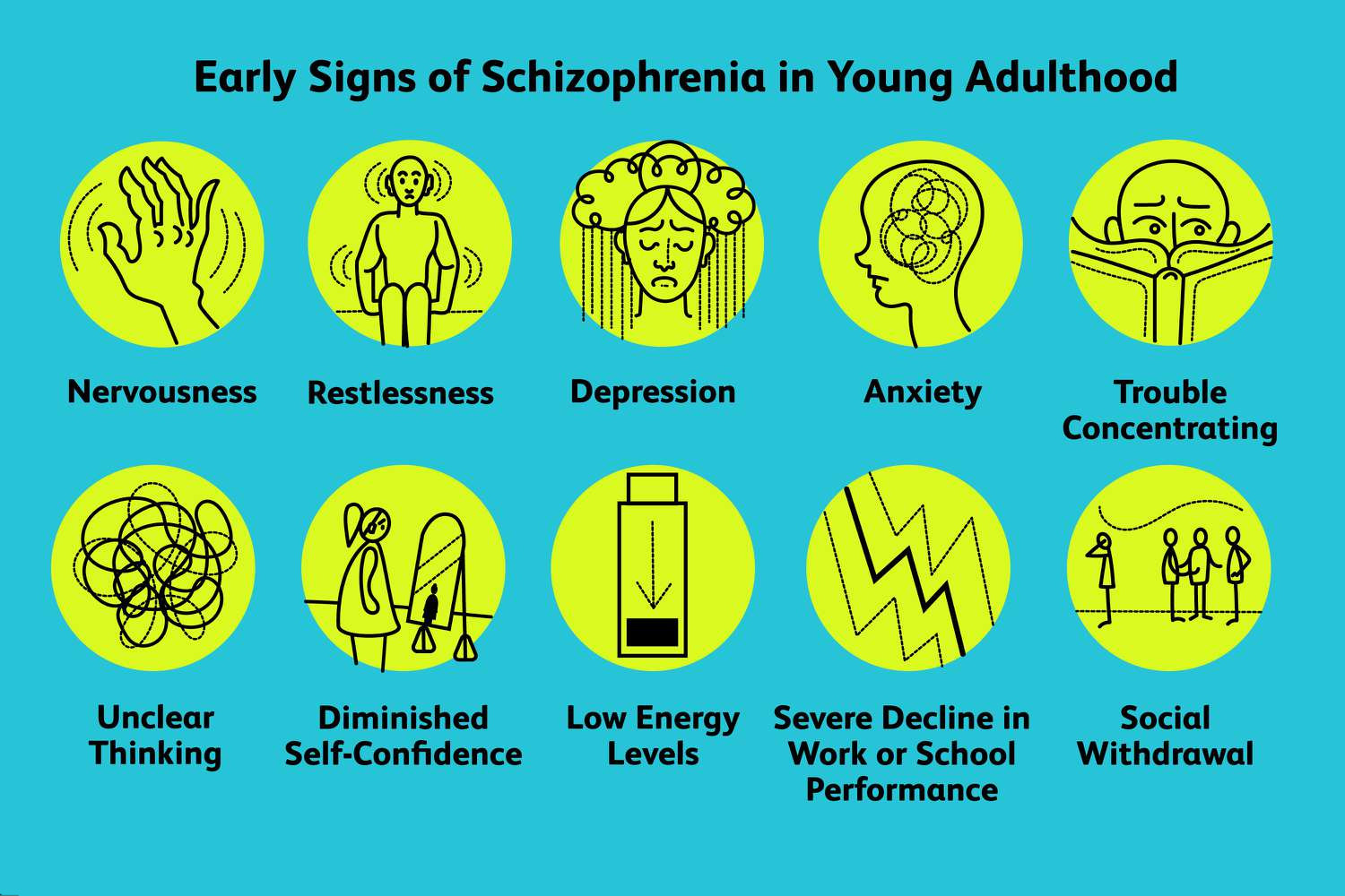
Nursing Paper Examples on Behcet’s Disease: Understanding a Complex Disorder

Nursing Paper Examples on Behcet’s Disease: Understanding a Complex Disorder

Behcet’s Disease, also known as Behcet’s Syndrome, is a rare autoimmune disorder characterized by recurrent oral and genital ulcers, skin lesions, and inflammation of the eyes. First identified by the Turkish dermatologist Hulusi Behcet in 1937, this condition primarily affects individuals in the Mediterranean, Middle Eastern, and Asian regions. Despite decades of research, the exact cause of Behcet’s Disease remains elusive, with genetic predisposition and environmental triggers believed to play key roles. The disease’s pathophysiology involves systemic inflammation and vasculitis, leading to various manifestations across multiple organs and tissues. Diagnosis is based on clinical criteria, and treatment aims to manage symptoms, reduce inflammation, and prevent complications. Patient education is crucial for empowering individuals with Behcet’s Disease to understand their condition and effectively manage their health. This paper aims to explore the causes, signs and symptoms, etiology, pathophysiology, DSM-5 diagnosis, treatment regimens, and patient education strategies related to Behcet’s Disease. (Nursing Paper Examples on Behcet’s Disease: Understanding a Complex Disorder)

Nursing Paper Examples on Behcet's Disease: Understanding a Complex Disorder

Causes of Behcet’s Disease

Behcet’s Disease is a multifactorial condition with complex underlying causes. While the precise etiology remains uncertain, several factors are believed to contribute to the development and progression of the disease.

Genetic Predisposition: Genetic susceptibility plays a significant role in Behcet’s Disease, with certain genetic markers associated with increased susceptibility to the condition. Notably, the HLA-B51 gene has been identified as a potential genetic risk factor, particularly in populations with a higher prevalence of the disease. However, it is essential to recognize that genetic predisposition alone is not sufficient to cause Behcet’s Disease, indicating the involvement of additional factors in disease pathogenesis.

Environmental Triggers: Environmental factors are thought to trigger and exacerbate Behcet’s Disease in genetically susceptible individuals. Infections, particularly viral and bacterial pathogens, have been proposed as potential triggers due to their ability to stimulate the immune system and initiate inflammatory responses. Additionally, environmental factors such as exposure to certain chemicals, dietary components, and climatic conditions may influence disease susceptibility and severity.

Immune System Dysregulation: Behcet’s Disease is characterized by dysregulation of the immune system, leading to abnormal immune responses and inflammation. Dysfunction in immune pathways, including aberrant activation of T cells and cytokine imbalances, contributes to the chronic inflammatory process observed in the disease. Dysregulation of innate and adaptive immune responses further perpetuates the inflammatory cascade, resulting in tissue damage and organ involvement.

Abnormal Responses to Microorganisms: Some evidence suggests that Behcet’s Disease may result from abnormal immune responses to specific microorganisms. Molecular mimicry, where microbial antigens resemble self-antigens, may trigger autoimmune reactions, leading to tissue damage and inflammation. Furthermore, alterations in the microbiome composition and dysbiosis in the gut microbiota have been implicated in Behcet’s Disease pathogenesis, highlighting the potential role of microbial factors in disease development.

Overall, Behcet’s Disease is a complex disorder influenced by a combination of genetic predisposition, environmental triggers, immune system dysregulation, and abnormal responses to microorganisms. Further research is needed to elucidate the precise mechanisms underlying disease pathogenesis and identify targeted therapeutic approaches. (Nursing Paper Examples on Behcet’s Disease: Understanding a Complex Disorder)

Nursing Paper Examples on Behcet's Disease: Understanding a Complex Disorder

Signs and Symptoms

Behcet’s Disease is characterized by a wide range of signs and symptoms affecting various organs and tissues throughout the body. The severity and frequency of symptoms can vary among affected individuals and may fluctuate over time.

Recurrent Oral and Genital Ulcers: One of the hallmark features of Behcet’s Disease is the presence of recurrent oral ulcers, which are often painful and can affect the lips, tongue, and oral mucosa. Genital ulcers, occurring on the vulva or scrotum, are also common and may recur frequently, leading to discomfort and impaired quality of life.

Skin Lesions: Behcet’s Disease can cause a variety of skin lesions, including erythema nodosum-like lesions, papulopustular lesions resembling acne, and pathergy, which is an exaggerated skin reaction to minor trauma. These skin manifestations can vary in appearance and distribution but are typically inflammatory and may leave scars upon healing.

Eye Inflammation (Uveitis): Inflammation of the eyes, specifically uveitis, is a significant complication of Behcet’s Disease and can lead to vision impairment or blindness if left untreated. Uveitis may present with symptoms such as eye pain, redness, blurred vision, sensitivity to light (photophobia), and floaters.

Arthritis: Joint involvement is common in Behcet’s Disease and can manifest as arthritis, causing joint pain, swelling, and stiffness. The arthritis associated with Behcet’s Disease often affects large joints such as the knees, ankles, and wrists, but can also involve smaller joints.

Gastrointestinal Involvement: Behcet’s Disease can affect the gastrointestinal tract, leading to symptoms such as abdominal pain, diarrhea, and gastrointestinal bleeding. Inflammation of the intestines, known as intestinal Behcet’s Disease, can mimic inflammatory bowel diseases like Crohn’s disease or ulcerative colitis.

Neurological Symptoms: In rare cases, Behcet’s Disease can involve the nervous system, leading to neurological symptoms such as headaches, cognitive dysfunction, seizures, and movement disorders. Neurological involvement typically occurs as a result of inflammation of the brain or spinal cord.

Vascular Complications: Behcet’s Disease can affect blood vessels, leading to various vascular complications such as deep vein thrombosis (DVT), arterial thrombosis, and arterial aneurysms. These vascular manifestations can pose significant risks and may require urgent medical intervention to prevent complications such as stroke or pulmonary embolism.

Overall, Behcet’s Disease is characterized by a diverse array of signs and symptoms affecting multiple organ systems, highlighting the systemic nature of the condition. Early recognition and appropriate management of these manifestations are essential for improving patient outcomes and preventing long-term complications. (Nursing Paper Examples on Behcet’s Disease: Understanding a Complex Disorder)

Nursing Paper Examples on Behcet's Disease: Understanding a Complex Disorder

Etiology

Behcet’s Disease is a complex disorder with an intricate etiology involving a combination of genetic, environmental, and immunological factors. While the precise cause of Behcet’s Disease remains unclear, several hypotheses have been proposed to elucidate its underlying etiology.

Genetic Predisposition: Genetic factors play a significant role in Behcet’s Disease, with evidence suggesting a genetic predisposition to the condition. Certain genetic markers, particularly variations in the HLA-B51 gene, have been associated with increased susceptibility to Behcet’s Disease, particularly in populations with a higher prevalence of the disorder. However, the inheritance pattern of Behcet’s Disease is complex and likely involves multiple genetic factors interacting with environmental triggers.

Environmental Triggers: Environmental factors are thought to contribute to the development and progression of Behcet’s Disease by triggering immune dysregulation and inflammatory responses in genetically susceptible individuals. Infections, particularly viral and bacterial pathogens, have been proposed as potential environmental triggers due to their ability to stimulate the immune system and initiate inflammatory cascades. Additionally, environmental factors such as dietary components, smoking, and climatic conditions may influence disease susceptibility and severity.

Immune Dysregulation: Behcet’s Disease is characterized by dysregulation of the immune system, leading to abnormal immune responses and chronic inflammation. Dysfunction in immune pathways, including aberrant activation of T cells, dysregulated cytokine production, and impaired regulation of inflammatory responses, contributes to the pathogenesis of the disease. These immunological abnormalities result in systemic inflammation and tissue damage, leading to the characteristic manifestations of Behcet’s Disease across multiple organ systems.

Microbial Factors: Some evidence suggests that Behcet’s Disease may result from abnormal immune responses to specific microbial antigens. Molecular mimicry, where microbial antigens resemble self-antigens, may trigger autoimmune reactions, leading to chronic inflammation and tissue damage. Furthermore, alterations in the composition of the microbiome and dysbiosis in the gut microbiota have been implicated in Behcet’s Disease pathogenesis, suggesting a potential role for microbial factors in disease development.

Behcet’s Disease is a complex disorder with a multifactorial etiology involving genetic predisposition, environmental triggers, immune dysregulation, and abnormal responses to microbial factors. Further research is needed to unravel the intricate interplay between these factors and identify targeted therapeutic approaches for Behcet’s Disease. (Nursing Paper Examples on Behcet’s Disease: Understanding a Complex Disorder)

Pathophysiology

Behcet’s Disease is characterized by systemic inflammation and vasculitis, leading to various manifestations across multiple organs and tissues. The pathophysiology of Behcet’s Disease involves a complex interplay of immune dysregulation, endothelial dysfunction, and inflammatory mediators.

Immune Dysregulation: Dysregulation of the immune system plays a central role in the pathogenesis of Behcet’s Disease. Abnormal activation of T cells, particularly CD4+ T cells, and dysregulated cytokine production contribute to the chronic inflammatory response observed in the disease. Elevated levels of pro-inflammatory cytokines, such as tumor necrosis factor-alpha (TNF-α), interleukin-1 (IL-1), and interleukin-6 (IL-6), further perpetuate the inflammatory cascade, leading to tissue damage and organ involvement.

Endothelial Dysfunction: Endothelial dysfunction, characterized by impaired endothelial cell function and integrity, is a key feature of Behcet’s Disease. Endothelial cells play a crucial role in maintaining vascular homeostasis and regulating inflammatory responses. In Behcet’s Disease, endothelial dysfunction leads to aberrant expression of adhesion molecules, increased vascular permeability, and enhanced leukocyte adhesion and migration into tissues. These alterations contribute to the development of vasculitis and tissue inflammation observed in Behcet’s Disease.

Vasculitis: Vasculitis, inflammation of blood vessels, is a hallmark feature of Behcet’s Disease and underlies many of its clinical manifestations. Vasculitis in Behcet’s Disease can affect blood vessels of all sizes, including arteries, veins, and capillaries, leading to a wide range of vascular complications such as thrombosis, aneurysms, and vessel occlusion. The inflammatory infiltrates in vessel walls, consisting of T cells, macrophages, and neutrophils, contribute to vascular damage and tissue injury, further perpetuating the inflammatory process.

Overall, Behcet’s Disease is characterized by immune dysregulation, endothelial dysfunction, and vasculitis, leading to systemic inflammation and tissue damage across multiple organ systems. Understanding the underlying pathophysiological mechanisms of Behcet’s Disease is crucial for developing targeted therapeutic strategies aimed at modulating the immune response and reducing inflammation to improve patient outcomes. (Nursing Paper Examples on Behcet’s Disease: Understanding a Complex Disorder)

DSM-5 Diagnosis

Behcet’s Disease is primarily diagnosed based on clinical criteria established by the International Study Group for Behcet’s Disease. According to the Diagnostic and Statistical Manual of Mental Disorders, Fifth Edition (DSM-5), the diagnosis of Behcet’s Disease requires the presence of recurrent oral ulcers plus any two of the following:

  1. Recurrent Genital Ulcers: The presence of recurrent genital ulcers, typically observed on the vulva or scrotum, is a common manifestation of Behcet’s Disease and is considered a diagnostic criterion.
  2. Eye Inflammation (Uveitis): Uveitis, characterized by inflammation of the uvea (middle layer of the eye), is a significant complication of Behcet’s Disease. Eye involvement, presenting as symptoms such as eye pain, redness, blurred vision, photophobia, or floaters, fulfills the diagnostic criteria.
  3. Skin Lesions: Various skin lesions, including erythema nodosum-like lesions, papulopustular lesions resembling acne, and pathergy (an exaggerated skin reaction to minor trauma), are characteristic of Behcet’s Disease and contribute to the diagnostic criteria.
  4. Positive Pathergy Test: The pathergy test is a diagnostic procedure in which a small needle prick is made on the skin, typically on the forearm, and the reaction is observed. A positive pathergy test, defined as the development of a papule or pustule at the site of the needle prick within 24 to 48 hours, is considered indicative of Behcet’s Disease.

In addition to these clinical criteria, other diagnostic tests such as laboratory investigations (e.g., inflammatory markers, HLA-B51 genetic testing) and imaging studies (e.g., ocular examinations, MRI) may be performed to rule out other conditions and assess for complications associated with Behcet’s Disease. (Nursing Paper Examples on Behcet’s Disease: Understanding a Complex Disorder)

Treatment Regimens

Treatment for Behcet’s Disease aims to alleviate symptoms, reduce inflammation, prevent complications, and improve the quality of life for affected individuals. The choice of treatment depends on the severity and specific manifestations of the disease in each individual.

Nonsteroidal Anti-Inflammatory Drugs (NSAIDs): NSAIDs such as ibuprofen or naproxen may be used to manage pain, reduce inflammation, and relieve symptoms associated with Behcet’s Disease, particularly during mild flares.

Corticosteroids: Corticosteroids, such as prednisone or methylprednisolone, are often prescribed to suppress immune-mediated inflammation during acute flares of Behcet’s Disease. These medications can help alleviate symptoms and reduce the severity of inflammatory manifestations, but long-term use may be associated with significant side effects.

Immunomodulatory Agents: Immunomodulatory agents such as colchicine, azathioprine, methotrexate, cyclosporine, and mycophenolate mofetil may be used to control disease activity, prevent relapses, and reduce the need for long-term corticosteroid therapy. Biologic therapies targeting specific immune pathways, such as tumor necrosis factor-alpha (TNF-α) inhibitors or interleukin-1 (IL-1) inhibitors, may also be considered for refractory cases or severe manifestations of Behcet’s Disease.

Topical Treatments: Topical treatments such as corticosteroid creams or ointments may be used to manage oral and genital ulcers and skin lesions associated with Behcet’s Disease. These topical therapies can help reduce pain, promote healing, and improve local symptoms. (Nursing Paper Examples on Behcet’s Disease: Understanding a Complex Disorder)

Patient Education and Self-Management

Patient education is essential for empowering individuals with Behcet’s Disease to understand their condition, manage symptoms, and make informed decisions about their health. Key components of patient education and self-management include:

  1. Understanding the Disease: Educating patients about the nature of Behcet’s Disease, its chronicity, and the potential impact on various organ systems helps individuals develop realistic expectations and cope with the challenges associated with the condition.
  2. Medication Adherence: Emphasizing the importance of adhering to prescribed medications as directed by healthcare providers helps optimize treatment outcomes and reduce the risk of disease flares and complications.
  3. Lifestyle Modifications: Encouraging patients to adopt healthy lifestyle habits such as regular exercise, balanced nutrition, adequate sleep, stress management, and smoking cessation can help improve overall well-being and potentially reduce disease activity.
  4. Monitoring and Self-Assessment: Teaching patients how to monitor disease symptoms, recognize signs of flares or complications, and seek prompt medical attention when necessary empowers individuals to actively participate in their care and collaborate with healthcare providers to optimize treatment outcomes.
  5. Disease-Specific Education: Providing tailored education about specific manifestations of Behcet’s Disease, such as oral and genital ulcer management, eye care, skin lesion care, and joint protection strategies, helps individuals manage symptoms and minimize the impact of the disease on their daily lives.

By providing comprehensive education and support, healthcare providers can empower individuals with Behcet’s Disease to effectively manage their condition, improve their quality of life, and achieve better long-term outcomes. (Nursing Paper Examples on Behcet’s Disease: Understanding a Complex Disorder)

Conclusion

Behcet’s Disease is a complex autoimmune disorder characterized by recurrent oral and genital ulcers, skin lesions, and inflammation of the eyes. While the exact cause remains unknown, a multifactorial etiology involving genetic predisposition, environmental triggers, immune dysregulation, and abnormal responses to microorganisms is implicated. The pathophysiology of Behcet’s Disease is characterized by systemic inflammation, vasculitis, and endothelial dysfunction. Diagnosis is based on clinical criteria, and treatment aims to alleviate symptoms, reduce inflammation, and prevent complications through a combination of NSAIDs, corticosteroids, immunomodulatory agents, and biological therapies. Patient education is crucial for empowering individuals to understand their condition, adhere to treatment regimens, and adopt self-management strategies to improve their quality of life. By providing comprehensive education and support, healthcare providers can help individuals with Behcet’s Disease effectively manage their condition and achieve better long-term outcomes. (Nursing Paper Examples on Behcet’s Disease: Understanding a Complex Disorder)

References

https://www.ncbi.nlm.nih.gov/books/NBK470257/

 
Do you need a similar assignment done for you from scratch? Order now!
Use Discount Code "Newclient" for a 15% Discount!

Nursing Paper Example on Understanding Barth Syndrome

Nursing Paper Example on Understanding Barth Syndrome

Barth Syndrome, a rare genetic disorder, presents complex challenges to affected individuals and their families. This condition, characterized by mutations in the TAZ gene, disrupts mitochondrial function, leading to a range of debilitating symptoms. Despite its rarity, Barth Syndrome profoundly impacts various bodily systems, particularly the heart, muscles, and immune system. Understanding the causes, signs, and symptoms of Barth Syndrome is essential for accurate diagnosis and effective management. This paper aims to explore the intricacies of Barth Syndrome, including its causes, signs, and symptoms, etiology, pathophysiology, DSM-5 diagnosis criteria, treatment regimens, patient education, and potential avenues for future research. By shedding light on this condition, we can enhance awareness, improve diagnostic accuracy, and provide better support for individuals living with Barth Syndrome. (Nursing Paper Example on Understanding Barth Syndrome)

Nursing Paper Example on Understanding Barth Syndrome

Causes

Barth Syndrome arises from mutations in the TAZ gene located on the X chromosome. These mutations disrupt the normal functioning of mitochondria, the cell’s powerhouse responsible for generating energy. The TAZ gene encodes a protein called tafazzin, essential for maintaining the structure and function of mitochondria. When mutations occur in the TAZ gene, tafazzin production is impaired, leading to mitochondrial dysfunction.

The exact mechanism by which TAZ gene mutations affect mitochondrial function is not fully understood. However, it is believed that tafazzin plays a crucial role in remodeling cardiolipin, a phospholipid found in the inner mitochondrial membrane. Cardiolipin is essential for various mitochondrial processes, including oxidative phosphorylation, the process by which cells generate energy.

Mutations in the TAZ gene disrupt cardiolipin remodeling, affecting mitochondrial function and energy production. As a result, cells, particularly those in tissues with high energy demands such as the heart, muscles, and immune system, are unable to function properly. This disruption in cellular energy metabolism contributes to the characteristic symptoms of Barth Syndrome, including cardiomyopathy, muscle weakness, growth delays, and neutropenia.

Since Barth Syndrome is an X-linked genetic disorder, it primarily affects males. Females who carry a single copy of the mutated TAZ gene are typically asymptomatic or may exhibit mild symptoms due to random X-chromosome inactivation.

Barth Syndrome is caused by mutations in the TAZ gene, leading to mitochondrial dysfunction and disruption of cellular energy metabolism. These mutations impair tafazzin production and cardiolipin remodeling, affecting various bodily systems and resulting in the characteristic symptoms of the disorder. (Nursing Paper Example on Understanding Barth Syndrome)

Nursing Paper Example on Understanding Barth Syndrome

Signs and Symptoms

Barth Syndrome presents a spectrum of symptoms that can vary in severity and may change over time. The hallmark features of Barth Syndrome include cardiomyopathy, muscle weakness, growth delays, and neutropenia.

Cardiomyopathy, a condition characterized by weakened heart muscles, is a common manifestation of Barth Syndrome. It can lead to symptoms such as fatigue, shortness of breath, chest pain, and an irregular heartbeat. In severe cases, cardiomyopathy can result in heart failure, a life-threatening condition requiring immediate medical attention.

Muscle weakness is another prominent symptom of Barth Syndrome. Individuals may experience generalized muscle weakness, particularly affecting skeletal muscles used for movement. This can lead to difficulties with mobility, including walking and climbing stairs, as well as challenges with activities of daily living.

Growth delays are frequently observed in individuals with Barth Syndrome, manifesting as below-average height and weight for their age. These growth deficits may become apparent in infancy or early childhood and can persist into adulthood.

Neutropenia, a condition characterized by low levels of neutrophils, a type of white blood cell, is a common hematological feature of Barth Syndrome. Neutrophils play a crucial role in the immune system’s defense against infections. Therefore, individuals with neutropenia are at an increased risk of recurrent bacterial infections, particularly of the skin, respiratory tract, and mucous membranes.

In addition to these hallmark symptoms, individuals with Barth Syndrome may experience other health issues, including feeding difficulties, developmental delays, gastrointestinal problems, and exercise intolerance. The severity and combination of symptoms can vary among affected individuals, making the clinical presentation of Barth Syndrome highly variable. Early recognition and diagnosis of these signs and symptoms are crucial for initiating appropriate medical management and supportive care. (Nursing Paper Example on Understanding Barth Syndrome)

Etiology

The etiology of Barth Syndrome lies in genetic mutations affecting the TAZ gene located on the X chromosome. This gene encodes a protein called tafazzin, which plays a vital role in maintaining the integrity of mitochondrial membranes, particularly through the remodeling of cardiolipin, a phospholipid crucial for mitochondrial function.

The mutations in the TAZ gene result in dysfunctional tafazzin, leading to abnormalities in cardiolipin composition and structure within the inner mitochondrial membrane. This disruption impairs the efficiency of oxidative phosphorylation, the process by which cells generate energy in the form of adenosine triphosphate (ATP). Consequently, affected cells experience energy deficits, leading to the characteristic symptoms of Barth Syndrome.

The inheritance pattern of Barth Syndrome follows an X-linked recessive pattern. Since the TAZ gene is located on the X chromosome, the condition predominantly affects males. Females have two X chromosomes and therefore, if they inherit a mutated TAZ gene on one chromosome, the normal gene on the other chromosome may compensate, resulting in milder or asymptomatic presentation. However, some carrier females may exhibit mild symptoms due to random X-chromosome inactivation.

While most cases of Barth Syndrome are caused by mutations in the TAZ gene, there is significant genetic heterogeneity observed among affected individuals. Variations in the specific mutations and their locations within the TAZ gene can influence the severity and presentation of the disorder. Additionally, other genetic and environmental factors may modulate the clinical features of Barth Syndrome, contributing to its variable expressivity and phenotypic spectrum.

Overall, the etiology of Barth Syndrome is rooted in genetic mutations affecting mitochondrial function, particularly involving the TAZ gene and its role in cardiolipin remodeling. Understanding these underlying genetic mechanisms is essential for accurate diagnosis, genetic counseling, and potential therapeutic interventions for individuals affected by Barth Syndrome. (Nursing Paper Example on Understanding Barth Syndrome)

Nursing Paper Example on Understanding Barth Syndrome

Pathophysiology

Barth Syndrome’s pathophysiology primarily revolves around mitochondrial dysfunction resulting from mutations in the TAZ gene. Tafazzin, the protein encoded by the TAZ gene, plays a crucial role in cardiolipin remodeling within the inner mitochondrial membrane. Cardiolipin is essential for maintaining mitochondrial structure and function, particularly in oxidative phosphorylation, the process by which cells generate ATP, the energy currency of the cell.

In individuals with Barth Syndrome, mutations in the TAZ gene lead to dysfunctional tafazzin, disrupting cardiolipin remodeling. This disruption affects the stability and fluidity of the mitochondrial membrane, impairing its ability to carry out oxidative phosphorylation efficiently. As a result, affected cells experience energy deficits, leading to various clinical manifestations observed in Barth Syndrome.

Mitochondrial dysfunction in Barth Syndrome primarily affects tissues with high energy demands, such as the heart, skeletal muscles, and immune system. Cardiomyocytes, the cells responsible for heart contraction, are particularly susceptible to mitochondrial dysfunction due to their continuous need for energy to maintain cardiac function. Consequently, individuals with Barth Syndrome often develop cardiomyopathy, characterized by weakened heart muscles and impaired cardiac function.

Skeletal muscles, which rely on oxidative phosphorylation for energy during physical activity, also exhibit dysfunction in Barth Syndrome. This leads to muscle weakness and fatigue, impacting mobility and physical functioning in affected individuals.

Additionally, mitochondrial dysfunction in Barth Syndrome affects the immune system, leading to neutropenia, a condition characterized by low levels of neutrophils, a type of white blood cell crucial for fighting infections. This renders individuals more susceptible to recurrent bacterial infections, further contributing to the clinical manifestations of the disorder.

Overall, the pathophysiology of Barth Syndrome involves mitochondrial dysfunction resulting from mutations in the TAZ gene, leading to energy deficits and cellular dysfunction, particularly in tissues with high energy demands. Understanding these underlying mechanisms is crucial for developing targeted interventions to manage and potentially mitigate the impact of Barth Syndrome on affected individuals.

DMS-5 Diagnosis

Diagnosing Barth Syndrome involves a comprehensive evaluation following the criteria outlined in the Diagnostic and Statistical Manual of Mental Disorders, Fifth Edition (DSM-5). The diagnostic process typically begins with a thorough medical history and physical examination to assess the presence of characteristic signs and symptoms associated with the disorder.

Genetic testing plays a crucial role in confirming the diagnosis of Barth Syndrome. Analysis of the TAZ gene for mutations is essential to identify specific genetic abnormalities associated with the condition. Identification of mutations in the TAZ gene confirms the diagnosis of Barth Syndrome, particularly in individuals presenting with characteristic clinical features.

In addition to genetic testing, clinical evaluation of symptoms is essential for diagnosing Barth Syndrome. Common manifestations such as cardiomyopathy, muscle weakness, growth delays, and neutropenia are carefully assessed to support the diagnosis. Laboratory tests may also be performed to evaluate cardiac function, muscle strength, growth parameters, and blood counts.

Diagnostic criteria outlined in the DSM-5 help clinicians establish a clear diagnosis of Barth Syndrome based on the presence of characteristic signs and symptoms, supported by genetic testing and clinical evaluation. The multidisciplinary approach involving medical history, physical examination, genetic testing, and laboratory investigations ensures accurate diagnosis and appropriate management of individuals with Barth Syndrome.

Overall, the DSM-5 criteria provide a standardized framework for diagnosing Barth Syndrome, enabling clinicians to identify affected individuals based on specific clinical features and genetic abnormalities. Early recognition and diagnosis are crucial for initiating timely interventions and providing appropriate medical care to improve outcomes for individuals with Barth Syndrome. (Nursing Paper Example on Understanding Barth Syndrome)

Treatment Regimens and Patient Education

The management of Barth Syndrome involves a multidisciplinary approach aimed at addressing the various symptoms and complications associated with the disorder. Treatment regimens focus on symptom management, supportive care, and promoting overall well-being in affected individuals. Patient education plays a crucial role in empowering patients and their families to understand the condition and actively participate in their care.

Medications: Pharmacological interventions are tailored to address specific symptoms and complications of Barth Syndrome. For individuals with cardiomyopathy, medications such as angiotensin-converting enzyme (ACE) inhibitors and beta-blockers may be prescribed to improve cardiac function and manage heart failure symptoms. Other medications may be used to address muscle weakness, growth delays, and neutropenia as needed.

Nutritional Support: Nutritional interventions are essential for individuals with Barth Syndrome to support growth and development, optimize energy levels, and maintain overall health. A balanced diet rich in nutrients, including proteins, carbohydrates, fats, vitamins, and minerals, is recommended. In some cases, dietary supplements or specialized formulas may be prescribed to address specific nutritional deficiencies or challenges.

Physical Therapy: Physical therapy plays a vital role in managing muscle weakness and promoting mobility and functional independence in individuals with Barth Syndrome. Tailored exercise programs, stretching exercises, and muscle-strengthening activities help improve muscle tone, flexibility, and overall physical function. Physical therapists also guide adaptive equipment and assistive devices to enhance mobility and facilitate activities of daily living.

Patient Education: Patient education is essential for individuals with Barth Syndrome and their families to understand the nature of the disorder, its potential complications, and the importance of adherence to treatment regimens. Education sessions provide information on symptom management, medication administration, dietary recommendations, and strategies to optimize overall health and well-being. Patients and families are encouraged to actively participate in treatment decisions, communicate openly with healthcare providers, and seek support from patient advocacy groups and support networks.

Regular Monitoring: Regular follow-up visits with healthcare providers are crucial for monitoring disease progression, assessing treatment efficacy, and addressing any emerging issues or complications. Monitoring may include cardiac evaluations, growth assessments, nutritional status evaluations, blood count monitoring, and other relevant tests as needed.

Overall, a comprehensive treatment approach, coupled with patient education and support, is essential for optimizing outcomes and enhancing the quality of life for individuals living with Barth Syndrome. By addressing symptoms, promoting healthy lifestyle habits, and fostering patient empowerment, healthcare providers can help individuals with Barth Syndrome thrive despite the challenges posed by this complex disorder. (Nursing Paper Example on Understanding Barth Syndrome)

Conclusion

Barth Syndrome is a rare genetic disorder characterized by mutations in the TAZ gene, leading to mitochondrial dysfunction and a range of debilitating symptoms. The causes, signs, and symptoms of Barth Syndrome, along with its etiology and pathophysiology, highlight the complexity of this condition. Accurate diagnosis according to DSM-5 criteria is crucial for appropriate management. Treatment regimens focus on symptom management, supportive care, and patient education. Patient education plays a pivotal role in empowering individuals and their families to understand the condition and actively participate in their care. By addressing symptoms, promoting healthy lifestyle habits, and fostering patient empowerment, healthcare providers can enhance the quality of life for individuals living with Barth Syndrome. Ongoing research offers hope for improved understanding and management of this complex disorder, paving the way for better outcomes in the future. (Nursing Paper Example on Understanding Barth Syndrome)

References

https://www.ncbi.nlm.nih.gov/books/NBK247162/

 
Do you need a similar assignment done for you from scratch? Order now!
Use Discount Code "Newclient" for a 15% Discount!

Nursing Paper Example on Alpers Disease: Understanding a Rare Neurological Disorder

Nursing Paper Example on Alpers Disease: Understanding a Rare Neurological Disorder

Alpers Disease, also recognized as Alpers-Huttenlocher syndrome, is an uncommon neurological disorder that profoundly impacts infants and young children, often presenting within the initial years of life. This condition, characterized by a trio of symptoms encompassing seizures, liver dysfunction, and progressive neurological degeneration, was first delineated by Bernard Jacob Alpers in 1931. Despite its rarity, Alpers Disease poses significant challenges to affected individuals and their families due to its severe and debilitating nature. Understanding the underlying causes, clinical manifestations, and management strategies for Alpers Disease is crucial for healthcare professionals and caregivers alike. In this paper, we will delve into the causes, signs and symptoms, etiology, pathophysiology, DMS-5 diagnosis criteria, treatment regimens and patient education, and conclude by highlighting the importance of comprehensive care for individuals grappling with this challenging condition. (Nursing Paper Example on Alpers Disease: Understanding a Rare Neurological Disorder)

Nursing Paper Example on Alpers Disease: Understanding a Rare Neurological Disorder

Causes

The exact cause of Alpers Disease remains enigmatic, yet it is predominantly attributed to genetic mutations affecting mitochondrial DNA (mtDNA). Mitochondria, revered as the powerhouse of the cell, play a pivotal role in energy production. The mutations implicated in Alpers Disease primarily target genes responsible for mitochondrial function, particularly those crucial for DNA replication and maintenance within mitochondria.

These genetic mutations engender mitochondrial dysfunction, thereby impeding energy metabolism, particularly in tissues with elevated energy requisites like the brain and liver. Consequently, affected individuals endure a cascade of physiological disturbances, culminating in the characteristic triad of symptoms associated with Alpers Disease.

Mitochondrial DNA mutations are typically inherited in an autosomal recessive pattern, signifying that both parents must carry a mutated gene for their offspring to manifest the disease. Occasionally, Alpers Disease can also result from spontaneous mutations occurring in the mtDNA during embryonic development.

It is essential to recognize that while genetic mutations serve as the primary precipitant for Alpers Disease, environmental factors, and additional genetic modifiers may influence the severity and clinical presentation of the disorder. Furthermore, the variable penetrance and expressivity of mitochondrial DNA mutations contribute to the heterogeneous nature of Alpers Disease, wherein affected individuals may exhibit a spectrum of symptoms ranging from mild to severe.

Given the intricate interplay between genetic predisposition and environmental factors in the pathogenesis of Alpers Disease, further research endeavors are imperative to elucidate the precise mechanisms underlying this debilitating disorder. Such insights hold the potential to inform the development of novel therapeutic interventions aimed at ameliorating the clinical course and enhancing the quality of life for individuals grappling with Alpers Disease. (Nursing Paper Example on Alpers Disease: Understanding a Rare Neurological Disorder)

Signs and Symptoms

Alpers Disease presents a constellation of symptoms that progressively worsen over time, severely impairing affected individuals’ quality of life. The clinical manifestations of this disorder are diverse, encompassing neurological, hepatic, and systemic abnormalities.

Nursing Paper Example on Alpers Disease: Understanding a Rare Neurological Disorder

Neurological Symptoms: Seizures represent a hallmark feature of Alpers Disease and often serve as the initial presenting symptom. These seizures typically commence in infancy or early childhood and tend to be refractory to conventional antiepileptic medications. As the disease progresses, affected individuals may experience various types of seizures, including focal, generalized, or myoclonic seizures.

Developmental Regression: Progressive developmental regression is a prominent feature of Alpers Disease, characterized by a loss of previously acquired developmental milestones. Affected individuals may exhibit a decline in motor skills, speech, and cognitive abilities, ultimately leading to profound intellectual disability.

Neurological Decline: Over time, individuals with Alpers Disease experience a relentless deterioration of neurological function, marked by a decline in motor coordination, muscle weakness, and spasticity. The progressive neurodegeneration may also manifest as movement disorders, including dystonia and ataxia.

Hepatic Dysfunction: Liver involvement is another cardinal feature of Alpers Disease, presenting as hepatomegaly, jaundice, and signs of hepatic failure. Liver dysfunction may manifest early in infancy or later in the disease course and can significantly impact overall prognosis.

Systemic Symptoms: In addition to neurological and hepatic manifestations, individuals with Alpers Disease may exhibit systemic symptoms, including failure to thrive, feeding difficulties, and metabolic disturbances.

Ophthalmological Abnormalities: Ophthalmological abnormalities, such as optic atrophy and retinitis pigmentosa, have also been reported in some individuals with Alpers Disease, further contributing to the multisystemic nature of the disorder.

The progressive nature and multisystemic involvement of Alpers Disease underscore the complex pathophysiology underlying this devastating disorder, necessitating a multidisciplinary approach to management and care. (Nursing Paper Example on Alpers Disease: Understanding a Rare Neurological Disorder)

Etiology

Alpers Disease is primarily rooted in genetic aberrations that disrupt mitochondrial function, leading to the characteristic clinical features observed in affected individuals. The genetic etiology of Alpers Disease predominantly revolves around mutations in genes encoding proteins essential for mitochondrial DNA (mtDNA) replication and maintenance.

Mitochondria play a pivotal role in cellular energy production through oxidative phosphorylation, a process that generates adenosine triphosphate (ATP). Mutations in genes encoding components of the mitochondrial DNA polymerase gamma (POLG) enzyme complex, such as the POLG gene itself, have been identified as major contributors to Alpers Disease. POLG is crucial for mtDNA replication and repair, and mutations in this gene impair mitochondrial DNA synthesis, leading to mitochondrial dysfunction.

Furthermore, mutations in other genes involved in mitochondrial DNA replication and maintenance, including TWNK (Twinkle) and DGUOK (Deoxyguanosine Kinase), have also been implicated in the pathogenesis of Alpers Disease. These genetic mutations disrupt mitochondrial DNA replication and repair processes, culminating in mitochondrial dysfunction and cellular energy depletion.

The inheritance pattern of Alpers Disease is typically autosomal recessive, necessitating the presence of mutations in both alleles of the causative gene for disease manifestation. However, in some cases, Alpers Disease can result from de novo mutations occurring sporadically during embryonic development.

Although the genetic etiology of Alpers Disease predominantly involves mutations in nuclear genes encoding mitochondrial proteins, mitochondrial DNA mutations can also contribute to disease pathogenesis. Mitochondrial DNA mutations can arise spontaneously or be maternally inherited and can further exacerbate mitochondrial dysfunction, amplifying the severity of clinical manifestations in affected individuals.

Understanding the underlying genetic basis of Alpers Disease is critical for accurate diagnosis, genetic counseling, and potential targeted therapeutic interventions aimed at mitigating the devastating consequences of this debilitating disorder. (Nursing Paper Example on Alpers Disease: Understanding a Rare Neurological Disorder)

Pathophysiology

The pathophysiology of Alpers Disease revolves around mitochondrial dysfunction, which disrupts cellular energy metabolism and leads to widespread neuronal and hepatic damage. Mitochondria, organelles responsible for generating cellular energy in the form of adenosine triphosphate (ATP), play a central role in the pathogenesis of this disorder.

Mutations in genes encoding proteins involved in mitochondrial DNA (mtDNA) replication and maintenance, such as POLG, TWNK, and DGUOK, impair mitochondrial DNA synthesis and repair processes. Consequently, mitochondrial dysfunction ensues, characterized by reduced ATP production and increased production of reactive oxygen species (ROS).

The impaired energy metabolism resulting from mitochondrial dysfunction particularly affects tissues with high energy demands, such as the brain and liver. Neurons are highly susceptible to mitochondrial dysfunction due to their reliance on ATP for synaptic transmission and neuronal signaling. The progressive neuronal degeneration observed in Alpers Disease is attributed to energy depletion, oxidative stress, and impaired mitochondrial function, leading to neuronal cell death.

Hepatic dysfunction in Alpers Disease stems from mitochondrial impairment and the resultant disruption of liver metabolism. Mitochondria play a crucial role in hepatic energy metabolism, and their dysfunction leads to hepatocyte injury, hepatomegaly, and liver failure.

The accumulation of reactive oxygen species (ROS) resulting from mitochondrial dysfunction exacerbates cellular damage, leading to oxidative stress and further amplifying tissue injury. Additionally, impaired mitochondrial function disrupts calcium homeostasis and alters cellular signaling pathways, contributing to the pathogenesis of Alpers Disease.

Overall, the pathophysiology of Alpers Disease is characterized by mitochondrial dysfunction, energy depletion, oxidative stress, and widespread cellular damage, particularly affecting the brain and liver. Understanding these underlying pathogenic mechanisms is crucial for developing targeted therapeutic interventions aimed at ameliorating mitochondrial dysfunction and mitigating the devastating consequences of this debilitating disorder. (Nursing Paper Example on Alpers Disease: Understanding a Rare Neurological Disorder)

DMS-5 Diagnosis

Diagnosing Alpers Disease typically involves a comprehensive clinical evaluation, neuroimaging studies, and genetic testing to ascertain the presence of characteristic symptoms and identify underlying genetic mutations. The Diagnostic and Statistical Manual of Mental Disorders, Fifth Edition (DSM-5), does not delineate specific diagnostic criteria for Alpers Disease but provides guidelines for assessing the clinical presentation and determining the appropriate diagnostic approach.

The DSM-5 criteria for epilepsy, a common feature of Alpers Disease, encompass recurrent, unprovoked seizures, which align with the clinical manifestation of seizures observed in affected individuals. Additionally, developmental regression, cognitive decline, and neurological abnormalities evident in Alpers Disease may fulfill the criteria for intellectual disability or other neurodevelopmental disorders outlined in the DSM-5.

Neuroimaging studies, particularly magnetic resonance imaging (MRI), play a crucial role in the diagnostic workup of Alpers Disease. MRI findings may reveal characteristic abnormalities, including cerebral atrophy, white matter changes, and signal abnormalities in the basal ganglia, which are consistent with the neurodegenerative nature of the disorder.

Genetic testing is paramount for confirming the diagnosis of Alpers Disease, as it enables the identification of mutations in genes associated with mitochondrial dysfunction, such as POLG, TWNK, and DGUOK. Targeted sequencing or whole-exome sequencing techniques are utilized to detect pathogenic variants in these genes, providing definitive confirmation of the diagnosis.

The diagnostic evaluation of Alpers Disease necessitates a multidisciplinary approach involving neurologists, geneticists, and other healthcare professionals. By integrating clinical assessment, neuroimaging studies, and genetic testing, healthcare providers can establish an accurate diagnosis and initiate appropriate management strategies tailored to the individual needs of patients with Alpers Disease. (Nursing Paper Example on Alpers Disease: Understanding a Rare Neurological Disorder)

Treatment Regimens and Patient Education

As of now, there is no cure for Alpers Disease, and treatment primarily focuses on managing symptoms and providing supportive care to improve the quality of life for affected individuals. Given the complex nature of this disorder, treatment regimens typically entail a multidisciplinary approach involving neurologists, hepatologists, geneticists, and other healthcare professionals.

Symptom Management: Antiepileptic drugs (AEDs) are commonly prescribed to manage seizures in individuals with Alpers Disease. However, seizures associated with this disorder are often refractory to conventional AEDs, necessitating a tailored approach to medication management. Adjunctive therapies, such as ketogenic diet therapy or other dietary interventions, may be considered in some cases to help control seizures.

Supportive Care: Supportive care plays a pivotal role in managing the diverse array of symptoms associated with Alpers Disease. Physical and occupational therapies are integral components of supportive care, aiming to optimize motor function, enhance mobility, and improve the overall quality of life for affected individuals. Speech therapy may also be beneficial in addressing communication difficulties and speech impairments.

Nutritional Support: Given the hepatic involvement observed in Alpers Disease, ensuring adequate nutritional support is essential to support overall health and well-being. Nutritional interventions may include specialized formulas, dietary modifications, or gastrostomy tube placement to facilitate adequate nutrient intake, particularly in individuals with feeding difficulties or failure to thrive.

Liver Transplantation: In cases of severe liver dysfunction or liver failure secondary to Alpers Disease, liver transplantation may be considered as a therapeutic option. Liver transplantation can provide a life-saving intervention by replacing the diseased liver with a healthy donor organ, thereby improving the overall prognosis and quality of life for affected individuals.

Patient Education: Patient education is integral to the management of Alpers Disease, as it empowers patients and their families to understand the nature of the disorder and actively participate in treatment decisions. Educating patients and caregivers about the signs and symptoms of Alpers Disease, available treatment options, and strategies for symptom management can help alleviate anxiety and improve coping mechanisms.

Additionally, providing information about available support services, such as patient advocacy groups and community resources, can facilitate access to additional support and assistance for affected individuals and their families. By fostering open communication and providing comprehensive education, healthcare providers can empower patients and caregivers to navigate the challenges associated with Alpers Disease effectively.

While there is currently no cure for Alpers Disease, a multidisciplinary approach to treatment focusing on symptom management, supportive care, and patient education can significantly improve the quality of life for affected individuals and their families. By addressing the diverse array of symptoms and providing comprehensive support, healthcare providers can optimize outcomes and enhance the overall well-being of individuals grappling with this rare and debilitating disorder. (Nursing Paper Example on Alpers Disease: Understanding a Rare Neurological Disorder)

Conclusion

Alpers Disease presents a significant challenge due to its rare occurrence and severe clinical manifestations affecting infants and young children. Through an exploration of its causes, signs and symptoms, etiology, pathophysiology, DSM-5 diagnosis, treatment regimens, and patient education, we have gained a deeper understanding of this debilitating disorder. While the exact cause remains elusive, genetic mutations disrupting mitochondrial function play a central role in its pathogenesis. Symptom management, supportive care, and patient education are paramount in addressing the diverse array of symptoms and optimizing outcomes for affected individuals and their families. Despite the absence of a cure, a multidisciplinary approach to treatment, including antiepileptic drugs, supportive therapies, and nutritional support, can significantly enhance the quality of life for those grappling with Alpers Disease. By fostering open communication and providing comprehensive education, healthcare providers can empower patients and caregivers to navigate the challenges associated with this rare and devastating disorder effectively. (Nursing Paper Example on Alpers Disease: Understanding a Rare Neurological Disorder)

References

https://www.ncbi.nlm.nih.gov/books/NBK540966/

 
Do you need a similar assignment done for you from scratch? Order now!
Use Discount Code "Newclient" for a 15% Discount!

Nursing Paper Example on The Absence of the Septum Pellucidum: Understanding a Rare Disorder

Nursing Paper Example on The Absence of the Septum Pellucidum: Understanding a Rare Disorder

The absence of the septum pellucidum is a rare condition where the structure that typically separates the two lateral ventricles of the brain is either partially or entirely missing. This disorder, although infrequent, presents significant challenges due to its association with various neurological and developmental abnormalities. Understanding the intricacies of this condition is essential for healthcare professionals to provide appropriate care and support to affected individuals and their families. Despite its rarity, the absence of the septum pellucidum underscores the complexity of brain development and highlights the need for further research to elucidate its underlying causes and improve diagnostic and therapeutic strategies. This paper explores the causes, signs, and symptoms, etiology, pathophysiology, DSM-5 diagnosis, treatment regimens, and patient education, and concludes with insights into the management and impact of this unique neurological disorder. (Nursing Paper Example on The Absence of the Septum Pellucidum: Understanding a Rare Disorder)

Nursing Paper Example on The Absence of the Septum Pellucidum: Understanding a Rare Disorder

Causes

The causes of the absence of the septum pellucidum are not yet fully understood, but researchers have identified several potential factors contributing to its development. One significant factor is believed to be abnormalities during fetal brain development. During the early stages of pregnancy, intricate processes guide the formation and organization of brain structures, including the septum pellucidum. Any disruption or deviation from these processes can lead to structural anomalies in the developing brain.

Genetic factors also play a role in predisposing individuals to this condition. Studies have suggested that certain genetic mutations or variations may increase the likelihood of developing the absence of the septum pellucidum. However, the specific genes involved and their precise contributions to the disorder remain areas of ongoing investigation.

Furthermore, environmental influences during pregnancy may contribute to the development of this condition. Factors such as maternal infections, exposure to toxins, or maternal health conditions can impact fetal brain development and increase the risk of structural abnormalities like the absence of the septum pellucidum.

Additionally, disruptions in the signaling pathways or molecular mechanisms involved in brain development may also contribute to the pathogenesis of this disorder. Research in this area aims to elucidate the intricate cellular and molecular processes underlying normal and abnormal brain development, providing valuable insights into the causes of conditions like the absence of the septum pellucidum.

Overall, the causes of the absence of the septum pellucidum are likely multifactorial, involving complex interactions between genetic predisposition and environmental influences during critical periods of fetal brain development. Further research is needed to unravel the precise mechanisms contributing to this condition and to develop targeted interventions for affected individuals. (Nursing Paper Example on The Absence of the Septum Pellucidum: Understanding a Rare Disorder)

Signs and Symptoms

The absence of the septum pellucidum presents a range of signs and symptoms that can vary in severity and presentation among affected individuals. Common manifestations of this disorder often become apparent during infancy, childhood, or adolescence.

Nursing Paper Example on The Absence of the Septum Pellucidum: Understanding a Rare Disorder

One of the primary symptoms associated with the absence of the septum pellucidum is cognitive deficits. Individuals affected by this condition may experience difficulties with learning, memory, attention, and problem-solving skills. These cognitive impairments can significantly impact academic performance and daily functioning.

Developmental delays are also commonly observed in individuals with the absence of the septum pellucidum. Motor skills, language development, and social-emotional milestones may be delayed compared to their peers. This can result in challenges with coordination, speech and language abilities, and social interactions.

Behavioral issues are another hallmark of the absence of the septum pellucidum. Individuals may exhibit behavioral problems such as impulsivity, hyperactivity, aggression, or emotional lability. These behavioral challenges can pose significant challenges for caregivers and may require behavioral interventions or management strategies.

Neurological abnormalities are frequently associated with the absence of the septum pellucidum. Seizures are a common neurological symptom, occurring in a significant portion of affected individuals. Visual impairments, including abnormalities in visual perception or processing, may also be present.

Additionally, some individuals with the absence of the septum pellucidum may experience other neurological symptoms such as headaches, balance problems, or sensory issues. These symptoms can further contribute to the overall impact of the disorder on an individual’s quality of life and daily functioning.

Overall, the signs and symptoms of the absence of the septum pellucidum encompass a wide range of cognitive, developmental, behavioral, and neurological manifestations that require comprehensive evaluation and management by healthcare professionals. Early recognition and intervention are essential for optimizing outcomes and supporting affected individuals in reaching their full potential. (Nursing Paper Example on The Absence of the Septum Pellucidum: Understanding a Rare Disorder)

Etiology

The etiology of the absence of the septum pellucidum is complex and involves multiple factors contributing to its development. While the precise cause remains unclear, researchers have identified several potential etiological factors that may play a role in the pathogenesis of this disorder.

Genetic factors are thought to contribute significantly to the development of the absence of the septum pellucidum. Studies have suggested a genetic predisposition, with certain gene mutations or variations increasing the susceptibility to structural abnormalities in the brain, including the absence of the septum pellucidum. However, the specific genes involved and their exact contributions to the disorder require further investigation.

Environmental influences during critical periods of fetal brain development are also believed to play a role in the etiology of the absence of the septum pellucidum. Factors such as maternal infections, exposure to toxins, nutritional deficiencies, or maternal health conditions can disrupt normal brain development and increase the risk of structural anomalies.

Furthermore, interactions between genetic and environmental factors may contribute to the pathogenesis of this disorder. Studies suggest that gene-environment interactions during prenatal development may influence the susceptibility to structural brain abnormalities like the absence of the septum pellucidum. These interactions may involve complex molecular mechanisms that impact neural development and organization.

Overall, the etiology of the absence of the septum pellucidum is likely multifactorial, involving a combination of genetic predisposition and environmental influences during critical periods of fetal brain development. Further research is needed to elucidate the specific genetic and environmental factors contributing to this disorder and to improve our understanding of its underlying mechanisms. Such insights are essential for the development of targeted interventions and management strategies for affected individuals. (Nursing Paper Example on The Absence of the Septum Pellucidum: Understanding a Rare Disorder)

Pathophysiology

The pathophysiology of the absence of the septum pellucidum involves disruptions in the normal development and organization of the brain, leading to structural abnormalities and functional deficits in affected individuals.

During fetal brain development, the septum pellucidum forms a thin membrane that separates the two lateral ventricles of the brain. In individuals with the absence of the septum pellucidum, this membrane fails to develop properly or is completely absent, resulting in a lack of separation between the ventricles.

This structural anomaly disrupts the normal architecture of the brain and affects the connectivity between different regions, leading to functional impairments. The absence of the septum pellucidum may disrupt neural pathways involved in cognitive processing, sensory perception, motor coordination, and emotional regulation.

Furthermore, the absence of the septum pellucidum can impact the flow of cerebrospinal fluid (CSF) within the brain. Normally, the septum pellucidum helps regulate the flow of CSF between the lateral ventricles. In its absence, CSF circulation may be altered, leading to changes in intracranial pressure and potentially contributing to symptoms such as headaches or neurological deficits.

The absence of the septum pellucidum is often associated with other structural brain abnormalities, such as agenesis of the corpus callosum or abnormalities in the development of the hippocampus. These additional anomalies further exacerbate the neurological and cognitive impairments observed in affected individuals.

Overall, the pathophysiology of the absence of the septum pellucidum involves disruptions in brain development, structural abnormalities, altered neural connectivity, and potential changes in CSF dynamics. These pathophysiological changes contribute to the wide range of symptoms and functional deficits observed in individuals with this disorder. Further research is needed to elucidate the underlying mechanisms and identify potential targets for therapeutic interventions. (Nursing Paper Example on The Absence of the Septum Pellucidum: Understanding a Rare Disorder)

DSM-5 Diagnosis

According to the Diagnostic and Statistical Manual of Mental Disorders, Fifth Edition (DSM-5), the absence of the septum pellucidum is not listed as a specific diagnostic entity. However, this condition may be recognized within the context of related disorders and associated symptoms.

Structural brain abnormalities, including the absence of the septum pellucidum, may be considered in the diagnosis of intellectual disabilities or developmental disorders. These disorders are characterized by significant limitations in intellectual functioning and adaptive behavior, affecting various domains of daily life.

In cases where the absence of the septum pellucidum is associated with neurological symptoms such as seizures, visual impairments, or other neurological deficits, additional diagnostic considerations may be warranted. Depending on the specific clinical presentation, the absence of the septum pellucidum may be identified as a contributing factor in the diagnosis of neurological conditions or disorders.

Furthermore, if the absence of the septum pellucidum is associated with behavioral disturbances or psychiatric symptoms, it may be considered within the diagnostic criteria for psychiatric disorders such as attention-deficit/hyperactivity disorder (ADHD), autism spectrum disorder (ASD), or mood disorders.

Overall, while the absence of the septum pellucidum is not specifically listed as a standalone diagnosis in the DSM-5, it may be recognized and considered within the context of associated symptoms and related disorders. Accurate diagnosis and comprehensive evaluation by healthcare professionals are essential for identifying and addressing the complex clinical presentations associated with this structural brain abnormality. (Nursing Paper Example on The Absence of the Septum Pellucidum: Understanding a Rare Disorder)

Treatment Regimens and Patient Education

Management of the absence of the septum pellucidum focuses on addressing symptoms, providing supportive care, and optimizing the overall well-being of affected individuals. Treatment regimens are tailored to address the specific needs and challenges associated with this rare neurological disorder.

1. Symptom Management:

  • Cognitive deficits: Educational interventions and specialized programs can help address learning difficulties and promote cognitive development. Individualized educational plans (IEPs) and accommodations in academic settings may be implemented.
  • Behavioral issues: Behavioral therapy, counseling, and psychosocial interventions can help manage behavioral challenges such as impulsivity, aggression, and emotional lability. Parent training programs may also be beneficial.
  • Neurological symptoms: Medications may be prescribed to manage seizures or other neurological symptoms. Close monitoring and adjustment of medication regimens are essential for optimal symptom control.

2. Supportive Care:

  • Speech and language therapy: For individuals with speech and language delays or difficulties, speech therapy can improve communication skills and enhance social interactions.
  • Occupational therapy: Occupational therapy focuses on improving motor skills, coordination, and activities of daily living to promote independence and functional abilities.
  • Physical therapy: Physical therapy may be beneficial for individuals with motor impairments, helping to improve mobility, strength, and coordination.

3. Patient Education:

  • Understanding the disorder: Educating patients and their families about the nature of the absence of the septum pellucidum, its potential impact on various aspects of life, and available resources for support is crucial.
  • Treatment options: Providing information about treatment options, including therapies, medications, and supportive services, empowers patients and families to make informed decisions about their care.
  • Monitoring and follow-up: Emphasizing the importance of regular monitoring, follow-up appointments, and collaboration with healthcare providers ensures ongoing management and adjustments to treatment regimens as needed.
  • Community resources: Connecting patients and families with community resources, support groups, and advocacy organizations can provide additional support and opportunities for networking and sharing experiences.

Patient education plays a vital role in promoting self-management, enhancing coping skills, and improving the overall quality of life for individuals with the absence of the septum pellucidum. By providing comprehensive information and support, healthcare professionals empower patients and families to navigate the challenges associated with this rare neurological disorder effectively. (Nursing Paper Example on The Absence of the Septum Pellucidum: Understanding a Rare Disorder)

Conclusion

The absence of the septum pellucidum presents a rare yet significant challenge in neurological and developmental disorders. Through a comprehensive understanding of its causes, signs, and symptoms, along with its complex etiology and pathophysiology, healthcare professionals can better diagnose and manage this condition. While not specifically listed in the DSM-5, recognition within related disorders facilitates tailored treatment approaches focused on symptom management and supportive care. Patient education plays a pivotal role in empowering individuals and families to navigate the challenges associated with the absence of the septum pellucidum, facilitating informed decision-making, and accessing appropriate resources. By implementing treatment regimens addressing cognitive, behavioral, and neurological symptoms, along with providing supportive services and community resources, healthcare professionals can enhance the overall well-being and quality of life of individuals affected by this disorder. Ongoing research and collaboration aim to advance our understanding and management of this unique neurological condition, ensuring optimal outcomes for affected individuals. (Nursing Paper Example on The Absence of the Septum Pellucidum: Understanding a Rare Disorder)

References

https://www.ncbi.nlm.nih.gov/pmc/articles/PMC7069873/

 
Do you need a similar assignment done for you from scratch? Order now!
Use Discount Code "Newclient" for a 15% Discount!

Nursing Paper Example on Agraphia: Understanding a Neurological Disorder

Nursing Paper Example on Agraphia: Understanding a Neurological Disorder

Agraphia, a neurological disorder characterized by impaired writing abilities, presents unique challenges to affected individuals. This condition, often arising from various underlying causes, significantly impacts communication and daily functioning. Understanding the etiology, signs, and symptoms of agraphia is crucial for accurate diagnosis and effective treatment. From strokes to traumatic brain injuries and neurodegenerative diseases, the causes of agraphia vary, highlighting the complexity of this condition. The pathophysiology involves disruptions in the brain’s language processing regions, leading to difficulties in forming letters, words, and coherent sentences. Diagnosing agraphia follows criteria outlined in the Diagnostic and Statistical Manual of Mental Disorders (DSM-5), ensuring standardized assessment and intervention approaches. Treatment regimens typically involve a multidisciplinary approach, focusing on improving writing skills and addressing underlying conditions. Patient education plays a vital role in empowering individuals to cope with challenges and maximize their quality of life despite the impact of agraphia. (Nursing Paper Example on Agraphia: Understanding a Neurological Disorder)

Nursing Paper Example on Agraphia: Understanding a Neurological Disorder

Causes of Agraphia

Agraphia stems from various underlying causes, each contributing to the impairment of writing abilities. One common cause is stroke, where disruption of blood flow to the brain leads to damage in areas responsible for language processing and production. Specifically, damage to regions such as Broca’s area in the left hemisphere can result in agraphia. Traumatic brain injury is another significant cause, often affecting neural pathways involved in writing due to physical trauma or shearing forces. Neurodegenerative diseases, such as Alzheimer’s, gradually deteriorate cognitive functions, including writing skills, as the disease progresses.

In addition to these primary causes, other factors can contribute to the development of agraphia. Brain tumors may exert pressure on brain structures responsible for language processing, leading to impairments in writing abilities. Infections affecting the brain, such as encephalitis or meningitis, can also result in agraphia through inflammation and damage to neural tissue. Furthermore, psychiatric disorders like schizophrenia or bipolar disorder may manifest with symptoms of agraphia, highlighting the complex interplay between mental health and cognitive functions.

In some cases, the exact cause of agraphia remains unknown, posing challenges in diagnosis and treatment. These idiopathic cases underscore the need for comprehensive evaluation and ongoing research to elucidate underlying mechanisms.

Overall, the causes of agraphia encompass a wide range of neurological, neurodegenerative, traumatic, and sometimes idiopathic factors. Understanding the specific cause is crucial for tailoring treatment approaches and addressing underlying conditions to improve writing skills and overall functional abilities in individuals affected by agraphia. (Nursing Paper Example on Agraphia: Understanding a Neurological Disorder)

Signs and Symptoms

Agraphia manifests through distinct signs and symptoms that affect an individual’s ability to write coherently. One prominent sign is impaired handwriting, characterized by illegible or disjointed writing that may be difficult to decipher. Individuals with agraphia often struggle to form letters and words correctly, resulting in irregular or distorted scripts.

Nursing Paper Example on Agraphia: Understanding a Neurological Disorder

Spelling difficulties are another common symptom of agraphia, wherein individuals may have trouble spelling words correctly or consistently. This difficulty extends beyond mere typographical errors and may involve fundamental challenges in recalling and representing letter sequences accurately.

In addition to handwriting and spelling impairments, individuals with agraphia may experience difficulty composing coherent sentences. This symptom manifests as disjointed or fragmented written expression, with sentences lacking proper syntax and organization. Consequently, written communication may be challenging to comprehend and may lack clarity and cohesion.

Moreover, agraphia can impact both written and oral language abilities, affecting overall communication skills. Individuals may struggle to express themselves effectively through writing or verbal communication, leading to frustration and communication breakdowns.

Furthermore, the severity of symptoms can vary widely among individuals with agraphia, ranging from mild to profound impairment. Some individuals may experience relatively mild difficulties with occasional spelling errors or handwriting inconsistencies, while others may struggle significantly with fundamental aspects of writing and language production.

Overall, recognizing these signs and symptoms is essential for timely diagnosis and intervention. Healthcare professionals rely on thorough assessments of writing skills, including handwriting samples and spelling tests, to evaluate the presence and severity of agraphia. By identifying and addressing these symptoms, individuals with agraphia can receive appropriate support and interventions to improve their writing abilities and enhance overall communication skills. (Nursing Paper Example on Agraphia: Understanding a Neurological Disorder)

Etiology of Agraphia

The etiology of agraphia encompasses various underlying factors that contribute to the impairment of writing abilities. One primary cause is stroke, which occurs when disruption of blood flow to the brain leads to damage in regions crucial for language processing and production. Ischemic strokes, resulting from blockages in blood vessels supplying the brain, and hemorrhagic strokes, caused by bleeding into the brain tissue, can both precipitate agraphia.

Traumatic brain injury (TBI) represents another significant etiological factor, often resulting from physical trauma or shearing forces affecting the brain. TBI can disrupt neural pathways involved in writing, leading to agraphia. The severity and location of the injury influence the extent of impairment and recovery potential.

Neurodegenerative diseases, such as Alzheimer’s disease, progressively deteriorate cognitive functions, including writing skills, as the disease advances. These conditions involve the accumulation of abnormal proteins and neuronal loss, particularly in brain regions crucial for language processing.

Additionally, brain tumors can contribute to the development of agraphia by exerting pressure on or infiltrating brain structures responsible for language production. The location and size of the tumor determine the specific impairments observed.

Infections affecting the brain, such as encephalitis or meningitis, can lead to agraphia through inflammation and damage to neural tissue. Psychiatric disorders, including schizophrenia and bipolar disorder, may also manifest with symptoms of agraphia, highlighting the intricate interplay between mental health and cognitive functions.

In some cases, the etiology of agraphia remains idiopathic, with no identifiable cause despite thorough evaluation. These cases underscore the complexity of the disorder and the need for ongoing research to elucidate underlying mechanisms and inform treatment approaches. Understanding the specific etiological factors contributing to agraphia is crucial for tailoring interventions and addressing underlying conditions to improve writing skills and overall functional abilities in affected individuals. (Nursing Paper Example on Agraphia: Understanding a Neurological Disorder)

Pathophysiology

The pathophysiology of agraphia involves disruptions in the brain’s intricate network of language-processing regions, leading to difficulties in writing and related language functions. Damage to specific brain areas crucial for language production and coordination, such as Broca’s area in the left hemisphere, plays a central role in the development of agraphia.

In cases of stroke-induced agraphia, ischemic or hemorrhagic events disrupt blood flow to the brain, resulting in localized damage to language centers. Ischemic strokes, caused by arterial blockages, deprive brain regions of oxygen and nutrients, leading to neuronal dysfunction and cell death. Hemorrhagic strokes, characterized by bleeding into brain tissue, exert pressure on surrounding structures, disrupting neural connectivity and function.

Traumatic brain injury (TBI) disrupts neural pathways involved in writing due to physical trauma or shearing forces affecting the brain. TBI can result in diffuse axonal injury, neuronal loss, and glial scarring, impairing communication between brain regions responsible for language processing and production.

Neurodegenerative diseases, such as Alzheimer’s, involve the progressive accumulation of abnormal proteins and neuronal loss, particularly in brain regions crucial for language processing. These pathological changes disrupt synaptic transmission and neural communication, impairing writing skills as the disease advances.

Furthermore, alterations in neurotransmitter systems, including acetylcholine and dopamine, may contribute to the pathophysiology of agraphia. Imbalances in these neurotransmitters disrupt neuronal signaling and synaptic plasticity, affecting cognitive functions such as language processing and writing abilities.

Overall, the pathophysiology of agraphia involves complex interactions between structural brain damage, neural connectivity disruptions, and neurotransmitter imbalances. Understanding these underlying mechanisms is crucial for developing targeted treatment approaches and addressing the specific needs of individuals with agraphia. (Nursing Paper Example on Agraphia: Understanding a Neurological Disorder)

DSM-5 Diagnosis

The Diagnostic and Statistical Manual of Mental Disorders, Fifth Edition (DSM-5), provides criteria for diagnosing agraphia as a neurocognitive disorder. To receive a diagnosis of agraphia, individuals must exhibit significant impairment in writing abilities that cannot be attributed to intellectual disabilities or other neurological conditions. The impairment must cause clinically significant distress or functional impairment in daily life activities.

According to DSM-5 criteria, the diagnosis of agraphia requires the presence of specific symptoms, including impaired handwriting, spelling difficulties, and difficulty composing coherent sentences. These symptoms must persist despite efforts to improve writing skills, such as remedial education or cognitive rehabilitation.

Additionally, the DSM-5 emphasizes the importance of ruling out other potential causes of writing impairments, such as intellectual disabilities, language disorders, or motor coordination deficits. A comprehensive evaluation, including standardized assessments of writing skills, neurological examinations, and medical history review, is essential to differentiate agraphia from other conditions.

Furthermore, the diagnosis of agraphia considers the severity and impact of symptoms on daily functioning. Clinicians assess the extent to which impaired writing abilities interfere with communication, academic or occupational performance, and social interactions.

Overall, the DSM-5 diagnosis of agraphia requires careful consideration of specific criteria and comprehensive evaluation to differentiate it from other neurocognitive disorders. By adhering to standardized diagnostic criteria, healthcare professionals can accurately identify agraphia and develop tailored treatment plans to address the unique needs of individuals affected by this condition. (Nursing Paper Example on Agraphia: Understanding a Neurological Disorder)

Treatment Regimens and Patient Education

Treatment regimens for agraphia typically involve a multidisciplinary approach tailored to individual needs. Speech therapists play a crucial role in implementing structured writing exercises and strategies to improve handwriting, spelling, and sentence construction. These interventions may include practicing letter formation, word recognition, and sentence composition through repetitive exercises and task-specific training. Additionally, speech therapists employ techniques such as modeling, cueing, and feedback to facilitate learning and reinforce correct writing behaviors.

Nursing Paper Example on Agraphia: Understanding a Neurological Disorder

Occupational therapists may also be involved in the treatment of agraphia, focusing on fine motor skills and coordination to enhance writing proficiency. Occupational therapy interventions may include activities to improve grip strength, hand-eye coordination, and motor planning necessary for handwriting. Furthermore, adaptive equipment and assistive devices, such as specialized writing utensils or keyboard adaptations, may be recommended to accommodate individual needs and enhance writing performance.

Cognitive rehabilitation techniques, including memory training and attentional exercises, may complement speech and occupational therapy interventions to address underlying cognitive deficits contributing to agraphia. These interventions aim to improve cognitive functions such as attention, working memory, and executive functioning, which are essential for effective writing and language processing.

In addition to formal therapy sessions, individuals with agraphia benefit from practicing writing skills in daily life activities and real-world contexts. Encouraging individuals to engage in writing tasks relevant to their personal interests and daily routines promotes skill generalization and functional independence.

Patient education plays a vital role in empowering individuals with agraphia to understand their condition and actively participate in treatment. Educating individuals and their families about the nature of agraphia, its underlying causes, and available treatment options fosters understanding and collaboration in the rehabilitation process. Providing strategies and resources to support writing skills, such as visual aids, mnemonic devices, and assistive technologies, enables individuals to overcome challenges associated with agraphia and maximize their functional abilities.

Moreover, educating individuals about compensatory strategies, such as breaking down writing tasks into smaller components or using alternative communication methods, empowers them to navigate daily life with confidence and adapt to their unique needs. By equipping individuals with knowledge and resources, patient education facilitates active participation in treatment and enhances the overall quality of life for individuals with agraphia. (Nursing Paper Example on Agraphia: Understanding a Neurological Disorder)

Conclusion

Agraphia poses significant challenges to individuals affected by this neurological disorder, impacting their ability to write coherently and communicate effectively. Understanding the multifaceted etiology, signs, and symptoms of agraphia is essential for accurate diagnosis and tailored treatment interventions. The Diagnostic and Statistical Manual of Mental Disorders (DSM-5) provides criteria for diagnosing agraphia, guiding healthcare professionals in evaluating and managing this condition effectively. Treatment regimens for agraphia involve a multidisciplinary approach, incorporating speech therapy, occupational therapy, and cognitive rehabilitation to address underlying impairments and improve writing skills. Additionally, patient education plays a crucial role in empowering individuals with agraphia to understand their condition, learn compensatory strategies, and actively participate in treatment. By combining clinical expertise with patient-centered care and support, healthcare professionals can help individuals with agraphia navigate challenges and maximize their quality of life despite the impact of this disorder. (Nursing Paper Example on Agraphia: Understanding a Neurological Disorder)

References

https://www.ncbi.nlm.nih.gov/books/NBK560722/

 
Do you need a similar assignment done for you from scratch? Order now!
Use Discount Code "Newclient" for a 15% Discount!

Nursing Paper Example on Understanding Agnosia: An Overview of a Neurological Disorder

Nursing Paper Example on Understanding Agnosia: An Overview of a Neurological Disorder

Agnosia, a neurological condition shrouded in mystery, presents a perplexing phenomenon where individuals struggle to recognize and interpret sensory stimuli despite intact sensory organs. This enigmatic disorder challenges the conventional understanding of sensory perception, highlighting the intricacies of the human brain’s processing capabilities. While rare, Agnosia’s impact on affected individuals is profound, disrupting fundamental aspects of daily life such as recognizing familiar faces, objects, or sounds. Understanding the complexities of Agnosia is essential for healthcare professionals tasked with diagnosis and management, as well as for individuals and their caregivers navigating the challenges it presents. This paper endeavors to delve into the depths of Agnosia, exploring its causes, signs and symptoms, etiology, pathophysiology, DSM-5 diagnosis, treatment regimens, patient education, and offering insights into this intricate neurological condition. Through unraveling the mysteries of Agnosia, we aim to shed light on its complexities and pave the way for improved understanding and management strategies. (Nursing Paper Example on Understanding Agnosia: An Overview of a Neurological Disorder)

Nursing Paper Example on Understanding Agnosia: An Overview of a Neurological Disorder

Causes of Agnosia

Agnosia arises from various neurological disruptions that interfere with the brain’s ability to process sensory information accurately. One primary cause is brain injury, often resulting from traumatic incidents such as car accidents, falls, or sports-related injuries. In these cases, the impact can lead to damage in specific brain regions responsible for sensory perception, triggering Agnosia.

Another contributing factor is cerebrovascular accidents, commonly known as strokes, which disrupt blood flow to the brain, leading to tissue damage. Depending on the affected area, strokes can impair sensory processing regions, causing Agnosia.

Furthermore, brain tumors can exert pressure on brain structures, causing compression or damage that disrupts sensory pathways, resulting in Agnosia. Neurodegenerative diseases such as Alzheimer’s disease, which progressively damages brain cells, can also contribute to Agnosia by affecting regions crucial for sensory processing.

Additionally, infectious diseases like encephalitis or meningitis can lead to inflammation of the brain, causing damage to sensory processing areas and subsequent Agnosia. Genetic factors may also play a role, as certain hereditary conditions predispose individuals to neurological abnormalities that can manifest as Agnosia.

Moreover, toxins or chemicals, whether ingested or environmental, can damage the brain and disrupt sensory processing, contributing to Agnosia. Drug abuse, particularly substances that affect neurotransmitter function, can also lead to neurological impairments and Agnosia.

Agnosia can stem from various causes, including brain injury, stroke, tumors, neurodegenerative diseases, infectious diseases, genetic factors, toxins, and drug abuse. Understanding these diverse etiological factors is crucial for diagnosing and managing Agnosia effectively, as each case may require specific approaches to treatment and intervention. (Nursing Paper Example on Understanding Agnosia: An Overview of a Neurological Disorder)

Signs and Symptoms

Agnosia presents a spectrum of signs and symptoms that vary depending on the type and severity of the condition. Visual Agnosia, for instance, may manifest as difficulty recognizing familiar objects, faces, or places despite intact vision. Individuals may struggle to identify common items such as keys, utensils, or household items, leading to challenges in daily activities.

Auditory Agnosia, on the other hand, affects the interpretation of sounds and speech. Affected individuals may have difficulty understanding spoken language, distinguishing between voices, or recognizing familiar tunes or melodies. This can lead to communication difficulties and social isolation.

Tactile Agnosia involves impaired recognition of textures, temperatures, or shapes through touch. Individuals may struggle to identify objects by touch alone, such as distinguishing between fabrics or perceiving the shape of items placed in their hands.

Furthermore, there are specific subtypes of Agnosia, such as Prosopagnosia, characterized by the inability to recognize faces, even those of close friends or family members. This can lead to social awkwardness and challenges in interpersonal relationships.

In some cases, individuals with Agnosia may exhibit anosognosia, a lack of awareness or recognition of their sensory deficits. They may deny or minimize their difficulties, leading to frustration and misunderstandings with others.

Overall, the signs and symptoms of Agnosia can significantly impact daily functioning, interpersonal relationships, and overall quality of life. Understanding these manifestations is crucial for accurate diagnosis and tailored interventions to help affected individuals cope with their sensory perception deficits effectively. (Nursing Paper Example on Understanding Agnosia: An Overview of a Neurological Disorder)

Etiology of Agnosia

The etiology of Agnosia is multifaceted, involving a complex interplay of neurological factors that disrupt sensory processing pathways in the brain. One primary contributor to Agnosia is brain injury, which can result from traumatic incidents such as falls, sports-related injuries, or motor vehicle accidents. These injuries can lead to structural damage or lesions in specific brain regions responsible for sensory perception, causing Agnosia.

Cerebrovascular accidents, commonly known as strokes, represent another significant etiological factor in Agnosia. Strokes disrupt blood flow to the brain, leading to tissue damage in critical sensory processing areas, thereby impairing sensory recognition and interpretation.

Moreover, brain tumors can exert pressure on brain structures, causing compression or damage that disrupts sensory pathways, leading to Agnosia. Neurodegenerative diseases, such as Alzheimer’s disease, Parkinson’s disease, or dementia, can also contribute to Agnosia by progressively damaging brain cells and affecting regions crucial for sensory processing.

Infectious diseases, including encephalitis or meningitis, can lead to inflammation of the brain, causing damage to sensory processing areas and subsequent Agnosia. Additionally, genetic factors may play a role in predisposing individuals to neurological abnormalities that manifest as Agnosia.

Furthermore, exposure to toxins or chemicals, whether ingested or environmental, can damage the brain and disrupt sensory processing, contributing to Agnosia. Drug abuse, particularly substances that affect neurotransmitter function, can also lead to neurological impairments and Agnosia.

Understanding the diverse etiological factors underlying Agnosia is crucial for accurate diagnosis and targeted interventions to address the underlying causes and mitigate its impact on affected individuals’ sensory perception and daily functioning. (Nursing Paper Example on Understanding Agnosia: An Overview of a Neurological Disorder)

Pathophysiology of Agnosia

Agnosia stems from disruptions in the neural pathways responsible for processing sensory information in the brain. These disruptions can arise from various etiological factors, including brain injury, stroke, tumors, neurodegenerative diseases, and genetic predispositions.

The pathophysiology of Agnosia involves abnormalities in specific brain regions involved in sensory perception, including the primary sensory cortices and associated higher-order processing areas. Structural damage or lesions in these regions impede the transmission and interpretation of sensory signals, leading to impaired recognition and interpretation of sensory stimuli.

In cases of visual Agnosia, for example, lesions in the occipital and temporal lobes, particularly the ventral visual pathway, disrupt the processing of visual information essential for object recognition. Similarly, lesions affecting the auditory cortex or associated pathways can result in auditory Agnosia, impairing the interpretation of sounds and speech.

Tactile Agnosia may arise from lesions in somatosensory processing areas, such as the parietal lobe, hindering the recognition of textures, shapes, or temperatures through touch. The pathophysiology of Agnosia varies depending on the type and location of brain damage, with specific regions implicated in each subtype.

Furthermore, neurodegenerative diseases like Alzheimer’s disease or Parkinson’s disease contribute to Agnosia through progressive damage to brain cells and disruption of neural pathways involved in sensory processing. Genetic factors may also influence the pathophysiology of Agnosia, predisposing individuals to structural or functional abnormalities in sensory processing regions.

Overall, the pathophysiology of Agnosia involves complex disruptions in neural pathways and brain regions responsible for sensory perception, highlighting the intricate nature of this neurological disorder and the diverse mechanisms underlying its manifestation. Understanding these pathophysiological mechanisms is essential for guiding diagnostic and therapeutic approaches to manage Agnosia effectively. (Nursing Paper Example on Understanding Agnosia: An Overview of a Neurological Disorder)

DSM-5 Diagnosis

The Diagnostic and Statistical Manual of Mental Disorders, Fifth Edition (DSM-5), classifies Agnosia under the broader category of Neurocognitive Disorders. Diagnosis of Agnosia involves a comprehensive assessment to evaluate sensory perception deficits and their impact on daily functioning.

Nursing Paper Example on Understanding Agnosia: An Overview of a Neurological Disorder

Clinical interviews play a crucial role in gathering information about the onset, duration, and progression of sensory recognition difficulties. Healthcare professionals also conduct neurological examinations to assess sensory modalities and identify any associated neurological deficits.

Neuropsychological testing is an essential component of the diagnostic process, involving specialized assessments to evaluate sensory recognition abilities across different modalities. These tests may include tasks to assess visual object recognition, auditory discrimination, tactile perception, and other sensory processing abilities.

The DSM-5 criteria for Agnosia emphasize the presence of significant impairment in recognizing or identifying familiar objects, faces, sounds, or other sensory stimuli despite intact sensory organs. The deficits must cause clinically significant distress or impairment in social, occupational, or other important areas of functioning.

Additionally, the diagnosis of Agnosia requires ruling out other potential etiologies for sensory recognition difficulties, such as sensory deficits due to primary sensory organ dysfunction or cognitive impairments unrelated to sensory processing.

Overall, the DSM-5 diagnosis of Agnosia involves a comprehensive evaluation of sensory perception deficits and their impact on daily functioning, guided by specific criteria outlined in the diagnostic manual. Accurate diagnosis is essential for implementing targeted interventions and support strategies to help individuals with Agnosia cope with their sensory recognition difficulties effectively. (Nursing Paper Example on Understanding Agnosia: An Overview of a Neurological Disorder)

Treatment Regimens and Patient Education for Agnosia

Effective management of Agnosia involves a multidisciplinary approach encompassing various treatment regimens and patient education strategies to address the complex nature of sensory recognition deficits.

Nursing Paper Example on Understanding Agnosia: An Overview of a Neurological Disorder

Treatment Regimens:

  1. Sensory Rehabilitation Techniques: Occupational therapists and rehabilitation specialists employ sensory retraining techniques to improve sensory perception and recognition abilities. These may include repetitive exposure to sensory stimuli, sensory discrimination exercises, and multisensory integration activities to enhance sensory processing skills.
  2. Assistive Devices: Utilizing assistive devices such as magnifiers, auditory aids, or tactile cueing devices can facilitate sensory recognition and compensate for deficits. These devices aim to enhance sensory input and improve functional independence in daily activities.
  3. Cognitive-Behavioral Therapies: Psychologists or cognitive therapists may employ cognitive-behavioral techniques to address emotional and psychological challenges associated with Agnosia. Therapy sessions focus on coping strategies, stress management, and cognitive restructuring to alleviate distress and enhance coping skills.
  4. Environmental Modifications: Modifying the environment to reduce sensory overload and enhance accessibility can improve functional abilities for individuals with Agnosia. This may involve simplifying surroundings, organizing objects, and providing clear cues to facilitate recognition and navigation.
  5. Medication: In some cases, medication may be prescribed to manage underlying conditions contributing to Agnosia, such as neurodegenerative diseases or neurological disorders. Pharmacological interventions aim to alleviate symptoms and slow disease progression, although their efficacy in treating Agnosia specifically may vary.

Patient Education:

  1. Understanding Agnosia: Providing comprehensive education about Agnosia helps individuals and their caregivers understand the nature of the condition, its causes, and implications. Education sessions include information about sensory recognition deficits, associated challenges, and available treatment options.
  2. Compensatory Strategies: Educating individuals about compensatory strategies and adaptive techniques to cope with sensory recognition difficulties is essential. This includes teaching techniques to enhance sensory perception, improve daily functioning, and maintain independence.
  3. Support Networks: Encouraging individuals to engage with support networks, such as support groups or online communities, can provide emotional support, practical advice, and share experiences with others facing similar challenges.
  4. Advocacy and Resources: Providing information about advocacy resources, disability services, and community support organizations helps individuals access necessary resources and navigate healthcare and social systems effectively.
  5. Regular Follow-Up: Emphasizing the importance of regular follow-up appointments with healthcare providers ensures ongoing monitoring of Agnosia symptoms, adjustment of treatment regimens as needed, and support for individuals and their caregivers throughout their journey with the condition.

By implementing comprehensive treatment regimens and patient education strategies, healthcare professionals can support individuals with Agnosia in managing their sensory recognition deficits, improving functional abilities, and enhancing overall quality of life. (Nursing Paper Example on Understanding Agnosia: An Overview of a Neurological Disorder)

Conclusion

Agnosia presents a complex neurological challenge characterized by impaired sensory recognition despite intact sensory organs. Through exploring its causes, signs and symptoms, etiology, pathophysiology, DSM-5 diagnosis, treatment regimens, and patient education, we gain valuable insights into this enigmatic disorder. The causes of Agnosia encompass diverse neurological factors such as brain injury, stroke, tumors, and neurodegenerative diseases. Its signs and symptoms manifest across various sensory modalities, affecting daily functioning and interpersonal relationships. Understanding the etiology and pathophysiology of Agnosia guides accurate diagnosis and targeted interventions. The DSM-5 criteria assist in identifying sensory recognition deficits, while treatment regimens encompass sensory rehabilitation, assistive devices, cognitive-behavioral therapies, and environmental modifications. Patient education plays a pivotal role in empowering individuals and their caregivers to cope with Agnosia effectively, fostering adaptive strategies, accessing support networks, and advocating for resources. By addressing Agnosia comprehensively, healthcare professionals can improve outcomes and enhance the quality of life for individuals navigating this challenging neurological condition. (Nursing Paper Example on Understanding Agnosia: An Overview of a Neurological Disorder)

References

https://www.ncbi.nlm.nih.gov/books/NBK493156/

 
Do you need a similar assignment done for you from scratch? Order now!
Use Discount Code "Newclient" for a 15% Discount!
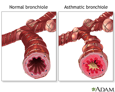
Nursing Paper Example on Understanding Encephalitis: Causes, Symptoms, Treatment, and Patient Care

Nursing Paper Example on Understanding Encephalitis: Causes, Symptoms, Treatment, and Patient Care

Encephalitis, a neurological disorder characterized by inflammation of the brain, affects millions worldwide, making it a significant public health concern. This essay aims to provide a comprehensive overview of encephalitis, focusing on its causes, symptoms, etiology, pathophysiology, DSM-5 diagnosis, treatment regimens, and patient education. Understanding encephalitis is vital due to its diverse etiologies and complex clinical presentations, which necessitate prompt diagnosis and appropriate management. By exploring the intricacies of this disorder, we can enhance our knowledge of its underlying mechanisms and improve therapeutic approaches. With a deeper understanding of encephalitis, healthcare professionals can better support patients in navigating their diagnosis, treatment, and recovery journey. Thus, this essay serves as a valuable resource for healthcare providers and individuals seeking to comprehend and address the challenges posed by encephalitis. (Nursing Paper Example on Understanding Encephalitis: Causes, Symptoms, Treatment, and Patient Care)

Nursing Paper Example on Understanding Encephalitis: Causes, Symptoms, Treatment, and Patient Care

Causes

Encephalitis has various causes, spanning infectious and non-infectious origins. Viral infections are predominant culprits, with herpes simplex virus (HSV) and varicella-zoster virus (VZV) being common offenders. These viruses can directly invade the brain, triggering an inflammatory response and leading to encephalitis. Additionally, other viral agents such as enteroviruses, arboviruses (e.g., West Nile virus), and influenza viruses can also induce encephalitis.

Bacterial infections like Lyme disease, caused by Borrelia burgdorferi, can result in neuroborreliosis, leading to encephalitis in some cases. Moreover, certain parasitic infections, such as toxoplasmosis and malaria, can cause encephalitis through indirect mechanisms.

Non-infectious triggers, including autoimmune reactions, can also contribute to encephalitis. Autoimmune encephalitis occurs when the body’s immune system mistakenly attacks healthy brain tissue, leading to inflammation and neurological dysfunction. Conditions like anti-NMDA receptor encephalitis and autoimmune limbic encephalitis exemplify this mechanism.

Environmental factors such as exposure to toxins or chemicals may also precipitate encephalitis. For instance, exposure to heavy metals like lead or mercury can trigger neuroinflammation, potentially leading to encephalitis.

Furthermore, immunocompromised individuals, such as those with HIV/AIDS or undergoing immunosuppressive therapy, are at higher risk of developing encephalitis due to their weakened immune defenses.

Understanding the diverse array of causes underlying encephalitis is crucial for accurate diagnosis and targeted treatment. Differentiating between infectious and non-infectious etiologies guides clinicians in selecting appropriate therapeutic interventions and optimizing patient outcomes. Thus, a comprehensive understanding of encephalitis causes is essential for effective management and improved prognosis.

Signs and Symptoms

Encephalitis manifests through a spectrum of signs and symptoms, often varying in severity and presentation. The hallmark features typically include fever, headache, and altered mental status, which may range from mild confusion to profound disorientation or coma. These cognitive changes often accompany behavioral alterations, including irritability, agitation, or personality changes.

Nursing Paper Example on Understanding Encephalitis: Causes, Symptoms, Treatment, and Patient Care

Neurological manifestations of encephalitis can encompass a wide array of symptoms, such as seizures, focal neurological deficits, and impaired consciousness. Seizures may manifest as generalized convulsions or focal motor seizures, further complicating the clinical picture.

In some cases, patients may exhibit psychiatric symptoms, including psychosis, hallucinations, or delusions, reflecting the profound impact of encephalitis on brain function. Additionally, movement disorders such as tremors, ataxia, or dyskinesias may arise due to disruption of neural circuits within the basal ganglia or cerebellum.

Furthermore, encephalitis can lead to autonomic dysfunction, presenting as fluctuations in blood pressure, heart rate, or temperature regulation. These autonomic disturbances contribute to the overall clinical complexity of encephalitis and may pose challenges in management.

Children with encephalitis may exhibit distinct symptoms, including irritability, lethargy, poor feeding, or developmental regression. Early recognition of these pediatric-specific signs is critical for prompt diagnosis and intervention.

It is essential to recognize the broad spectrum of signs and symptoms associated with encephalitis, as timely identification facilitates early initiation of appropriate treatment and improves clinical outcomes. Vigilance for subtle neurological changes, particularly in high-risk populations, is paramount for early intervention and preventing potential complications. Thus, a comprehensive understanding of encephalitis symptoms is essential for timely diagnosis and effective management. (Nursing Paper Example on Understanding Encephalitis: Causes, Symptoms, Treatment, and Patient Care)

Etiology

Encephalitis encompasses a diverse etiology, reflecting the multifaceted nature of this neurological disorder. Viral infections stand as predominant etiological factors, with herpes simplex virus (HSV) and varicella-zoster virus (VZV) accounting for a significant proportion of cases. These viruses gain access to the central nervous system (CNS) via neuronal pathways, causing direct neuronal injury and triggering an inflammatory response within the brain.

Other viral agents, including enteroviruses, arboviruses (e.g., West Nile virus), and influenza viruses, can also precipitate encephalitis through various mechanisms. These viruses may disseminate hematogenously or via peripheral nerves, leading to CNS invasion and subsequent neuroinflammation.

Bacterial infections represent another important etiological category, with pathogens such as Borrelia burgdorferi (causing Lyme disease) and Mycobacterium tuberculosis capable of inducing encephalitis. These bacteria may infiltrate the CNS through hematogenous spread or direct extension from adjacent structures, inciting an inflammatory cascade within the brain parenchyma.

Parasitic infections, though less common, can also contribute to encephalitis pathogenesis. Toxoplasma gondii and Plasmodium species (causing malaria) are notable examples, with encephalitis arising secondary to parasitic invasion of the CNS and subsequent inflammatory response.

Non-infectious etiologies, particularly autoimmune reactions, are increasingly recognized as significant contributors to encephalitis. Autoimmune encephalitis encompasses a spectrum of disorders characterized by immune-mediated attacks on neuronal antigens, leading to neuroinflammation and neurological dysfunction.

Furthermore, environmental factors such as exposure to toxins or chemicals may precipitate encephalitis by eliciting neuroinflammatory responses within the brain. Understanding the diverse etiological factors underlying encephalitis is crucial for accurate diagnosis and targeted therapeutic interventions, ultimately improving patient outcomes and guiding preventive strategies. (Nursing Paper Example on Understanding Encephalitis: Causes, Symptoms, Treatment, and Patient Care)

Nursing Paper Example on Understanding Encephalitis: Causes, Symptoms, Treatment, and Patient Care

Pathophysiology

The pathophysiology of encephalitis involves a complex interplay of inflammatory cascades, immune responses, and neuronal dysfunction, culminating in neurological impairment. Viral encephalitis typically begins with viral invasion of the central nervous system (CNS), facilitated by hematogenous dissemination or neuronal spread from peripheral sites of infection. Upon entering the CNS, viruses target neurons, glial cells, and endothelial cells, initiating a robust immune response characterized by cytokine release and activation of resident immune cells.

This inflammatory milieu triggers endothelial cell activation and disruption of the blood-brain barrier (BBB), allowing infiltration of immune cells into the brain parenchyma. Activated microglia and infiltrating macrophages release pro-inflammatory mediators, exacerbating neuroinflammation and promoting neuronal injury.

In addition to direct viral-induced damage, immune-mediated mechanisms contribute to neuronal dysfunction in encephalitis. Autoimmune encephalitis, for instance, results from antibodies targeting neuronal antigens, leading to synaptic dysfunction, neuronal excitotoxicity, and impaired neurotransmission.

The ensuing neuronal injury and inflammation disrupt neural circuits and neurotransmitter pathways, manifesting clinically as altered mental status, seizures, and focal neurological deficits. Moreover, disruption of the BBB facilitates the entry of neurotoxic substances and exacerbates neuroinflammation, further exacerbating neuronal injury.

The pathophysiological processes underlying encephalitis are dynamic and multifactorial, involving a delicate balance between viral replication, immune responses, and neuronal damage. Understanding these mechanisms is crucial for developing targeted therapeutic strategies aimed at mitigating neuroinflammation, preserving neuronal function, and optimizing patient outcomes. Further research into the pathophysiology of encephalitis is warranted to unravel its complexities and identify novel therapeutic targets for intervention. (Nursing Paper Example on Understanding Encephalitis: Causes, Symptoms, Treatment, and Patient Care)

DSM-5 Diagnosis

Diagnosing encephalitis entails a comprehensive evaluation based on clinical presentation, laboratory findings, and neuroimaging studies, as outlined in the Diagnostic and Statistical Manual of Mental Disorders, Fifth Edition (DSM-5). The DSM-5 provides criteria for diagnosing neurocognitive disorders, including those resulting from infectious or inflammatory etiologies such as encephalitis.

The DSM-5 criteria emphasize the presence of significant cognitive decline from a previous level of functioning, which may manifest as impaired memory, executive function, attention, or language skills. Additionally, individuals with encephalitis may exhibit disturbances in consciousness, orientation, perception, or behavior, reflecting the multifaceted nature of the disorder.

Laboratory investigations play a crucial role in confirming the diagnosis of encephalitis. Cerebrospinal fluid (CSF) analysis reveals lymphocytic pleocytosis, elevated protein levels, and sometimes the presence of specific pathogens or antibodies indicative of viral or autoimmune etiologies. Serological tests for viral antibodies and polymerase chain reaction (PCR) assays can identify viral pathogens circulating in the CNS.

Neuroimaging studies, particularly magnetic resonance imaging (MRI) of the brain, may demonstrate characteristic findings such as focal or diffuse signal abnormalities in the affected brain regions. These imaging findings complement clinical and laboratory assessments, aiding in confirming the diagnosis of encephalitis and guiding treatment decisions.

The DSM-5 diagnosis of encephalitis underscores the importance of a multidisciplinary approach involving neurologists, infectious disease specialists, and psychiatrists. By integrating clinical, laboratory, and imaging data, healthcare professionals can accurately diagnose encephalitis and initiate timely interventions to optimize patient outcomes. Moreover, adherence to DSM-5 criteria ensures consistency and precision in diagnosing encephalitis across diverse clinical settings, facilitating effective management and support for affected individuals. (Nursing Paper Example on Understanding Encephalitis: Causes, Symptoms, Treatment, and Patient Care)

Treatment Regimens and Patient Education

Treatment of encephalitis necessitates a multifaceted approach aimed at addressing the underlying cause, managing symptoms, and preventing complications. Antiviral therapy is the mainstay of treatment for viral encephalitis, targeting specific viral pathogens such as herpes simplex virus (HSV) or varicella-zoster virus (VZV). Intravenous administration of antiviral medications like acyclovir or ganciclovir is initiated promptly upon suspicion of viral encephalitis to mitigate viral replication and reduce neuronal damage.

In cases of bacterial encephalitis, antibiotic therapy targeting the causative pathogen is imperative. Prompt initiation of antibiotics like ceftriaxone or penicillin G is crucial to combat bacterial invasion of the central nervous system (CNS) and prevent systemic complications.

In autoimmune encephalitis, immunomodulatory therapy aims to suppress the aberrant immune response directed against neuronal antigens. Treatment may involve corticosteroids, intravenous immunoglobulin (IVIG), or immunosuppressive agents like rituximab or cyclophosphamide to attenuate neuroinflammation and preserve neuronal function.

Supportive care plays a pivotal role in managing complications and promoting recovery in patients with encephalitis. This includes close monitoring of vital signs, maintenance of adequate hydration, seizure management, and addressing nutritional needs. In severe cases with neurological sequelae, rehabilitation therapy may be necessary to optimize functional outcomes and enhance quality of life.

Patient education is integral to the management of encephalitis, empowering individuals and their caregivers to navigate the challenges associated with the condition. Patients should be educated about the importance of adhering to prescribed medications, attending follow-up appointments, and recognizing warning signs of disease progression or relapse. Furthermore, patients and caregivers should receive guidance on strategies to optimize brain health, including adequate rest, nutrition, and cognitive stimulation.

Moreover, raising awareness about encephalitis within the community is crucial to promoting early recognition and timely intervention. Educational initiatives aimed at healthcare providers, schools, and the general public can facilitate prompt referral to specialized care centers and improve outcomes for individuals affected by encephalitis.

The treatment of encephalitis involves a comprehensive approach encompassing specific antiviral or antibiotic therapy, immunomodulatory interventions, and supportive care measures. Patient education is paramount in empowering individuals and caregivers to actively participate in disease management and promote optimal outcomes. By addressing both the medical and educational aspects of encephalitis, healthcare providers can enhance the quality of care and support for affected individuals and their families. (Nursing Paper Example on Understanding Encephalitis: Causes, Symptoms, Treatment, and Patient Care)

Conclusion

Encephalitis is a complex neurological disorder with diverse causes, symptoms, and treatment regimens. This essay has provided a comprehensive overview, emphasizing the importance of understanding its multifaceted etiology, including viral, bacterial, autoimmune, and environmental factors. The discussion of signs and symptoms underscored the varied clinical presentations, highlighting the need for early recognition and intervention. Additionally, the exploration of pathophysiology elucidated the intricate interplay of inflammatory processes and neuronal dysfunction underlying encephalitis. The DSM-5 diagnosis section outlined the criteria for accurate identification, guiding clinicians in diagnostic evaluation and treatment planning. Furthermore, the treatment regimens and patient education section emphasized the multidisciplinary approach to management, incorporating antiviral, antibiotic, and immunomodulatory therapies alongside supportive care measures. By addressing both the medical and educational aspects, healthcare providers can optimize outcomes for individuals affected by encephalitis, underscoring the importance of timely intervention and comprehensive support. (Nursing Paper Example on Understanding Encephalitis: Causes, Symptoms, Treatment, and Patient Care)

References

https://www.ncbi.nlm.nih.gov/books/NBK470162/

 
Do you need a similar assignment done for you from scratch? Order now!
Use Discount Code "Newclient" for a 15% Discount!

Nursing Paper Example on Septicemia: A Neurological Disorder

Nursing Paper Example on Septicemia: A Neurological Disorder

Septicemia, also known as sepsis, is a critical neurological disorder that arises from the body’s exaggerated response to infection. It is a life-threatening condition that demands immediate medical attention due to its potential to cause severe complications and mortality. This disorder occurs when pathogens, such as bacteria, viruses, or fungi, enter the bloodstream, triggering a systemic inflammatory response. Despite advances in medical science, septicemia remains a significant public health concern globally, contributing to a substantial burden of morbidity and mortality. Understanding the causes, signs, and symptoms, as well as the etiology and pathophysiology of septicemia, is crucial for effective diagnosis and management. This paper explores the multifaceted aspects of septicemia, including its causes, clinical manifestations, diagnostic criteria, treatment regimens, patient education, and concludes with insights into ongoing challenges and future directions in managing this neurological disorder. (Nursing Paper Example on Septicemia: A Neurological Disorder)

Nursing Paper Example on Septicemia: A Neurological Disorder

Causes of Septicemia

Septicemia stems from various infections infiltrating the bloodstream, leading to a systemic inflammatory response. Bacterial infections are the primary culprits, with gram-positive bacteria like Staphylococcus aureus and Streptococcus pneumoniae being common offenders. Gram-negative bacteria such as Escherichia coli and Pseudomonas aeruginosa also contribute significantly to septicemia cases. Additionally, viral infections, including influenza and herpes, and fungal infections like Candida albicans can provoke septicemia, albeit less frequently.

The source of infection varies, encompassing a spectrum of conditions ranging from respiratory tract infections like pneumonia and urinary tract infections to abdominal infections such as appendicitis and peritonitis. Even seemingly innocuous skin infections, if not adequately treated, can escalate into septicemia.

Moreover, invasive medical procedures and devices, such as urinary catheters, intravenous lines, and surgical interventions, pose a risk of introducing pathogens into the bloodstream, precipitating septicemia. Immunocompromised individuals, including those with HIV/AIDS, cancer undergoing chemotherapy, or recipients of organ transplants, are particularly susceptible to developing septicemia due to their compromised immune systems.

Furthermore, certain underlying medical conditions can predispose individuals to septicemia. Chronic diseases like diabetes, kidney disease, and liver cirrhosis impair the body’s ability to fight infections, making affected individuals more prone to developing septicemia.

Septicemia arises from diverse sources of infection, predominantly bacterial, but also viral and fungal. Respiratory, urinary, and abdominal infections are common origins, along with compromised skin barriers and invasive medical procedures. Additionally, underlying medical conditions and immunocompromised states increase susceptibility to septicemia. Understanding these multifaceted causes is vital for early recognition, prompt treatment, and effective management of this neurological disorder. (Nursing Paper Example on Septicemia: A Neurological Disorder)

Signs and Symptoms

Septicemia manifests through a constellation of signs and symptoms, reflecting the body’s systemic inflammatory response to infection. The presentation can vary widely, ranging from subtle early indicators to severe, life-threatening manifestations.

Nursing Paper Example on Septicemia: A Neurological Disorder

Early signs often include fever, characterized by an elevated body temperature above 100.4°F (38°C), accompanied by chills and rigors. The heart rate accelerates, a condition known as tachycardia, as the body attempts to compensate for decreased blood pressure. Similarly, rapid breathing, or tachypnea, occurs in response to the increased metabolic demands and decreased oxygen levels.

As septicemia progresses, patients may experience altered mental status, ranging from confusion and disorientation to lethargy and coma. This neurological impairment stems from inadequate oxygen delivery to the brain due to compromised blood flow.

The circulatory system undergoes significant changes, leading to low blood pressure, or hypotension, which can manifest as dizziness, light-headedness, and fainting. Additionally, peripheral vasoconstriction occurs, causing cool extremities and reduced urine output due to decreased renal perfusion.

Furthermore, patients may exhibit gastrointestinal symptoms such as nausea, vomiting, and abdominal pain. The liver and spleen may become enlarged as part of the immune response, contributing to discomfort in the upper abdomen.

In severe cases, septicemia progresses to septic shock, characterized by profound hypotension and organ dysfunction, including acute kidney injury, liver failure, and respiratory failure. Septic shock is a medical emergency requiring immediate intervention to prevent irreversible organ damage and death.

The signs and symptoms of septicemia encompass a wide array of manifestations, including fever, tachycardia, altered mental status, hypotension, gastrointestinal symptoms, and ultimately, septic shock. Recognizing these clinical features promptly is essential for initiating timely treatment and improving patient outcomes in this neurological disorder. (Nursing Paper Example on Septicemia: A Neurological Disorder)

Etiology of Septicemia

The etiology of septicemia revolves around the intricate interplay between infectious agents, the immune system, and various predisposing factors. Septicemia primarily originates from bacterial, viral, or fungal infections infiltrating the bloodstream, triggering a dysregulated immune response.

Bacterial infections are the most common etiological agents of septicemia. Gram-positive bacteria, including Staphylococcus aureus and Streptococcus pneumoniae, are frequently implicated, along with gram-negative bacteria like Escherichia coli and Pseudomonas aeruginosa. These pathogens possess virulence factors that enable them to evade host defenses and disseminate into the bloodstream, initiating the cascade of events leading to septicemia.

Viral infections, although less common, can also precipitate septicemia. Influenza viruses, herpes simplex viruses, and human immunodeficiency virus (HIV) are among the viral pathogens associated with septicemia. These viruses can directly infect immune cells or induce a cytokine storm, exacerbating the systemic inflammatory response.

Fungal infections, particularly those caused by Candida species, represent another etiological factor contributing to septicemia, especially in immunocompromised individuals. Candida albicans, in particular, can colonize indwelling medical devices like urinary catheters and intravenous lines, serving as a nidus for bloodstream invasion.

Moreover, certain host factors predispose individuals to septicemia. Immunocompromised states, such as HIV/AIDS, cancer chemotherapy, and immunosuppressive therapy post-organ transplantation, impair the body’s ability to mount an effective immune response against invading pathogens. Additionally, chronic medical conditions like diabetes mellitus, chronic kidney disease, and liver cirrhosis compromise host defenses, increasing susceptibility to septicemia.

The etiology of septicemia encompasses various infectious agents, primarily bacteria, followed by viruses and fungi. Understanding these underlying factors is crucial for targeted interventions aimed at preventing, diagnosing, and managing septicemia in this neurological disorder. (Nursing Paper Example on Septicemia: A Neurological Disorder)

Pathophysiology of Septicemia

The pathophysiology of septicemia involves a complex cascade of events orchestrated by the host immune system in response to invading pathogens in the bloodstream. This dysregulated immune response leads to widespread inflammation and organ dysfunction, culminating in the clinical manifestations of septicemia.

The initial phase of septicemia begins with the invasion of pathogens into the bloodstream, often originating from localized infections in various body sites. These pathogens release pathogen-associated molecular patterns (PAMPs) and toxins, triggering the activation of pattern recognition receptors (PRRs) on immune cells such as macrophages and neutrophils.

Subsequently, a robust immune response ensues, characterized by the release of pro-inflammatory cytokines, including tumor necrosis factor-alpha (TNF-α), interleukin-1 (IL-1), and interleukin-6 (IL-6). These cytokines amplify the inflammatory cascade, recruiting more immune cells to the site of infection and promoting vascular permeability.

The ensuing endothelial dysfunction and increased vascular permeability lead to systemic microvascular leakage, impairing tissue perfusion and oxygen delivery. Concurrently, activation of the coagulation cascade occurs, resulting in disseminated intravascular coagulation (DIC), a hallmark feature of severe sepsis.

As septicemia progresses, the dysregulated immune response transitions from a pro-inflammatory to an anti-inflammatory state, characterized by the release of anti-inflammatory cytokines like interleukin-10 (IL-10). This immunosuppressive phase contributes to immune paralysis and secondary infections, further exacerbating organ dysfunction.

Ultimately, the combined effects of widespread inflammation, microvascular dysfunction, coagulopathy, and immunosuppression culminate in multi-organ dysfunction syndrome (MODS) and septic shock. This life-threatening condition requires prompt recognition and aggressive management to mitigate organ damage and improve patient outcomes in septicemia, a critical neurological disorder. (Nursing Paper Example on Septicemia: A Neurological Disorder)

DMS-5 Diagnosis

The Diagnostic and Statistical Manual of Mental Disorders, Fifth Edition (DSM-5), does not explicitly categorize septicemia as a neurological disorder. However, it recognizes the cognitive and neurological manifestations that may occur in severe cases of sepsis, a condition closely related to septicemia.

In the DSM-5, sepsis-related encephalopathy is characterized by alterations in consciousness, attention, cognition, or perception resulting from sepsis-induced systemic inflammation. These cognitive changes can range from mild confusion and disorientation to delirium, coma, and even death in severe cases.

The diagnosis of sepsis-related encephalopathy is typically made based on clinical assessment, which includes evaluating the patient’s level of consciousness, cognitive function, and neurological signs. Laboratory tests, such as blood cultures to identify the causative pathogen and inflammatory markers like C-reactive protein (CRP) and procalcitonin, may support the diagnosis.

Neuroimaging studies, such as computed tomography (CT) or magnetic resonance imaging (MRI) of the brain, may be performed to rule out other neurological conditions or identify complications of sepsis, such as cerebral edema or infarction.

Additionally, electroencephalography (EEG) may be utilized to assess for abnormal electrical activity in the brain, which can occur in severe cases of sepsis-related encephalopathy.

Overall, while septicemia itself is not a formal diagnosis in the DSM-5, the cognitive and neurological sequelae of sepsis-related encephalopathy are recognized within the diagnostic framework of the manual. Early recognition and appropriate management of sepsis-related encephalopathy are crucial for optimizing patient outcomes in this neurological disorder. (Nursing Paper Example on Septicemia: A Neurological Disorder)

Treatment Regimens and Patient Education

The management of septicemia requires a comprehensive approach aimed at eradicating the underlying infection, stabilizing hemodynamics, and providing supportive care to prevent organ dysfunction and complications. Additionally, patient education plays a crucial role in empowering individuals to recognize early signs of infection, seek prompt medical attention, and adhere to prescribed treatment regimens.

Treatment Regimens:

  1. Antibiotic Therapy: Prompt initiation of broad-spectrum antibiotics is paramount in treating septicemia. Empirical antibiotic therapy is initiated based on the suspected source of infection and local antimicrobial resistance patterns. Once the causative pathogen is identified through blood cultures, antibiotic therapy is adjusted accordingly to target the specific organism.
  2. Fluid Resuscitation: Intravenous fluid administration is essential to restore intravascular volume and improve tissue perfusion. Balanced crystalloids are preferred for fluid resuscitation, while caution is exercised to avoid fluid overload, especially in patients with pre-existing cardiac or renal conditions.
  3. Vasopressor Therapy: In patients with persistent hypotension despite fluid resuscitation, vasopressor agents such as norepinephrine or vasopressin may be administered to maintain adequate mean arterial pressure and tissue perfusion.
  4. Supportive Care: Patients with septicemia often require intensive care unit (ICU) admission for close monitoring and supportive care. This may include mechanical ventilation for respiratory support, renal replacement therapy for acute kidney injury, and monitoring of hemodynamic parameters.
  5. Source Control: Surgical intervention may be necessary to remove the source of infection, such as drainage of abscesses or debridement of infected tissue.

Patient Education:

  1. Recognition of Symptoms: Educating patients about the signs and symptoms of infection, including fever, chills, rapid heart rate, and confusion, enables early recognition and timely medical intervention.
  2. Importance of Antibiotic Adherence: Emphasizing the importance of completing the full course of antibiotics as prescribed to eradicate the infection and prevent recurrence or antibiotic resistance.
  3. Follow-Up Care: Encouraging patients to follow up with healthcare providers for ongoing monitoring of their condition, including repeat blood cultures and assessment of organ function.
  4. Preventive Measures: Advising patients on preventive measures to reduce the risk of infection, such as hand hygiene, vaccination, and avoiding known sources of infection.
  5. Awareness of Complications: Educating patients about the potential complications of septicemia, including organ dysfunction and long-term sequelae, promotes early recognition of worsening symptoms and prompt medical intervention.

A multidisciplinary approach to the treatment of septicemia, including antibiotic therapy, fluid resuscitation, and supportive care, is essential for optimizing patient outcomes. Equally important is patient education, which empowers individuals to recognize symptoms, adhere to treatment regimens, and adopt preventive measures to mitigate the risk of recurrent infections in this neurological disorder. (Nursing Paper Example on Septicemia: A Neurological Disorder)

Conclusion

Septicemia, a neurological disorder triggered by systemic infection, presents a significant medical challenge requiring prompt recognition and intervention. This essay has highlighted the multifaceted nature of septicemia, exploring its causes, signs and symptoms, etiology, pathophysiology, DSM-5 diagnosis, treatment regimens, and patient education. By elucidating the complex interplay between infectious agents, immune responses, and predisposing factors, a deeper understanding of septicemia’s pathophysiology emerges. Moreover, the importance of early recognition and aggressive management, including antibiotic therapy, fluid resuscitation, and supportive care, cannot be overstated. Furthermore, patient education plays a crucial role in empowering individuals to recognize symptoms, adhere to treatment regimens, and adopt preventive measures. Through a comprehensive approach encompassing both medical interventions and patient education, healthcare professionals can effectively manage septicemia, thereby improving patient outcomes and reducing the burden of this neurological disorder. (Nursing Paper Example on Septicemia: A Neurological Disorder)

References

https://www.ncbi.nlm.nih.gov/books/NBK537054/

 
Do you need a similar assignment done for you from scratch? Order now!
Use Discount Code "Newclient" for a 15% Discount!
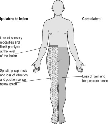
Nursing Paper Example on Myasthenia Gravis: Understanding a Neurological Disorder

Nursing Paper Example on Myasthenia Gravis: Understanding a Neurological Disorder

Myasthenia Gravis (MG) stands as a perplexing neurological ailment, disrupting neuromuscular communication and inducing muscle weakness. This chronic condition poses significant challenges to those affected, impacting their daily activities and overall quality of life. Despite its prevalence, MG remains enigmatic in terms of its precise etiology and optimal management strategies. Understanding this disorder is essential, not only for healthcare professionals but also for patients and their caregivers, to navigate its complexities effectively. In this essay, we delve into the intricacies of Myasthenia Gravis, exploring its causes, signs and symptoms, etiology, pathophysiology, DSM-5 diagnosis, treatment regimens, and the crucial aspect of patient education. By comprehensively examining each aspect, we aim to shed light on this condition, offering insights into its management and fostering a better understanding among those affected and the broader community. (Nursing Paper Example on Myasthenia Gravis: Understanding a Neurological Disorder)

Nursing Paper Example on Myasthenia Gravis: Understanding a Neurological Disorder

Causes

The exact cause of Myasthenia Gravis (MG) remains elusive, but it is primarily attributed to autoimmune dysfunction. In MG, the immune system mistakenly targets the acetylcholine receptors at neuromuscular junctions, disrupting the transmission of signals between nerves and muscles. This autoimmune response leads to the production of antibodies that either block or destroy these receptors, impairing their function.

Several factors may contribute to the development of MG. Genetic predisposition plays a role, as individuals with a family history of autoimmune disorders are more susceptible. Additionally, environmental factors such as infections, particularly respiratory or viral illnesses, can trigger the onset or exacerbation of MG symptoms.

Furthermore, certain medications, such as antibiotics, quinidine, and beta-blockers, have been associated with the development of MG or worsening of symptoms in individuals predisposed to the disorder. Moreover, hormonal changes, such as those occurring during pregnancy or menstrual cycles, may influence MG symptoms.

Thymus gland abnormalities are also linked to MG, with around 15% of individuals with MG having a thymoma (tumor of the thymus gland), and a higher percentage showing thymic hyperplasia (enlargement of the thymus gland). The thymus gland plays a role in the development and maturation of certain immune cells, and its abnormalities are thought to contribute to the autoimmune response seen in MG.

While these factors are associated with the development or exacerbation of MG, the precise interplay between genetic predisposition, environmental triggers, and immune system dysfunction remains the subject of ongoing research. Understanding the underlying causes of MG is crucial for developing targeted therapeutic approaches and improving outcomes for individuals affected by this debilitating neurological disorder. (Nursing Paper Example on Myasthenia Gravis: Understanding a Neurological Disorder)

Signs and Symptoms

Myasthenia Gravis (MG) manifests through a variety of signs and symptoms, primarily stemming from muscle weakness and fatigue. These symptoms can vary widely among individuals and may fluctuate in severity over time.

One of the hallmark symptoms of MG is muscle weakness, which typically worsens with activity and improves with rest. This weakness commonly affects muscles that control eye movements and eyelid function, leading to drooping eyelids (ptosis) and double vision (diplopia). Ptosis can impair vision and cause discomfort, while diplopia can significantly impact daily activities such as reading and driving.

Nursing Paper Example on Myasthenia Gravis: Understanding a Neurological Disorder

Additionally, individuals with MG may experience weakness in other facial muscles, leading to difficulty in facial expressions, chewing, and swallowing. Weakness in the muscles of the throat and neck can result in dysphagia (difficulty swallowing) and dysarthria (difficulty speaking), which can affect nutrition and communication.

Muscle weakness in MG is not limited to the face and neck but can also affect the limbs, leading to difficulty in activities such as lifting objects, climbing stairs, or walking long distances. The weakness tends to be more pronounced after repetitive use of muscles and may improve with rest.

Fatigue is another common symptom of MG, often accompanying muscle weakness. This fatigue can be both physical and mental, impacting activities that require sustained effort or concentration.

In some cases, MG can also affect respiratory muscles, leading to respiratory distress, shortness of breath, or difficulty breathing, particularly during physical exertion or at night.

Overall, the signs and symptoms of MG can significantly impair daily functioning and quality of life. Prompt recognition and management of these symptoms are crucial for improving outcomes and enhancing the well-being of individuals living with this condition. (Nursing Paper Example on Myasthenia Gravis: Understanding a Neurological Disorder)

Etiology

Myasthenia Gravis (MG) is primarily characterized by autoimmune dysfunction, where the body’s immune system erroneously targets components of the neuromuscular junction, leading to muscle weakness. The exact etiology of MG remains elusive, but several factors contribute to its development and progression.

Genetic predisposition plays a significant role in the etiology of MG, as individuals with a family history of autoimmune disorders are more susceptible to developing the condition. Certain genetic variations may predispose individuals to an abnormal immune response against the neuromuscular junction components, increasing their risk of MG.

Environmental factors also contribute to the etiology of MG. Infections, particularly respiratory or viral illnesses, have been implicated as potential triggers for the onset or exacerbation of MG symptoms. These infections can stimulate the immune system and precipitate an autoimmune response against the neuromuscular junction.

Furthermore, certain medications have been associated with the development or worsening of MG symptoms. Antibiotics, quinidine, and beta-blockers are among the medications that can exacerbate MG or trigger its onset, especially in individuals predisposed to the disorder.

Thymus gland abnormalities are also linked to the etiology of MG. Approximately 15% of individuals with MG have a thymoma (tumor of the thymus gland), while a higher percentage exhibit thymic hyperplasia (enlargement of the thymus gland). The thymus gland plays a crucial role in the maturation of immune cells, and abnormalities in this gland are thought to contribute to the autoimmune response seen in MG.

Overall, the etiology of MG is multifactorial, involving a complex interplay between genetic predisposition, environmental triggers, and immune system dysfunction. Understanding these factors is essential for elucidating the underlying mechanisms of MG and developing targeted therapeutic approaches to improve outcomes for affected individuals. (Nursing Paper Example on Myasthenia Gravis: Understanding a Neurological Disorder)

Pathophysiology

Myasthenia Gravis (MG) arises from a disruption in neuromuscular transmission due to autoimmune dysfunction. The pathophysiology of MG involves an intricate interplay of immune-mediated processes that target the neuromuscular junction (NMJ), impairing communication between nerves and muscles.

In MG, the immune system produces autoantibodies, primarily immunoglobulin G (IgG), that target key components of the NMJ, particularly the acetylcholine receptors (AChR). These autoantibodies bind to AChR, leading to receptor blockade or destruction through complement-mediated mechanisms. Consequently, the binding of acetylcholine (ACh) to its receptors on the postsynaptic membrane is inhibited, disrupting the transmission of nerve impulses across the NMJ.

Additionally, some individuals with MG produce autoantibodies against muscle-specific kinase (MuSK), another protein crucial for NMJ function. These MuSK autoantibodies interfere with signaling pathways involved in AChR clustering and maintenance, further exacerbating neuromuscular dysfunction.

The resulting reduction in AChR density and impaired AChR function at the NMJ leads to muscle weakness and fatigue, characteristic of MG. The severity of symptoms correlates with the extent of AChR loss and dysfunction, as well as the degree of NMJ remodeling.

Furthermore, the thymus gland plays a significant role in the pathophysiology of MG, particularly in individuals with thymoma or thymic hyperplasia. Abnormalities in the thymus gland, such as follicular hyperplasia or germinal center formation, contribute to the breakdown of self-tolerance and the generation of autoreactive T cells, perpetuating the autoimmune response against the NMJ.

Overall, the pathophysiology of MG involves a complex cascade of immune-mediated events that disrupt neuromuscular transmission, resulting in muscle weakness and fatigue. Understanding these underlying mechanisms is crucial for developing targeted therapies to alleviate symptoms and improve outcomes for individuals with MG. (Nursing Paper Example on Myasthenia Gravis: Understanding a Neurological Disorder)

DSM-5 Diagnosis

Diagnosing Myasthenia Gravis (MG) involves a comprehensive evaluation based on clinical presentation, electromyography (EMG), serological tests, and imaging studies. While the Diagnostic and Statistical Manual of Mental Disorders, Fifth Edition (DSM-5), primarily focuses on mental health conditions, the diagnosis of MG typically follows guidelines established by medical associations and neurology societies.

Clinical assessment is fundamental in diagnosing MG, with emphasis on characteristic symptoms such as muscle weakness, ptosis, diplopia, dysphagia, and dysarthria. The fluctuating nature of symptoms and their exacerbation with activity aid in distinguishing MG from other neuromuscular disorders.

Electromyography (EMG) plays a crucial role in confirming the diagnosis of MG by assessing neuromuscular transmission. EMG findings typically reveal a decremental response to repetitive nerve stimulation, reflecting impaired neuromuscular transmission characteristic of MG.

Serological tests are utilized to detect autoantibodies against acetylcholine receptors (AChR) or muscle-specific kinase (MuSK), which are present in the majority of individuals with MG. A positive serological result, in conjunction with clinical findings, further supports the diagnosis of MG.

Imaging studies, particularly computed tomography (CT) or magnetic resonance imaging (MRI) of the chest, are performed to assess the thymus gland in individuals suspected of having thymoma or thymic hyperplasia associated with MG.

Overall, the diagnosis of MG is based on a combination of clinical features, electromyography findings, serological tests for autoantibodies, and imaging studies to evaluate thymic abnormalities. A comprehensive diagnostic approach ensures accurate identification of MG, enabling timely initiation of appropriate management strategies to optimize patient outcomes. (Nursing Paper Example on Myasthenia Gravis: Understanding a Neurological Disorder)

Treatment Regimens and Patient Education

Management of Myasthenia Gravis (MG) involves a multifaceted approach aimed at alleviating symptoms, improving neuromuscular transmission, and suppressing the autoimmune response. Additionally, patient education plays a crucial role in empowering individuals with MG to actively participate in their care and optimize treatment outcomes.

Treatment Regimens:

  1. Medications: Pharmacotherapy is the cornerstone of MG management. Acetylcholinesterase inhibitors such as pyridostigmine improve neuromuscular transmission by inhibiting the breakdown of acetylcholine. These medications help alleviate muscle weakness and fatigue in many individuals with MG.
  2. Immunosuppressants: For individuals with moderate to severe MG or those who do not respond adequately to acetylcholinesterase inhibitors, immunosuppressive agents such as corticosteroids, azathioprine, mycophenolate mofetil, or rituximab may be prescribed. These medications help suppress the autoimmune response, thereby reducing the production of autoantibodies and mitigating disease progression.
  3. Plasmapheresis and Intravenous Immunoglobulin (IVIg): In acute exacerbations of MG or when rapid symptom relief is needed, plasmapheresis or IVIg therapy may be employed to remove circulating autoantibodies and modulate immune function, respectively.
  4. Thymectomy: Surgical removal of the thymus gland (thymectomy) is recommended for individuals with thymoma or thymic hyperplasia associated with MG. Thymectomy may result in disease remission or reduction in disease severity, particularly in younger individuals with early-stage MG.
  5. Symptomatic Management: Adjunctive therapies such as ocular lubricants for dry eyes, speech therapy for dysarthria, and dietary modifications for dysphagia may be recommended to address specific symptoms and improve quality of life.

Patient Education:

  1. Medication Adherence: Patients must understand the importance of adhering to their prescribed medication regimen, including dosing schedules and potential side effects.
  2. Recognition of Exacerbations: Educating patients about the signs and symptoms of MG exacerbations, such as worsening muscle weakness or respiratory distress, enables prompt medical intervention and prevents complications.
  3. Avoidance of Triggers: Patients should be advised to avoid factors known to exacerbate MG symptoms, such as stress, fatigue, certain medications, and infections.
  4. Regular Follow-up: Encouraging patients to attend regular follow-up appointments with their healthcare providers ensures ongoing monitoring of disease progression and treatment efficacy.
  5. Lifestyle Modifications: Providing guidance on energy conservation strategies, adaptive devices, and support services helps individuals with MG optimize their daily activities and maintain independence.

By employing a comprehensive treatment approach and empowering patients with education and support, the management of Myasthenia Gravis can be tailored to individual needs, leading to improved symptom control and enhanced quality of life. (Nursing Paper Example on Myasthenia Gravis: Understanding a Neurological Disorder)

Conclusion

Myasthenia Gravis (MG) presents a complex clinical challenge, characterized by muscle weakness and fatigue due to autoimmune dysfunction at the neuromuscular junction. In this essay, we explored the multifaceted aspects of MG, including its causes, signs and symptoms, etiology, pathophysiology, DSM-5 diagnosis, treatment regimens, and patient education. By employing a concise yet comprehensive approach, we elucidated the intricate interplay of immune-mediated processes underlying MG and highlighted the importance of accurate diagnosis and tailored treatment strategies. Furthermore, the inclusion of patient education as a pivotal component of MG management emphasizes the significance of empowering individuals with knowledge and support to actively participate in their care. Overall, by understanding the complexities of MG and implementing a holistic approach encompassing both medical interventions and patient education, healthcare providers can optimize outcomes and enhance the quality of life for individuals living with this challenging neurological disorder.(Nursing Paper Example on Myasthenia Gravis: Understanding a Neurological Disorder)

References

https://www.ncbi.nlm.nih.gov/books/NBK559331/

 
Do you need a similar assignment done for you from scratch? Order now!
Use Discount Code "Newclient" for a 15% Discount!