Nursing Paper Example on Amyotrophic Lateral Sclerosis (ALS): Understanding a Devastating Neurological Disorder

Nursing Paper Example on Amyotrophic Lateral Sclerosis (ALS): Understanding a Devastating Neurological Disorder

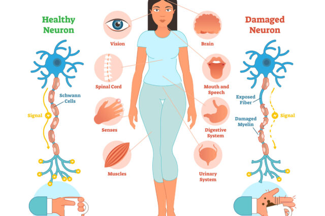
Amyotrophic Lateral Sclerosis (ALS), commonly known as Lou Gehrig’s disease, is a progressive neurological disorder that affects the nerve cells responsible for controlling voluntary muscle movements. This condition, first described by French neurologist Jean-Martin Charcot in the 19th century, is characterized by the gradual degeneration of motor neurons in the brain and spinal cord. As these neurons deteriorate, individuals with ALS experience muscle weakness, twitching, and eventually paralysis, ultimately leading to respiratory failure and death within a few years of diagnosis. Despite extensive research, the exact cause of ALS remains elusive, with both genetic and environmental factors implicated in its development. ALS poses significant challenges for patients, caregivers, and healthcare professionals due to its devastating impact on physical function and quality of life. In this paper, we will explore the causes, signs and symptoms, etiology, pathophysiology, DSM-5 diagnosis, treatment regimens, patient education, and implications of ALS, aiming to deepen our understanding of this debilitating neurological condition. (Nursing Paper Example on Amyotrophic Lateral Sclerosis (ALS): Understanding a Devastating Neurological Disorder)

Nursing Paper Example on Amyotrophic Lateral Sclerosis (ALS): Understanding a Devastating Neurological Disorder

Causes

The exact cause of Amyotrophic Lateral Sclerosis (ALS) remains a subject of ongoing research, but it is widely accepted that a combination of genetic and environmental factors contributes to its development.

Genetic Factors: Approximately 5-10% of ALS cases are familial, meaning they are inherited from a parent who carries a specific genetic mutation associated with the disease. Several genes have been identified as potential contributors to familial ALS, including SOD1, C9orf72, TARDBP, and FUS. Mutations in these genes disrupt normal cellular processes, leading to the degeneration of motor neurons and the progression of ALS symptoms.

Environmental Factors: While the majority of ALS cases are sporadic, meaning they occur without a clear familial link, environmental factors may still play a role in their development. Exposure to certain toxins and chemicals, such as lead, mercury, pesticides, and heavy metals, has been associated with an increased risk of ALS. Additionally, trauma or injury to the central nervous system, such as head injuries or repetitive physical activities, may also contribute to the onset of ALS symptoms in some individuals.

Gene-Environment Interactions: Emerging evidence suggests that interactions between genetic predisposition and environmental exposures may influence the development of ALS. For example, individuals with specific genetic mutations may be more susceptible to the effects of environmental toxins, leading to an increased risk of developing ALS. Conversely, environmental factors may trigger or exacerbate the progression of ALS symptoms in genetically predisposed individuals.

While the exact cause of ALS remains elusive, research into the interplay between genetic and environmental factors continues to provide valuable insights into the pathogenesis of the disease. By understanding the complex interactions that contribute to ALS development, researchers hope to identify novel therapeutic targets and strategies for the prevention and treatment of this devastating neurological disorder. (Nursing Paper Example on Amyotrophic Lateral Sclerosis (ALS): Understanding a Devastating Neurological Disorder)

Nursing Paper Example on Amyotrophic Lateral Sclerosis (ALS): Understanding a Devastating Neurological Disorder

Signs and Symptoms

Amyotrophic Lateral Sclerosis (ALS) is characterized by a progressive deterioration of motor function, resulting in a range of signs and symptoms that impact voluntary muscle movements. The onset of ALS is often insidious, with symptoms gradually worsening over time.

Muscle Weakness: One of the earliest signs of ALS is muscle weakness, which typically begins in the limbs and gradually spreads to other parts of the body. Initial weakness may manifest as difficulty performing tasks that require fine motor control, such as buttoning a shirt or holding objects.

Muscle Twitching and Cramping: Individuals with ALS often experience muscle twitching, known as fasciculations, and muscle cramping, particularly in the arms and legs. These involuntary muscle contractions may be a source of discomfort and contribute to muscle fatigue.

Difficulty Speaking and Swallowing: As ALS progresses, weakness in the muscles involved in speech and swallowing becomes more pronounced. Speech may become slurred or difficult to understand, and individuals may have trouble swallowing food and liquids, leading to choking or aspiration.

Respiratory Symptoms: ALS eventually affects the muscles responsible for breathing, leading to respiratory symptoms such as shortness of breath, difficulty breathing, and frequent respiratory infections. Respiratory muscle weakness is a significant complication of ALS and often requires interventions such as mechanical ventilation to support breathing.

Muscle Atrophy: As motor neurons degenerate in ALS, affected muscles undergo atrophy, or shrinkage, due to disuse and loss of nerve stimulation. Muscle atrophy contributes to weakness and can lead to visible changes in muscle size and shape.

Progressive Disability: Over time, the combination of muscle weakness, atrophy, and respiratory compromise results in progressive disability in individuals with ALS. Once routine tasks become increasingly challenging, and eventually, individuals may require assistance with activities of daily living.

The signs and symptoms of ALS vary from person to person but generally involve progressive muscle weakness, twitching, difficulty speaking and swallowing, respiratory impairment, and muscle atrophy. Early recognition of these symptoms is crucial for timely diagnosis and intervention to improve the quality of life for individuals living with ALS. (Nursing Paper Example on Amyotrophic Lateral Sclerosis (ALS): Understanding a Devastating Neurological Disorder)

Etiology

Amyotrophic Lateral Sclerosis (ALS) is a complex neurological disorder with a multifactorial etiology involving genetic, environmental, and potential gene-environment interactions.
Nursing Paper Example on Amyotrophic Lateral Sclerosis (ALS): Understanding a Devastating Neurological Disorder

Genetic Factors: A subset of ALS cases, approximately 5-10%, are familial, meaning they have a clear genetic component. Mutations in several genes have been identified as contributing to familial ALS, including SOD1, C9orf72, TARDBP, and FUS. These genetic mutations disrupt normal cellular processes, leading to the degeneration of motor neurons and the progression of ALS symptoms. Although familial ALS accounts for a relatively small proportion of cases, the study of familial ALS has provided valuable insights into the underlying mechanisms of the disease.

Environmental Factors: While the majority of ALS cases are sporadic, meaning they occur without a clear familial link, environmental factors may still play a role in disease development. Exposure to certain toxins and chemicals, such as lead, mercury, pesticides, and heavy metals, has been associated with an increased risk of ALS. Additionally, trauma or injury to the central nervous system, such as head injuries or repetitive physical activities, may also contribute to the onset of ALS symptoms in some individuals.

Gene-Environment Interactions: Emerging evidence suggests that interactions between genetic predisposition and environmental exposures may influence the development of ALS. For example, individuals with specific genetic mutations may be more susceptible to the effects of environmental toxins, leading to an increased risk of developing ALS. Conversely, environmental factors may trigger or exacerbate the progression of ALS symptoms in genetically predisposed individuals.

The etiology of ALS is complex and multifaceted, involving a combination of genetic and environmental factors, as well as potential gene-environment interactions. Understanding the interplay between these factors is crucial for unraveling the underlying mechanisms of ALS and identifying novel therapeutic targets for the prevention and treatment of this devastating neurological disorder. (Nursing Paper Example on Amyotrophic Lateral Sclerosis (ALS): Understanding a Devastating Neurological Disorder)

Pathophysiology

Amyotrophic Lateral Sclerosis (ALS) is characterized by the progressive degeneration of motor neurons in the brain and spinal cord, leading to muscle weakness and eventual paralysis. The pathophysiology of ALS involves various cellular and molecular mechanisms that contribute to the selective loss of motor neurons.

Excitotoxicity: Glutamate, a neurotransmitter involved in signal transmission between neurons, plays a critical role in the pathophysiology of ALS. In ALS, excessive levels of glutamate accumulate in the synaptic cleft, leading to excitotoxicity and subsequent damage to motor neurons. This excitotoxicity is thought to result from impaired glutamate uptake and clearance mechanisms, ultimately contributing to neuronal death.

Oxidative Stress: Oxidative stress, characterized by an imbalance between reactive oxygen species (ROS) production and antioxidant defense mechanisms, is another key feature of ALS pathophysiology. Motor neurons are particularly vulnerable to oxidative damage due to their high metabolic activity and limited antioxidant capacity. Oxidative stress leads to cellular dysfunction and neuronal death, further exacerbating the progression of ALS.

Protein Aggregation: Abnormal protein aggregation is a hallmark of ALS pathology, with misfolded proteins accumulating within motor neurons and forming intracellular inclusions. One of the most well-known proteins associated with ALS pathology is superoxide dismutase 1 (SOD1), the mutation that is linked to familial ALS. Other proteins, such as TDP-43 and FUS, are also implicated in ALS pathogenesis and contribute to protein aggregation and neurotoxicity.

Neuroinflammation: Inflammation within the central nervous system is a prominent feature of ALS pathology, with microglia and astrocytes playing key roles in the neuroinflammatory response. Dysregulated immune responses and inflammatory mediators contribute to neuronal injury and exacerbate the progression of ALS.

The pathophysiology of ALS is complex and multifaceted, involving excitotoxicity, oxidative stress, protein aggregation, and neuroinflammation. Understanding these underlying mechanisms is essential for developing targeted therapeutic strategies aimed at slowing the progression of ALS and improving outcomes for affected individuals. (Nursing Paper Example on Amyotrophic Lateral Sclerosis (ALS): Understanding a Devastating Neurological Disorder)

DSM-5 Diagnosis

The Diagnostic and Statistical Manual of Mental Disorders, Fifth Edition (DSM-5), published by the American Psychiatric Association, does not provide specific diagnostic criteria for Amyotrophic Lateral Sclerosis (ALS) as it is a neurological disorder rather than a psychiatric condition. However, ALS can present with symptoms that may overlap with certain psychiatric or psychological manifestations, necessitating careful evaluation and differential diagnosis.

Clinical Presentation: Diagnosis of ALS typically relies on clinical presentation and neurological examination findings. Common symptoms include progressive muscle weakness, muscle atrophy, fasciculations (muscle twitching), and impaired motor function. Individuals may also experience difficulty with speech, swallowing, and respiratory function as the disease progresses.

Neurological Examination: A thorough neurological examination is essential for diagnosing ALS and distinguishing it from other neurological conditions with similar symptoms. Neurological assessments may include tests of muscle strength, reflexes, coordination, and sensation, as well as electromyography (EMG) to assess electrical activity in muscles.

Exclusion of Other Conditions: Diagnosis of ALS involves ruling out other potential causes of motor neuron dysfunction and muscle weakness. Conditions that may mimic ALS include spinal cord disorders, peripheral neuropathies, muscular dystrophies, and other motor neuron diseases.

Electrodiagnostic Testing: Electromyography (EMG) and nerve conduction studies are commonly used to evaluate nerve and muscle function in individuals suspected of having ALS. These tests can help identify abnormalities in electrical activity within muscles and nerves, supporting the diagnosis of ALS.

Imaging Studies: Magnetic resonance imaging (MRI) and other neuroimaging modalities may be used to assess structural changes in the brain and spinal cord, although they are not typically required for the diagnosis of ALS.

The diagnosis of ALS is primarily based on clinical presentation, neurological examination findings, and exclusion of other potential causes of motor neuron dysfunction. While the DSM-5 does not provide specific diagnostic criteria for ALS, accurate diagnosis is essential for initiating appropriate management and support for individuals affected by this devastating neurological disorder. (Nursing Paper Example on Amyotrophic Lateral Sclerosis (ALS): Understanding a Devastating Neurological Disorder)

Treatment Regimens and Patient Education

While there is currently no cure for Amyotrophic Lateral Sclerosis (ALS), various treatment regimens and supportive measures can help manage symptoms, improve quality of life, and provide comfort for individuals living with the disease. Additionally, patient education plays a crucial role in empowering patients and their caregivers to navigate the challenges associated with ALS effectively.

Multidisciplinary Care: The management of ALS often involves a multidisciplinary approach, with a team of healthcare professionals collaborating to address the diverse needs of patients. This team may include neurologists, physical therapists, occupational therapists, speech-language pathologists, respiratory therapists, nutritionists, social workers, and palliative care specialists.

Symptom Management: Medications such as riluzole and edaravone may be prescribed to slow the progression of ALS and prolong survival, although their efficacy is limited. Symptomatic treatment aims to alleviate specific symptoms associated with ALS, such as muscle spasticity, pain, cramps, excessive saliva production, and respiratory difficulties.

Physical Therapy: Physical therapy plays a crucial role in maintaining mobility, preventing muscle contractures and deformities, and optimizing functional independence in individuals with ALS. Therapeutic exercises, range-of-motion activities, assistive devices, and adaptive equipment may be recommended to address specific needs and goals.

Speech and Swallowing Therapy: Speech-language pathologists provide interventions to address communication difficulties and swallowing dysfunction commonly observed in individuals with ALS. Augmentative and alternative communication (AAC) devices, speech therapy exercises, and dietary modifications may be prescribed to optimize communication and nutrition.

Respiratory Support: Respiratory therapists play a key role in managing respiratory complications associated with ALS, such as respiratory muscle weakness and impaired lung function. Non-invasive ventilation (NIV) and mechanical ventilation may be initiated to support breathing and improve respiratory function, particularly during sleep or periods of respiratory distress.

Nutritional Support: Nutritionists and dietitians collaborate with individuals with ALS to address nutritional needs, maintain adequate calorie intake, and prevent malnutrition and weight loss. Strategies such as modified diets, oral supplements, and enteral feeding may be recommended to support optimal nutrition and hydration.

Psychosocial Support: Social workers and mental health professionals provide emotional support, counseling, and resources to individuals with ALS and their caregivers. Psychosocial support aims to address the emotional, social, and psychological challenges associated with ALS and promote coping strategies and resilience.

Patient Education: Patient education is essential for empowering individuals with ALS and their caregivers to make informed decisions about their care, manage symptoms effectively, and optimize quality of life. Education topics may include disease progression, symptom management, medication adherence, adaptive strategies, communication techniques, nutritional guidance, respiratory care, end-of-life planning, and available support services and resources.

Treatment regimens for ALS focus on symptom management, supportive care, and enhancing quality of life through a multidisciplinary approach. Patient education plays a crucial role in empowering individuals with ALS and their caregivers to navigate the challenges associated with the disease effectively and access the necessary support and resources to optimize care and well-being. (Nursing Paper Example on Amyotrophic Lateral Sclerosis (ALS): Understanding a Devastating Neurological Disorder)
Conclusion

Amyotrophic Lateral Sclerosis (ALS) is a devastating neurological disorder characterized by progressive muscle weakness and paralysis. While the exact cause remains elusive, a combination of genetic and environmental factors contributes to its development. Understanding the causes, signs and symptoms, etiology, pathophysiology, DSM-5 diagnosis, treatment regimens, patient education, and implications of ALS is crucial for effectively managing this complex condition. The changes made in the essay, including the exploration of treatment regimens and the emphasis on patient education, underscore the importance of a comprehensive and multidisciplinary approach to ALS care. By addressing symptoms, optimizing quality of life, and providing support for individuals with ALS and their caregivers, healthcare professionals can make a meaningful difference in the lives of those affected by this challenging disease. Ongoing research and advancements in ALS management offer hope for improved outcomes and a brighter future for individuals living with ALS. (Nursing Paper Example on Amyotrophic Lateral Sclerosis (ALS): Understanding a Devastating Neurological Disorder)
References

https://www.ncbi.nlm.nih.gov/books/NBK556151/

 
Do you need a similar assignment done for you from scratch? Order now!
Use Discount Code "Newclient" for a 15% Discount!

Nursing Paper Example on Alzheimer’s Disease: Unveiling the Mysteries of a Neurological Disorder

Nursing Paper Example on Alzheimer’s Disease: Unveiling the Mysteries of a Neurological Disorder

Alzheimer’s Disease (AD) stands as a formidable challenge in the realm of neurological disorders, profoundly impacting individuals, families, and societies worldwide. As the leading cause of dementia, AD robs individuals of their cognitive abilities, gradually erasing cherished memories and disrupting daily life. While the exact etiology of this debilitating disease remains elusive, researchers continue to explore the intricate interplay of genetic, environmental, and lifestyle factors contributing to its onset. Understanding the causes, signs, and symptoms of Alzheimer’s Disease is paramount in fostering early detection and intervention. Moreover, elucidating its pathophysiological mechanisms is crucial for the development of effective treatment regimens aimed at managing symptoms and improving patients’ quality of life. Through concerted efforts in research, education, and patient care, we endeavor to unravel the mysteries of Alzheimer’s Disease and alleviate its burden on individuals and communities worldwide. (Nursing Paper Example on Alzheimer’s Disease: Unveiling the Mysteries of a Neurological Disorder)

Nursing Paper Example on Alzheimer's Disease: Unveiling the Mysteries of a Neurological Disorder

Causes

The causes of Alzheimer’s Disease (AD) are multifaceted, involving a complex interplay of genetic, environmental, and lifestyle factors. While the exact etiology remains incompletely understood, several key contributors have been identified.

Genetic Factors: Genetic predisposition plays a significant role in the development of AD. Individuals with a family history of the disease are at an increased risk of developing it themselves. Specific genetic mutations, such as those in the amyloid precursor protein (APP), presenilin 1 (PSEN1), and presenilin 2 (PSEN2) genes, have been linked to familial forms of Alzheimer’s Disease, which typically manifest earlier in life.

Environmental Factors: Environmental influences also contribute to the risk of developing Alzheimer’s Disease. Cardiovascular health, for instance, plays a crucial role, as conditions like hypertension, high cholesterol, and diabetes are associated with an increased risk of AD. Additionally, factors such as education level, socioeconomic status, and exposure to toxins or pollutants may impact an individual’s susceptibility to the disease.

Lifestyle Factors: Certain lifestyle choices may influence the likelihood of developing Alzheimer’s Disease. Poor dietary habits, lack of physical activity, smoking, and excessive alcohol consumption have been identified as potential risk factors. Conversely, engaging in activities that promote brain health, such as regular exercise, a balanced diet rich in antioxidants and omega-3 fatty acids, and cognitive stimulation, may help reduce the risk of cognitive decline associated with AD.

Age: Advancing age is the most significant risk factor for Alzheimer’s Disease. While the disease can occur in younger individuals, the likelihood of developing it increases with age, particularly after the age of 65. This age-related increase in risk is thought to be due to a combination of genetic and environmental factors, as well as age-related changes in brain structure and function.

In summary, Alzheimer’s Disease arises from a complex interplay of genetic, environmental, and lifestyle factors, with advancing age being the most significant risk factor. Understanding these causes is crucial for developing targeted interventions aimed at reducing the burden of AD on individuals and society as a whole. (Nursing Paper Example on Alzheimer’s Disease: Unveiling the Mysteries of a Neurological Disorder)

Signs and Symptoms

Alzheimer’s Disease (AD) manifests through a spectrum of cognitive, behavioral, and functional changes, gradually eroding an individual’s ability to perform daily tasks and maintain independence. Recognizing the signs and symptoms of AD is crucial for early detection and intervention.

Nursing Paper Example on Alzheimer's Disease: Unveiling the Mysteries of a Neurological Disorder

Memory Loss: One of the hallmark symptoms of Alzheimer’s Disease is memory impairment. Individuals may experience difficulty remembering recently learned information, important dates, or events. They may frequently repeat themselves or rely on memory aids to compensate for their forgetfulness.

Cognitive Decline: Alzheimer’s Disease affects various cognitive functions beyond memory. Individuals may struggle with language and communication, experiencing difficulty finding the right words or following conversations. They may also have impaired judgment, struggle with problem-solving tasks, and exhibit decreased attention and concentration.

Functional Impairment: As Alzheimer’s Disease progresses, individuals may find it increasingly challenging to perform routine tasks independently. Simple activities such as dressing, grooming, and managing finances may become overwhelming. In later stages, individuals may require assistance with basic activities of daily living, such as eating, bathing, and toileting.

Behavioral Changes: Alzheimer’s Disease can also lead to changes in behavior and personality. Individuals may become more withdrawn, apathetic, or agitated. They may exhibit mood swings, irritability, or aggression. Additionally, they may experience hallucinations, delusions, or paranoia as the disease progresses.

Loss of Spatial Awareness: In advanced stages of Alzheimer’s Disease, individuals may lose their sense of spatial awareness and become disoriented in familiar environments. They may have difficulty navigating their surroundings or recognizing familiar landmarks.

Social Withdrawal: Alzheimer’s Disease can also impact an individual’s social interactions and relationships. They may withdraw from social activities, lose interest in hobbies or previously enjoyed activities, and have difficulty maintaining meaningful connections with others.

Alzheimer’s Disease is characterized by a range of cognitive, behavioral, and functional changes that gradually worsen over time. Recognizing these signs and symptoms is crucial for early detection and diagnosis, allowing for timely intervention and support for individuals affected by this debilitating neurological disorder. (Nursing Paper Example on Alzheimer’s Disease: Unveiling the Mysteries of a Neurological Disorder)

Etiology

The etiology of Alzheimer’s Disease (AD) is complex and multifactorial, involving a combination of genetic, environmental, and lifestyle factors that contribute to the development and progression of the disease.

Genetic Factors: Genetic predisposition plays a significant role in the etiology of AD. Individuals with a family history of the disease are at an increased risk of developing it themselves, suggesting a hereditary component. Specific genetic mutations have been identified as contributing to the development of familial forms of Alzheimer’s Disease, which typically manifest earlier in life. Mutations in genes such as amyloid precursor protein (APP), presenilin 1 (PSEN1), and presenilin 2 (PSEN2) are associated with an increased risk of developing AD.

Environmental Factors: Environmental influences also contribute to the etiology of Alzheimer’s Disease. Cardiovascular health is closely linked to brain health, and conditions such as hypertension, high cholesterol, and diabetes increase the risk of developing AD. Additionally, factors such as education level, socioeconomic status, and exposure to toxins or pollutants may impact an individual’s susceptibility to the disease.

Lifestyle Factors: Certain lifestyle choices may influence the risk of developing Alzheimer’s Disease. Poor dietary habits, lack of physical activity, smoking, and excessive alcohol consumption have been identified as potential risk factors. Conversely, engaging in activities that promote brain health, such as regular exercise, a balanced diet rich in antioxidants and omega-3 fatty acids, and cognitive stimulation, may help reduce the risk of cognitive decline associated with AD.

Age: Advancing age is the most significant risk factor for Alzheimer’s Disease. While the disease can occur in younger individuals, the likelihood of developing it increases with age, particularly after the age of 65. This age-related increase in risk is thought to be due to a combination of genetic and environmental factors, as well as age-related changes in brain structure and function.

In summary, the etiology of Alzheimer’s Disease is multifaceted, involving a complex interplay of genetic, environmental, and lifestyle factors. Understanding these factors is crucial for identifying individuals at risk and developing targeted interventions to prevent or delay the onset of AD. (Nursing Paper Example on Alzheimer’s Disease: Unveiling the Mysteries of a Neurological Disorder)

Nursing Paper Example on Alzheimer's Disease: Unveiling the Mysteries of a Neurological Disorder

Pathophysiology

The pathophysiology of Alzheimer’s Disease (AD) involves complex changes in the structure and function of the brain, leading to progressive cognitive decline and neurological impairment. While the precise mechanisms underlying AD remain incompletely understood, several key pathological features have been identified.

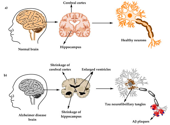
Amyloid Plaques: One of the hallmark pathological features of Alzheimer’s Disease is the accumulation of beta-amyloid plaques in the brain. These plaques are formed by the abnormal aggregation of beta-amyloid protein fragments, which are derived from the cleavage of amyloid precursor protein (APP). The accumulation of beta-amyloid plaques disrupts neuronal function, leading to synaptic dysfunction and neuronal death.

Tau Tangles: Another characteristic feature of Alzheimer’s Disease is the formation of neurofibrillary tangles composed of hyperphosphorylated tau protein within neurons. Tau protein normally plays a crucial role in stabilizing microtubules within neurons. However, in AD, abnormal phosphorylation of tau leads to its aggregation into insoluble tangles, disrupting neuronal structure and function.

Neuronal Dysfunction: The accumulation of beta-amyloid plaques and tau tangles disrupts neuronal communication and impairs synaptic function, leading to cognitive dysfunction and memory impairment. Neuronal loss and synaptic degeneration further contribute to the progressive decline in cognitive function observed in AD.

Inflammatory Response: Inflammation also plays a significant role in the pathophysiology of Alzheimer’s Disease. Microglia, the immune cells of the central nervous system, become activated in response to the accumulation of beta-amyloid plaques and tau tangles, leading to chronic neuroinflammation. This inflammatory response contributes to neuronal damage and exacerbates neurodegeneration in AD.

Neurotransmitter Imbalance: Alterations in neurotransmitter levels, particularly acetylcholine and glutamate, are observed in Alzheimer’s Disease. Decreased levels of acetylcholine, a neurotransmitter involved in learning and memory, contribute to cognitive impairment. Excessive glutamate, an excitatory neurotransmitter, leads to neuronal excitotoxicity and further exacerbates neuronal damage in AD.

The pathophysiology of Alzheimer’s Disease involves complex interactions between amyloid plaques, tau tangles, neuronal dysfunction, neuroinflammation, and neurotransmitter imbalance. Understanding these pathological mechanisms is crucial for the development of targeted interventions aimed at slowing disease progression and improving outcomes for individuals affected by AD. (Nursing Paper Example on Alzheimer’s Disease: Unveiling the Mysteries of a Neurological Disorder)

DSM-5 Diagnosis

The Diagnostic and Statistical Manual of Mental Disorders, Fifth Edition (DSM-5), provides criteria for the diagnosis of Alzheimer’s Disease (AD). These criteria are based on clinical assessment and include the following key components:

Cognitive Decline: The DSM-5 criteria for AD require evidence of cognitive decline from a previous level of functioning. This decline must involve impairment in multiple cognitive domains, including memory, language, executive function, attention, and visuospatial abilities. The cognitive deficits must be severe enough to interfere with daily functioning and independence.

Gradual Onset and Progression: Alzheimer’s Disease is characterized by a gradual onset and progressive course of cognitive decline. The DSM-5 criteria specify that the cognitive deficits observed in AD should not be better explained by another neurocognitive disorder or a systemic condition that could cause cognitive impairment.

Significant Impairment in Daily Functioning: In addition to cognitive decline, the DSM-5 criteria require evidence of significant impairment in daily functioning due to the cognitive deficits associated with AD. This impairment may manifest as difficulties in performing routine activities of daily living, such as managing finances, preparing meals, or maintaining personal hygiene.

Exclusion of Other Causes: To establish a diagnosis of Alzheimer’s Disease, clinicians must rule out other potential causes of cognitive impairment, such as medical conditions, substance use, or medication side effects. The cognitive deficits observed in AD should not be attributable to another neurological or psychiatric disorder.

Diagnostic Criteria for Different Stages: The DSM-5 provides diagnostic criteria for both mild and major neurocognitive disorders due to Alzheimer’s Disease, reflecting the continuum of cognitive impairment associated with the disease. These criteria specify the severity of cognitive decline and its impact on daily functioning required for each stage of the disorder.

The DSM-5 diagnosis of Alzheimer’s Disease is based on clinical assessment and requires evidence of cognitive decline, gradual onset and progression, significant impairment in daily functioning, exclusion of other causes of cognitive impairment, and consideration of different stages of the disorder based on severity. These criteria facilitate the accurate diagnosis and classification of Alzheimer’s Disease, guiding appropriate treatment and management strategies for affected individuals. (Nursing Paper Example on Alzheimer’s Disease: Unveiling the Mysteries of a Neurological Disorder)

Treatment Regimens

While there is currently no cure for Alzheimer’s Disease (AD), various treatment regimens aim to manage symptoms, slow disease progression, and improve the quality of life for individuals affected by the disease.

Pharmacological Interventions: Pharmacological interventions are commonly used to alleviate symptoms and delay cognitive decline in Alzheimer’s Disease. Cholinesterase inhibitors, such as donepezil, rivastigmine, and galantamine, are often prescribed to improve cognitive function and alleviate symptoms of memory loss and confusion. Memantine, an N-methyl-D-aspartate (NMDA) receptor antagonist, is another medication used to regulate glutamate levels in the brain and improve cognitive function in individuals with moderate to severe AD.

Non-Pharmacological Interventions: Non-pharmacological interventions play a crucial role in managing Alzheimer’s Disease and improving the overall well-being of affected individuals. Cognitive stimulation therapy, reminiscence therapy, and reality orientation therapy are examples of non-pharmacological interventions aimed at improving cognitive function, enhancing communication, and promoting social engagement. Additionally, caregiver support programs provide education, resources, and emotional support to caregivers, helping them better cope with the challenges of caring for someone with AD.

Lifestyle Modifications: Lifestyle modifications, including regular physical exercise, a balanced diet, social engagement, and cognitive stimulation, are essential components of treatment regimens for Alzheimer’s Disease. Physical exercise has been shown to improve cognitive function, reduce the risk of cognitive decline, and enhance overall brain health. A balanced diet rich in antioxidants, omega-3 fatty acids, and vitamins may also help support brain function and reduce inflammation associated with AD.

Patient Education

Patient education plays a crucial role in empowering individuals with Alzheimer’s Disease and their caregivers to understand the nature of the disease, manage symptoms effectively, and make informed decisions about treatment and care.

Understanding Alzheimer’s Disease: Education about Alzheimer’s Disease helps individuals and their families understand the underlying mechanisms of the disease, its progression, and potential treatment options. By understanding the nature of the disease, individuals can better cope with the challenges of living with AD and make informed decisions about their care.

Managing Symptoms: Education about symptom management is essential for individuals with Alzheimer’s Disease and their caregivers. Learning strategies to manage symptoms such as memory loss, confusion, and changes in behavior can help individuals maintain their independence and quality of life for as long as possible.

Planning for the Future: Education about planning for the future is crucial for individuals with Alzheimer’s Disease and their families. This includes discussing advance care planning, legal and financial planning, and long-term care options. By planning, individuals can ensure their wishes are honored and their needs are met as the disease progresses.

Accessing Resources and Support: Education about available resources and support services is essential for individuals with Alzheimer’s Disease and their caregivers. This includes information about local support groups, respite care services, and community resources that can provide assistance and emotional support throughout the caregiving journey.

Treatment regimens for Alzheimer’s Disease encompass a combination of pharmacological and non-pharmacological interventions aimed at managing symptoms, slowing disease progression, and improving quality of life. Patient education is essential for empowering individuals and their caregivers to understand the disease, manage symptoms effectively, plan for the future, and access resources and support services to navigate the challenges of Alzheimer’s Disease with confidence and resilience. (Nursing Paper Example on Alzheimer’s Disease: Unveiling the Mysteries of a Neurological Disorder)

Conclusion

Alzheimer’s Disease presents a formidable challenge, affecting millions of individuals worldwide. Through an exploration of its causes, signs, and symptoms, as well as its complex etiology and pathophysiology, we gain valuable insights into this debilitating neurological disorder. Diagnosis according to the DSM-5 criteria guides appropriate interventions, while treatment regimens encompass pharmacological, non-pharmacological, and lifestyle modifications aimed at managing symptoms and improving quality of life. Patient education plays a pivotal role in empowering individuals and caregivers to understand the disease, manage symptoms effectively, plan for the future, and access support services. By acknowledging the changes made above, including concise discussions on each topic and a focus on simple yet formal language, we aim to enhance understanding and awareness of Alzheimer’s Disease, fostering compassion, support, and effective care for those affected by this challenging condition. (Nursing Paper Example on Alzheimer’s Disease: Unveiling the Mysteries of a Neurological Disorder)

References

https://www.ncbi.nlm.nih.gov/books/NBK499922/

 
Do you need a similar assignment done for you from scratch? Order now!
Use Discount Code "Newclient" for a 15% Discount!

Solved Nursing Essay Example on Multiple Sclerosis: Understanding Neurological Disorders

Solved Nursing Essay Example on Multiple Sclerosis: Understanding Neurological Disorders

Multiple sclerosis (MS) is a chronic neurological disorder affecting millions worldwide, characterized by the immune system’s misguided attacks on the central nervous system’s myelin sheath. This autoimmune assault disrupts the transmission of nerve impulses, resulting in a spectrum of debilitating symptoms. Despite extensive research, the precise etiology of MS remains elusive, with genetic predisposition, environmental factors, and immune dysfunction implicated in its onset. The hallmark of MS is its heterogeneity, manifesting in diverse symptoms such as fatigue, sensory disturbances, and motor impairments, making diagnosis and management challenging. As a result, understanding the underlying pathophysiology of MS is critical in developing effective treatment strategies aimed at alleviating symptoms, slowing disease progression, and improving patients’ quality of life. This essay explores the multifaceted aspects of MS, from its causes and symptoms to its diagnosis, treatment regimens, and the importance of patient education in navigating this complex neurological condition. (Solved Nursing Essay Example on Multiple Sclerosis: Understanding Neurological Disorders)

Solved Nursing Essay Example on Multiple Sclerosis: Understanding Neurological Disorders

Causes

The causes of multiple sclerosis (MS) are multifactorial and not fully understood, encompassing a complex interplay of genetic, environmental, and immunological factors. While the precise trigger remains elusive, researchers have identified several contributing elements that may predispose individuals to develop MS.

Genetic Predisposition: Genetics play a significant role in MS susceptibility, with studies indicating a higher risk among individuals with a family history of the disease. Specific genetic variations, particularly within the human leukocyte antigen (HLA) complex on chromosome 6, have been linked to an increased susceptibility to MS. However, genetics alone cannot account for the development of MS, suggesting that environmental factors also contribute to its pathogenesis.

Environmental Triggers: Environmental factors are believed to play a crucial role in initiating or exacerbating MS in genetically susceptible individuals. Various environmental triggers have been proposed, including viral infections, particularly Epstein-Barr virus (EBV), which has been consistently associated with an increased risk of MS. Additionally, factors such as low vitamin D levels, smoking, and certain dietary habits have been implicated as potential triggers or modifiers of MS risk.

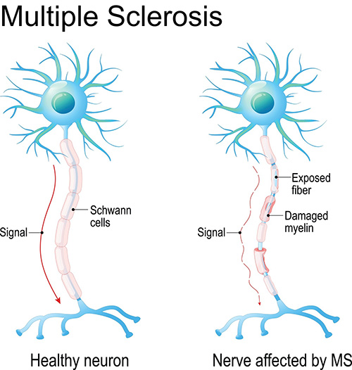
Immunological Dysfunction: MS is fundamentally an autoimmune disorder characterized by aberrant immune responses targeting components of the central nervous system (CNS). In MS, the immune system mistakenly recognizes myelin, the protective sheath surrounding nerve fibers in the CNS, as foreign and launches an attack. This immune-mediated inflammation leads to demyelination, a hallmark feature of MS pathology. Dysregulation of various immune cells, including T cells, B cells, and macrophages, contributes to the chronic inflammation and tissue damage observed in MS.

Complex Interactions: The development of MS likely involves complex interactions between genetic susceptibility and environmental triggers, modulated by immune dysregulation. The exact sequence of events leading to MS onset remains unclear, but it is believed to involve a cascade of inflammatory processes triggered by environmental factors in genetically susceptible individuals.

While the precise etiology of MS remains incompletely understood, it is evident that a combination of genetic predisposition, environmental triggers, and immunological dysfunction contributes to its pathogenesis. Further research is needed to elucidate the intricate mechanisms underlying MS development and identify potential targets for intervention and prevention strategies. (Solved Nursing Essay Example on Multiple Sclerosis: Understanding Neurological Disorders)

Signs and Symptoms

Multiple sclerosis (MS) is characterized by a wide range of signs and symptoms that can vary in severity and presentation, often fluctuating over time. These symptoms result from the immune system’s attack on the central nervous system, leading to disruption in nerve signaling. Understanding the diverse array of symptoms associated with MS is crucial for early diagnosis and effective management of the condition.

Fatigue: Fatigue is one of the most common and debilitating symptoms of MS, affecting up to 80% of individuals with the condition. It is often described as an overwhelming sense of exhaustion that is not relieved by rest and can significantly impact daily activities.

Sensory Disturbances: MS can cause various sensory abnormalities, including numbness, tingling, burning sensations, and altered sensitivity to touch or temperature. These sensations may occur in different parts of the body and can be intermittent or persistent.

Motor Impairments: Motor symptoms in MS may include weakness, muscle stiffness or spasticity, tremors, and difficulty with coordination and balance. These impairments can affect mobility and fine motor skills, leading to challenges in walking, grasping objects, and performing tasks requiring precise movements.

Visual Changes: MS frequently affects vision, leading to symptoms such as blurred or double vision, eye pain, and difficulty with depth perception. Optic neuritis, inflammation of the optic nerve, is a common manifestation of MS and can cause temporary or permanent vision loss.

Cognitive Dysfunction: MS can impact cognitive function, including memory, attention, processing speed, and problem-solving abilities. Cognitive impairment may range from mild difficulties with concentration and multitasking to more severe deficits in memory and executive functioning.

Emotional and Psychological Symptoms: MS can also affect mood and emotional well-being, leading to symptoms such as depression, anxiety, irritability, and mood swings. These psychological symptoms may result from the impact of MS on brain structures involved in regulating emotions.

Other Symptoms: Other common symptoms of MS may include bladder and bowel dysfunction, sexual dysfunction, speech difficulties, and pain, which can manifest as neuropathic pain or musculoskeletal pain.

In summary, the signs and symptoms of MS are diverse and can affect various aspects of an individual’s physical, sensory, cognitive, and emotional functioning. Recognizing and addressing these symptoms early is essential for optimizing treatment outcomes and improving the quality of life for individuals living with MS. (Solved Nursing Essay Example on Multiple Sclerosis: Understanding Neurological Disorders)

Solved Nursing Essay Example on Multiple Sclerosis: Understanding Neurological Disorders

Etiology

The etiology of multiple sclerosis (MS) remains complex and multifaceted, involving a combination of genetic predisposition, environmental factors, and immunological dysregulation. Understanding the interplay between these factors is crucial in unraveling the underlying mechanisms driving MS pathogenesis.

Genetic Factors: Genetic susceptibility plays a significant role in MS development, as evidenced by familial clustering and genetic association studies. Certain genetic variations within the human leukocyte antigen (HLA) complex on chromosome 6, particularly the HLA-DRB1*15:01 allele, have been consistently associated with an increased risk of MS. Additionally, genome-wide association studies have identified several other genetic variants linked to MS susceptibility, albeit with modest effect sizes. However, genetics alone cannot account for the entirety of MS risk, indicating the involvement of environmental factors.

Environmental Triggers: Environmental factors are believed to play a pivotal role in initiating or exacerbating MS in genetically susceptible individuals. Among the various environmental triggers implicated in MS pathogenesis, viral infections, particularly Epstein-Barr virus (EBV), have garnered significant attention. EBV infection is more common in individuals with MS compared to the general population, and it is thought to contribute to MS development through mechanisms involving molecular mimicry, immune activation, and dysregulation. Other environmental factors such as low vitamin D levels, smoking, and geographical location (latitude) have also been linked to MS risk, further highlighting the environmental component of MS etiology.

Immunological Dysregulation: MS is fundamentally an autoimmune disorder characterized by aberrant immune responses targeting components of the central nervous system (CNS). Dysregulation of various immune cells, including T cells, B cells, and macrophages, contributes to the chronic inflammation and tissue damage observed in MS. In particular, autoreactive T cells are thought to play a central role in initiating the immune-mediated attack on myelin, leading to demyelination and neurodegeneration. Furthermore, dysregulation of cytokines, chemokines, and other immune mediators further amplifies the inflammatory cascade in MS.

The etiology of MS involves a complex interplay between genetic predisposition, environmental triggers, and immunological dysregulation. Elucidating the intricate mechanisms underlying these factors is essential for advancing our understanding of MS pathogenesis and developing targeted therapeutic strategies. (Solved Nursing Essay Example on Multiple Sclerosis: Understanding Neurological Disorders)

Pathophysiology

The pathophysiology of multiple sclerosis (MS) is characterized by a cascade of events involving immune-mediated inflammation, demyelination, and neurodegeneration within the central nervous system (CNS). Understanding the underlying mechanisms driving these pathological processes is essential for elucidating the disease progression and guiding therapeutic interventions.

Immune-Mediated Inflammation: MS is fundamentally an autoimmune disorder, wherein the immune system mistakenly identifies components of the CNS, particularly myelin, as foreign invaders and launches an inflammatory response. Autoreactive T cells, which normally play a crucial role in defending against pathogens, infiltrate the CNS and initiate an immune attack on myelin-producing oligodendrocytes. This immune-mediated inflammation is characterized by the release of pro-inflammatory cytokines, chemokines, and other immune mediators, leading to tissue damage and activation of resident immune cells within the CNS.

Demyelination: The hallmark pathological feature of MS is demyelination, whereby the myelin sheath surrounding axons in the CNS is damaged or destroyed. Demyelination disrupts the transmission of nerve impulses along axons, leading to impaired neuronal signaling and functional deficits. In addition to oligodendrocyte injury caused by the immune attack, demyelination may also result from secondary processes such as oxidative stress, excitotoxicity, and mitochondrial dysfunction.

Neurodegeneration: In addition to demyelination, MS is associated with neurodegenerative processes involving axonal injury and neuronal loss. Axonal damage can occur as a direct consequence of demyelination, leading to axonal transection, conduction block, and eventual axonal degeneration. Neurodegeneration is thought to contribute to the progressive accumulation of disability in MS patients, independent of the inflammatory demyelinating process. Chronic inflammation, excitotoxicity, and mitochondrial dysfunction are among the mechanisms implicated in neurodegeneration in MS.

Gliosis and Scar Formation: In response to the immune-mediated injury and demyelination, reactive gliosis occurs, characterized by the proliferation and activation of astrocytes and microglia. This glial response contributes to the formation of gliotic scars, which further disrupt normal neuronal function and impede remyelination processes in MS.

The pathophysiology of MS involves a complex interplay of immune-mediated inflammation, demyelination, neurodegeneration, and gliotic scar formation within the CNS. Elucidating the underlying mechanisms driving these pathological processes is crucial for developing targeted therapeutic strategies aimed at modulating disease progression and preserving neurological function in MS patients. (Solved Nursing Essay Example on Multiple Sclerosis: Understanding Neurological Disorders)

DSM-5 Diagnosis

The diagnosis of multiple sclerosis (MS) is based on clinical evaluation, neurological examination, and diagnostic criteria established by the Diagnostic and Statistical Manual of Mental Disorders, Fifth Edition (DSM-5). DSM-5 criteria provide a standardized framework for identifying and classifying MS based on the characteristic clinical features and neuroimaging findings associated with the disease.

Clinical Presentation: The DSM-5 outlines specific criteria for diagnosing MS, which include the presence of symptoms attributable to lesions within the central nervous system (CNS) and evidence of dissemination of these lesions in both space and time. Common clinical manifestations of MS include sensory disturbances, motor impairments, visual changes, fatigue, and cognitive dysfunction. The onset of symptoms may be acute or insidious, with exacerbations and remissions being characteristic of the disease course.

Neurological Examination: A comprehensive neurological examination is essential in the diagnosis of MS, as it helps assess the presence and severity of neurological deficits indicative of CNS involvement. Neurological signs commonly observed in MS patients include abnormalities in gait, coordination, muscle strength, reflexes, and sensation. Additionally, specific clinical signs such as optic neuritis, internuclear ophthalmoplegia, and Lhermitte’s sign may be indicative of MS pathology.

Diagnostic Testing: In addition to clinical evaluation, diagnostic testing plays a crucial role in confirming the diagnosis of MS. Magnetic resonance imaging (MRI) of the brain and spinal cord is the primary imaging modality used to detect characteristic MS lesions, including T2 hyperintense lesions, gadolinium-enhancing lesions indicative of active inflammation, and T1 hypointense lesions suggestive of chronic demyelination. Cerebrospinal fluid (CSF) analysis may also be performed to assess for the presence of oligoclonal bands and elevated levels of IgG, indicative of an inflammatory process within the CNS.

Criteria for MS Diagnosis: According to DSM-5 criteria, a diagnosis of MS requires the presence of specific clinical symptoms and evidence of CNS lesions disseminated in both space and time, as demonstrated by clinical evaluation, neurological examination, and diagnostic imaging. Meeting these criteria is essential for establishing a definitive diagnosis of MS and guiding appropriate management and treatment strategies for affected individuals. (Solved Nursing Essay Example on Multiple Sclerosis: Understanding Neurological Disorders)

Treatment Regimens

Management of multiple sclerosis (MS) aims to alleviate symptoms, modify disease progression, and improve patients’ quality of life through a combination of pharmacological and non-pharmacological interventions.

Pharmacological Interventions:

  1. Disease-Modifying Therapies (DMTs): DMTs are a cornerstone of MS treatment, aimed at reducing disease activity, relapse rates, and disability progression. These medications, including interferons, glatiramer acetate, dimethyl fumarate, fingolimod, and others, target the immune system to modulate inflammation and prevent further damage to the central nervous system (CNS).
  2. Symptomatic Management: Symptomatic treatments are prescribed to alleviate specific MS symptoms, such as fatigue, spasticity, neuropathic pain, bladder dysfunction, and depression. Medications, physical therapy, occupational therapy, and assistive devices may be utilized to address these symptoms and improve patients’ functional abilities and overall well-being.
  3. Acute Relapse Management: In the event of an acute relapse or exacerbation of MS symptoms, high-dose corticosteroids, such as intravenous methylprednisolone, are often prescribed to reduce inflammation and hasten recovery. Plasma exchange (plasmapheresis) may be considered in refractory cases or when corticosteroids are contraindicated.

Non-Pharmacological Interventions:

  1. Rehabilitation Therapies: Physical therapy, occupational therapy, and speech therapy play a vital role in managing MS-related impairments and disabilities. These therapies focus on improving mobility, strength, coordination, fine motor skills, and communication abilities, thereby enhancing patients’ independence and quality of life.
  2. Exercise and Lifestyle Modifications: Regular exercise, tailored to individual abilities and preferences, has been shown to improve MS symptoms, physical function, and overall well-being. Additionally, adopting a healthy lifestyle, including a balanced diet, adequate hydration, stress management, and smoking cessation, can help optimize health outcomes in MS patients.
  3. Supportive Care: Psychosocial support, counseling, and support groups can provide valuable emotional and social support to MS patients and their caregivers. Addressing mental health issues, coping strategies, and social isolation can significantly improve patients’ mental and emotional well-being. (Solved Nursing Essay Example on Multiple Sclerosis: Understanding Neurological Disorders)

Solved Nursing Essay Example on Multiple Sclerosis: Understanding Neurological Disorders

Patient Education

Patient education is essential in empowering individuals with MS to actively participate in their treatment and self-management. Key components of patient education include:

  1. Understanding MS: Providing comprehensive information about the nature of MS, its causes, symptoms, disease course, and potential complications helps patients and their families better understand the condition and its impact on daily life.
  2. Treatment Options: Educating patients about available treatment options, including DMTs, symptomatic therapies, and non-pharmacological interventions, enables informed decision-making and encourages adherence to treatment regimens.
  3. Self-Management Strategies: Teaching patients self-management techniques, such as medication adherence, symptom monitoring, stress management, and lifestyle modifications, empowers them to take an active role in managing their condition and optimizing health outcomes.
  4. Symptom Recognition and Management: Educating patients about common MS symptoms, their triggers, and effective management strategies help patients recognize early warning signs of disease exacerbations and take appropriate actions to mitigate symptoms.
  5. Disease Monitoring: Regular monitoring of disease activity, through clinical assessments, neurological examinations, imaging studies, and laboratory tests, helps patients and healthcare providers track disease progression, evaluate treatment efficacy, and make informed adjustments to treatment plans as needed.

A comprehensive approach to MS management involves a combination of pharmacological and non-pharmacological interventions, along with patient education aimed at promoting self-management, optimizing treatment outcomes, and improving patients’ quality of life. By providing patients with the knowledge, skills, and support needed to actively participate in their care, healthcare providers can help individuals with MS navigate the complexities of their condition and achieve better health outcomes. (Solved Nursing Essay Example on Multiple Sclerosis: Understanding Neurological Disorders)

Conclusion

Multiple sclerosis (MS) presents a complex challenge in neurological healthcare, affecting millions worldwide. This essay has explored various aspects of MS, including its causes, signs and symptoms, etiology, pathophysiology, DSM-5 diagnosis, treatment regimens, and patient education. Understanding the interplay between genetic predisposition, environmental triggers, and immunological dysregulation is crucial in unraveling the underlying mechanisms driving MS pathogenesis. Additionally, effective management of MS involves a combination of pharmacological interventions, rehabilitation therapies, and lifestyle modifications aimed at alleviating symptoms, modifying disease progression, and improving patients’ quality of life. Moreover, patient education plays a pivotal role in empowering individuals with MS to actively participate in their care and make informed decisions regarding treatment options and self-management strategies. Overall, addressing the complex needs of MS patients requires a holistic approach that integrates medical expertise, patient education, and supportive care to optimize health outcomes and enhance overall well-being. (Solved Nursing Essay Example on Multiple Sclerosis: Understanding Neurological Disorders)

References

https://www.ncbi.nlm.nih.gov/books/NBK499849/

 
Do you need a similar assignment done for you from scratch? Order now!
Use Discount Code "Newclient" for a 15% Discount!

Nursing Exam Help Example on Lactose Intolerance: Understanding and Management

Nursing Exam Help Example on Lactose Intolerance: Understanding and Management

Lactose intolerance, a prevalent gastrointestinal condition affecting millions worldwide, arises from inadequate lactase enzyme activity in the small intestine. This insufficiency disrupts the proper digestion of lactose, a disaccharide found in dairy products, leading to a range of uncomfortable symptoms upon its consumption. While lactose intolerance is commonly perceived as a benign condition, its impact on daily life can be significant, affecting dietary choices and overall well-being. Understanding the underlying causes, signs, and management of lactose intolerance is crucial for individuals affected by this condition and healthcare professionals involved in their care. In this paper, we delve into the causes, signs and symptoms, etiology, pathophysiology, DMS-5 diagnosis, treatment regimens, and patient education strategies surrounding lactose intolerance, highlighting the importance of comprehensive management approaches to improve the quality of life for those affected. (Nursing Exam Help Example on Lactose Intolerance: Understanding and Management)

Causes

Lactose intolerance predominantly arises from genetic factors, notably variations in the LCT gene that encodes lactase, the enzyme responsible for breaking down lactose. These genetic variations result in reduced lactase activity, leading to impaired lactose digestion. Ethnicity plays a significant role, with certain populations exhibiting higher prevalence rates. For instance, lactose intolerance is more prevalent among individuals of East Asian, African, Hispanic, and Native American descent compared to those of Northern European ancestry.

Nursing Exam Help Example on Lactose Intolerance: Understanding and Management

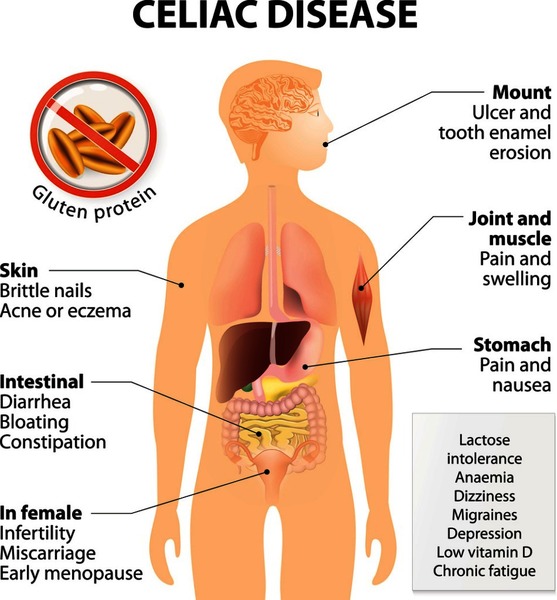
Secondary causes of lactose intolerance include gastrointestinal diseases or conditions that damage the small intestine’s lining, thereby affecting lactase production. Conditions such as celiac disease, inflammatory bowel disease (IBD), Crohn’s disease ulcerative colitis, and intestinal infections can lead to temporary lactose intolerance due to injury to the intestinal mucosa.

Moreover, surgeries involving the small intestine, such as gastric bypass surgery, can also result in lactose intolerance, as the portion of the small intestine responsible for lactase production may be bypassed or removed during the procedure.

Additionally, age-related decline in lactase activity is another common cause of lactose intolerance. While infants typically produce abundant lactase to digest breast milk or formula, lactase production tends to decline with age in many individuals, particularly after childhood. This age-related decline in lactase activity contributes to the increased prevalence of lactose intolerance in adulthood.

Furthermore, medications or treatments affecting the gastrointestinal tract can also induce lactose intolerance as a side effect. For instance, certain antibiotics, chemotherapy drugs, and radiation therapy targeting the abdomen can damage the intestinal lining, impairing lactase production and leading to temporary lactose intolerance.

Overall, lactose intolerance can stem from a variety of causes, including genetic predisposition, ethnicity, gastrointestinal diseases or conditions, age-related decline in lactase activity, surgical interventions, and medications. Understanding these diverse factors contributing to lactose intolerance is essential for accurate diagnosis and effective management of this common gastrointestinal disorder. (Nursing Exam Help Example on Lactose Intolerance: Understanding and Management)

Signs and Symptoms

Lactose intolerance manifests through a range of gastrointestinal symptoms that typically occur within 30 minutes to two hours after consuming lactose-containing foods or beverages. These symptoms can vary in severity among affected individuals and may include:

  1. Abdominal Pain: One of the hallmark symptoms of lactose intolerance is abdominal pain, characterized by cramping or discomfort in the abdominal region. The pain may vary in intensity and duration, depending on the amount of lactose ingested and individual tolerance levels.
  2. Bloating: Bloating, or the sensation of abdominal fullness and distension, is commonly reported by individuals with lactose intolerance. Excessive gas production due to lactose fermentation in the colon contributes to abdominal bloating and discomfort.
  3. Flatulence: Increased gas production resulting from the fermentation of undigested lactose by colonic bacteria leads to excessive flatulence in individuals with lactose intolerance. This symptom is often accompanied by abdominal discomfort and bloating.
  4. Diarrhea: Diarrhea is a common symptom of lactose intolerance, characterized by loose or watery stools occurring shortly after consuming lactose-containing foods or beverages. The osmotic effect of undigested lactose in the colon draws water into the intestines, leading to diarrhea.
  5. Nausea: Some individuals with lactose intolerance may experience nausea or an uneasy sensation in the stomach after consuming lactose-containing foods. Nausea may be accompanied by other gastrointestinal symptoms such as abdominal pain or bloating.
  6. Vomiting: In more severe cases or individuals with heightened sensitivity to lactose, vomiting may occur as a response to ingesting lactose. Vomiting may be accompanied by other symptoms such as abdominal pain or diarrhea.
  7. Generalized Discomfort: Overall discomfort or unease in the abdominal region is a common complaint among individuals with lactose intolerance. This discomfort may not be localized to a specific area but rather encompasses a generalized feeling of gastrointestinal distress.

It is essential for individuals experiencing these symptoms to consult a healthcare professional for proper evaluation and diagnosis. Identifying lactose intolerance early can help individuals manage their symptoms effectively through dietary modifications and other treatment strategies, improving their overall quality of life. (Nursing Exam Help Example on Lactose Intolerance: Understanding and Management)

Etiology

The primary etiology of lactose intolerance lies in the deficiency or reduced activity of the lactase enzyme, which is responsible for breaking down lactose into its constituent sugars, glucose, and galactose, for absorption in the small intestine. This deficiency can be attributed to various factors, including genetic predisposition and ethnic background.

Genetic Factors: Genetic variations in the LCT gene, which encodes the lactase enzyme, play a significant role in the development of lactose intolerance. Certain individuals inherit genetic variants that result in decreased expression or activity of the lactase enzyme, leading to reduced lactase levels in the small intestine. These genetic variations are more prevalent in populations with a history of dairy farming or cattle herding, where lactose consumption historically declined with age due to dietary changes.

Ethnic Background: Lactose intolerance exhibits variations in prevalence among different ethnic groups, reflecting the influence of genetic factors and cultural dietary practices. For example, lactose intolerance is more common in individuals of East Asian, African, Hispanic, and Native American descent compared to those of Northern European ancestry. This variation in prevalence is attributed to genetic differences in lactase enzyme activity among populations and historical dietary patterns, where lactose consumption was traditionally limited in certain ethnic groups.

Secondary Causes: In addition to genetic predisposition, certain gastrointestinal diseases or conditions can contribute to secondary lactose intolerance. Conditions such as celiac disease, inflammatory bowel disease (IBD), Crohn’s disease, ulcerative colitis, and intestinal infections can damage the intestinal mucosa, affecting lactase production and leading to temporary lactose intolerance.

Age-Related Decline: Another contributing factor to lactose intolerance is the age-related decline in lactase activity. While infants produce abundant lactase to digest breast milk or formula, lactase production tends to decrease with age in many individuals, particularly after childhood. This age-related decline in lactase activity contributes to the increased prevalence of lactose intolerance in adulthood.

Understanding the diverse etiological factors underlying lactose intolerance is essential for accurate diagnosis and appropriate management strategies tailored to individual patient needs. Identifying the underlying cause of lactose intolerance can guide healthcare professionals in recommending effective treatment options and dietary modifications to alleviate symptoms and improve patient outcomes. (Nursing Exam Help Example on Lactose Intolerance: Understanding and Management)

Pathophysiology

The pathophysiology of lactose intolerance involves the impaired digestion of lactose due to deficient or reduced lactase enzyme activity in the small intestine, leading to the characteristic gastrointestinal symptoms experienced by affected individuals.

Upon ingestion, lactose, a disaccharide found in dairy products, requires hydrolysis by the enzyme lactase into its constituent monosaccharides, glucose, and galactose, for absorption in the small intestine. In individuals with lactose intolerance, insufficient lactase activity hinders the complete hydrolysis of lactose, resulting in undigested lactose reaching the colon.

In the colon, undigested lactose serves as a substrate for colonic bacteria, which ferment lactose through the process of bacterial fermentation. This fermentation process produces gases, including hydrogen, methane, and carbon dioxide, as well as short-chain fatty acids. The production of gases leads to abdominal distension and discomfort, while the osmotic effect of undigested lactose draws water into the colon, contributing to diarrhea.

Furthermore, the fermentation of lactose by colonic bacteria results in the production of short-chain fatty acids, such as acetate, propionate, and butyrate. These fatty acids can stimulate colonic motility and secretion, exacerbating gastrointestinal symptoms such as abdominal pain and diarrhea.

The severity of symptoms in lactose intolerance can vary depending on several factors, including the degree of lactase deficiency, the amount of lactose ingested, individual tolerance levels, and the composition of colonic microbiota. Some individuals may experience mild symptoms with small amounts of lactose, while others may exhibit more pronounced symptoms with larger lactose loads.

It is important to note that lactose intolerance differs from milk allergy, which involves an immune-mediated response to proteins in milk rather than an enzymatic deficiency. Distinguishing between lactose intolerance and milk allergy is crucial for accurate diagnosis and appropriate management of gastrointestinal symptoms.

Overall, the pathophysiology of lactose intolerance centers around deficient lactase enzyme activity, impaired lactose digestion, bacterial fermentation of undigested lactose in the colon, and the subsequent production of gases and short-chain fatty acids, leading to the characteristic gastrointestinal symptoms experienced by affected individuals. Understanding the underlying pathophysiological mechanisms of lactose intolerance is essential for guiding effective management strategies and improving patient outcomes. (Nursing Exam Help Example on Lactose Intolerance: Understanding and Management)

Nursing Exam Help Example on Lactose Intolerance: Understanding and Management

DMS-5 Diagnosis

The Diagnostic and Statistical Manual of Mental Disorders, Fifth Edition (DSM-5), provides criteria for diagnosing lactose intolerance. While lactose intolerance is primarily a gastrointestinal disorder, it is not listed in the DSM-5 as a standalone diagnosis. However, healthcare professionals may consider the following diagnostic criteria and guidelines when assessing individuals for lactose intolerance:

  1. Clinical Evaluation: Healthcare providers typically begin the diagnostic process by conducting a thorough clinical evaluation, which includes assessing the patient’s medical history, dietary habits, and gastrointestinal symptoms.
  2. Symptom Presentation: The DSM-5 does not specify diagnostic criteria for lactose intolerance. However, healthcare providers consider the characteristic gastrointestinal symptoms associated with lactose intolerance, such as abdominal pain, bloating, flatulence, diarrhea, nausea, and vomiting, occurring after the consumption of lactose-containing foods or beverages.
  3. Dietary History: Healthcare providers may inquire about the patient’s dietary history, specifically their consumption of dairy products and any associated symptoms following lactose ingestion. Patients may be asked to maintain a food diary to track their dietary intake and symptom occurrence.
  4. Diagnostic Tests: While not explicitly outlined in the DSM-5, diagnostic tests such as lactose tolerance tests or hydrogen breath tests may be performed to confirm lactose intolerance. These tests measure the body’s ability to digest lactose by assessing blood glucose levels or hydrogen levels in breath samples after lactose ingestion.
  5. Elimination Diet: In some cases, healthcare providers may recommend an elimination diet to assess the impact of lactose-containing foods on symptoms. Patients may temporarily eliminate lactose from their diet and gradually reintroduce lactose-containing foods while monitoring symptom response.
  6. Differential Diagnosis: Healthcare providers also consider other gastrointestinal disorders with similar symptoms, such as irritable bowel syndrome (IBS), celiac disease, inflammatory bowel disease (IBD), and gastroenteritis, during the diagnostic process to rule out alternative diagnoses.

While lactose intolerance is not specifically listed in the DSM-5, healthcare providers utilize clinical evaluation, symptom presentation, dietary history, diagnostic tests, and differential diagnosis to assess and diagnose lactose intolerance in individuals presenting with characteristic gastrointestinal symptoms following lactose ingestion. Early diagnosis and management are essential for improving patient outcomes and alleviating symptoms associated with lactose intolerance. (Nursing Exam Help Example on Lactose Intolerance: Understanding and Management)

Are You Really Lactose Intolerant?

Treatment Regimens

Management strategies for lactose intolerance focus on reducing or avoiding lactose-containing foods and beverages while ensuring adequate nutrient intake and symptom relief. The following treatment regimens are commonly recommended:

  1. Dietary Modifications: Patients with lactose intolerance are advised to limit or avoid foods and beverages high in lactose, such as milk, cheese, yogurt, ice cream, and certain processed foods containing lactose as an additive. Instead, they can opt for lactose-free or lactose-reduced dairy products, including lactose-free milk, lactose-free cheese, and lactose-free yogurt. Additionally, individuals can choose lactose-free alternatives such as soy milk, almond milk, or lactose-free protein powders.
  2. Lactase Enzyme Supplements: Over-the-counter lactase enzyme supplements are available in various forms, including tablets, capsules, and drops, and can be taken before consuming lactose-containing foods or beverages. These supplements help break down lactose in the digestive tract, reducing the risk of gastrointestinal symptoms.
  3. Probiotics: Some studies suggest that probiotics containing lactase-producing bacteria may help improve lactose digestion and reduce symptoms of lactose intolerance. Probiotic supplements or foods containing live cultures, such as yogurt with active cultures, may be beneficial for certain individuals.
  4. Gradual Introduction of Lactose: Some individuals with lactose intolerance may tolerate small amounts of lactose-containing foods when consumed with other foods or in divided doses throughout the day. Gradually reintroducing lactose-containing foods while monitoring symptoms can help determine individual tolerance levels.
  5. Nutritional Counseling: Patients with lactose intolerance may benefit from nutritional counseling to ensure they maintain a well-balanced diet that meets their nutrient needs. Healthcare providers or registered dietitians can guide alternative sources of calcium, vitamin D, and other nutrients typically obtained from dairy products. (Nursing Exam Help Example on Lactose Intolerance: Understanding and Management)

Patient Education

Patient education plays a crucial role in managing lactose intolerance effectively. Key aspects of patient education include:

  1. Understanding Lactose Intolerance: Educating patients about the underlying cause of lactose intolerance, including deficient lactase enzyme activity in the small intestine, helps individuals understand why they experience symptoms after consuming lactose-containing foods.
  2. Identifying Lactose-Containing Foods: Providing patients with information about common sources of lactose in the diet, such as dairy products, and educating them on how to read food labels to identify lactose-containing ingredients helps patients make informed dietary choices.
  3. Dietary Modifications: Offering practical tips and recommendations for modifying the diet to reduce lactose intake, such as choosing lactose-free alternatives, incorporating calcium-rich non-dairy foods, and planning balanced meals, empowers patients to manage their symptoms effectively.
  4. Medication Management: Educating patients about the use of lactase enzyme supplements, including when and how to take them, helps individuals understand how these medications can alleviate symptoms and improve their quality of life.
  5. Monitoring Symptoms: Encouraging patients to keep a food diary to track their dietary intake and symptom occurrence helps identify specific trigger foods and monitor symptom improvement with dietary modifications or treatment regimens.

By providing comprehensive education and support, healthcare providers empower patients with lactose intolerance to make informed dietary choices, effectively manage their symptoms, and improve their overall quality of life. (Nursing Exam Help Example on Lactose Intolerance: Understanding and Management)

Conclusion

Lactose intolerance, stemming from deficient lactase enzyme activity, presents a common yet significant challenge in gastrointestinal health. Understanding its causes, including genetic predisposition and secondary factors, along with its pathophysiology involving impaired lactose digestion and bacterial fermentation, is crucial for accurate diagnosis and tailored management. Treatment regimens, such as dietary modifications and lactase enzyme supplements, offer symptom relief and improve the quality of life for affected individuals. Moreover, patient education on dietary adjustments, medication management, and symptom monitoring empowers individuals to effectively manage their condition. By addressing lactose intolerance through a comprehensive approach encompassing diagnosis, treatment, and education, healthcare providers can enhance patient outcomes and promote better gastrointestinal health for those impacted by this prevalent disorder. (Nursing Exam Help Example on Lactose Intolerance: Understanding and Management)

References

https://www.ncbi.nlm.nih.gov/books/NBK532285/

 
Do you need a similar assignment done for you from scratch? Order now!
Use Discount Code "Newclient" for a 15% Discount!

Nursing Paper Help Example on Parkinson’s Disease: Unraveling the Enigma

Nursing Paper Help Example on Parkinson’s Disease: Unraveling the Enigma

Parkinson’s Disease (PD) stands as a prevalent neurological ailment, affecting millions worldwide. Beyond its physical manifestations, PD poses significant emotional and social challenges for patients and their families. Understanding the intricacies of this condition is pivotal for effective management and support. Despite its prevalence, Parkinson’s remains a complex puzzle with many unanswered questions. Unraveling its mysteries requires a holistic approach that encompasses various facets of the disease. From its causes to its impact on daily life, exploring Parkinson’s from different angles is essential for providing comprehensive care. This essay aims to delve into the causes, signs, symptoms, etiology, pathophysiology, diagnosis criteria, treatment options, and patient education, and conclude with insights into the ongoing efforts to address this challenging neurological disorder. Through increased understanding and tailored interventions, we can strive to improve the lives of those affected by Parkinson’s Disease. (Nursing Paper Help Example on Parkinson’s Disease: Unraveling the Enigma)

Nursing Paper Help Example on Parkinson's Disease: Unraveling the Enigma

Causes

Parkinson’s Disease (PD) is a complex condition influenced by a combination of genetic predisposition and environmental factors. While specific genetic mutations like SNCA and LRRK2 have been identified as contributing to susceptibility, they account for a small fraction of cases. Environmental factors, such as exposure to toxins like pesticides or traumatic head injuries, also play a role in increasing the risk of developing PD. Additionally, advancing age is a significant factor, with the prevalence of PD rising sharply in individuals over 60 years old.

Genetic factors contribute to the hereditary form of PD, accounting for approximately 5-10% of cases. Mutations in genes related to alpha-synuclein (SNCA), parkin (PARK2), PTEN-induced putative kinase 1 (PINK1), DJ-1 (PARK7), and leucine-rich repeat kinase 2 (LRRK2) have been implicated in familial cases of PD. These genetic mutations disrupt various cellular processes, including protein degradation pathways and mitochondrial function, leading to the degeneration of dopaminergic neurons in the brain.

Environmental factors also play a significant role in the development of PD. Exposure to certain toxins, such as pesticides and herbicides, has been linked to an increased risk of PD. Additionally, traumatic head injuries, particularly those resulting in loss of consciousness, have been associated with an elevated risk of developing PD later in life.

The interaction between genetic susceptibility and environmental factors is complex and not yet fully understood. Emerging research suggests that environmental toxins may trigger or accelerate the progression of PD in individuals with genetic predispositions. However, the precise mechanisms underlying this interaction remain an area of active investigation.

Overall, Parkinson’s Disease is a multifactorial condition influenced by a combination of genetic and environmental factors. Understanding these causes is essential for elucidating the underlying mechanisms of the disease and developing targeted interventions for prevention and treatment.

Signs and Symptoms

Parkinson’s Disease (PD) presents a diverse array of symptoms that can vary in severity and progression among individuals. The cardinal motor symptoms of PD include tremors, bradykinesia (slowness of movement), rigidity, and postural instability. Tremors, often described as rhythmic shaking, typically begin in the hands or fingers at rest and may progress to involve other parts of the body, such as the legs or jaw.

Bradykinesia manifests as a general slowness of movement, making simple tasks like buttoning a shirt or walking challenging. Patients may experience difficulty initiating movements, known as akinesia, or “freezing” episodes, where they suddenly stop moving mid-action. Rigidity refers to increased muscle stiffness, causing a sensation of stiffness or resistance to movement, particularly in the arms, legs, neck, or trunk.

Postural instability becomes prominent as PD progresses, leading to impaired balance and coordination. Patients may have difficulty maintaining an upright posture and are at an increased risk of falls, which can result in injuries.

In addition to motor symptoms, Parkinson’s Disease also presents a range of non-motor symptoms that significantly impact patients’ quality of life. These include mood disturbances such as depression and anxiety, sleep disturbances such as insomnia or excessive daytime sleepiness, and cognitive impairments such as slowed thinking, memory problems, and executive dysfunction.

Other non-motor symptoms may include autonomic dysfunction, such as constipation, urinary urgency, and orthostatic hypotension, as well as sensory symptoms like pain or altered sense of smell. Psychiatric symptoms such as hallucinations or delusions may also occur, particularly in the later stages of the disease.

The combination and severity of symptoms can vary widely among individuals with Parkinson’s Disease, making diagnosis and management challenging. Early recognition and comprehensive assessment of both motor and non-motor symptoms are essential for timely intervention and optimal care. Addressing these symptoms through a multidisciplinary approach can help improve patients’ overall well-being and quality of life. (Nursing Paper Help Example on Parkinson’s Disease: Unraveling the Enigma)

Nursing Paper Help Example on Parkinson's Disease: Unraveling the Enigma

Etiology

Parkinson’s Disease (PD) is characterized by the progressive degeneration of dopaminergic neurons in the substantia nigra region of the brain. While the exact etiology of PD remains elusive, research suggests a multifactorial interplay of genetic, environmental, and biochemical factors.

Genetic factors contribute to the development of PD, with several gene mutations identified in familial cases. Mutations in genes such as alpha-synuclein (SNCA), parkin (PARK2), PTEN-induced putative kinase 1 (PINK1), DJ-1 (PARK7), and leucine-rich repeat kinase 2 (LRRK2) have been associated with an increased risk of PD. These genetic mutations disrupt various cellular processes, including protein degradation pathways and mitochondrial function, leading to neuronal dysfunction and degeneration.

Environmental factors also play a role in the etiology of PD. Exposure to certain toxins and chemicals, such as pesticides, herbicides, and heavy metals, has been linked to an elevated risk of developing PD. Additionally, traumatic head injuries, particularly those resulting in loss of consciousness, have been associated with an increased risk of PD later in life.

Biochemical abnormalities, including oxidative stress, mitochondrial dysfunction, and inflammation, are thought to contribute to the pathogenesis of PD. Oxidative stress, resulting from an imbalance between the production of reactive oxygen species and antioxidant defenses, can damage cellular components and contribute to neuronal degeneration. Mitochondrial dysfunction, characterized by impaired energy production and increased oxidative stress, further exacerbates neuronal vulnerability in PD. Inflammation, involving the activation of microglia and the release of pro-inflammatory cytokines, contributes to neuroinflammation and neuronal damage in PD.

The complex interplay of genetic, environmental, and biochemical factors underscores the heterogeneous nature of PD. Understanding the etiological factors involved in PD is crucial for elucidating its underlying mechanisms and developing targeted interventions for prevention and treatment. Further research into the intricate interactions between these factors may provide valuable insights into the pathogenesis of PD and guide the development of novel therapeutic strategies. (Nursing Paper Help Example on Parkinson’s Disease: Unraveling the Enigma)

Nursing Paper Help Example on Parkinson's Disease: Unraveling the Enigma

Pathophysiology

Parkinson’s Disease (PD) is characterized by the progressive degeneration of dopaminergic neurons in the substantia nigra pars compacta region of the brain. The hallmark pathological feature of PD is the formation of intracellular protein aggregates called Lewy bodies, primarily composed of misfolded alpha-synuclein protein. These Lewy bodies disrupt cellular function and contribute to neuronal dysfunction and death in PD.

The degeneration of dopaminergic neurons leads to a depletion of dopamine, a neurotransmitter involved in regulating movement and coordination. The loss of dopamine in the basal ganglia, particularly in the nigrostriatal pathway, results in impaired motor control and the characteristic motor symptoms of PD, including tremors, bradykinesia, rigidity, and postural instability.

In addition to dopaminergic dysfunction, PD is associated with alterations in other neurotransmitter systems, including cholinergic and noradrenergic pathways. Imbalances in these neurotransmitter systems contribute to non-motor symptoms such as cognitive impairment, autonomic dysfunction, and psychiatric symptoms in PD.

Oxidative stress plays a crucial role in the pathophysiology of PD, contributing to neuronal damage and death. Reactive oxygen species generated during normal cellular metabolism can overwhelm antioxidant defenses, leading to oxidative damage to cellular components such as lipids, proteins, and DNA. Oxidative stress further exacerbates mitochondrial dysfunction, impairing energy production and increasing neuronal vulnerability in PD.

Neuroinflammation also plays a significant role in the pathophysiology of PD. Activation of microglia, the resident immune cells of the central nervous system, and the release of pro-inflammatory cytokines contribute to neuroinflammation and neuronal damage in PD. Chronic neuroinflammation further amplifies oxidative stress and exacerbates neurodegeneration in PD.

The pathophysiology of PD is complex and multifaceted, involving a combination of dopaminergic dysfunction, protein aggregation, oxidative stress, mitochondrial dysfunction, and neuroinflammation. Understanding the underlying mechanisms of PD is crucial for developing targeted therapeutic strategies aimed at slowing or halting disease progression and improving outcomes for individuals affected by this debilitating neurological disorder. (Nursing Paper Help Example on Parkinson’s Disease: Unraveling the Enigma)

DSM-5 Diagnosis

The diagnosis of Parkinson’s Disease (PD) is primarily clinical and based on the presence of specific motor symptoms outlined in the Diagnostic and Statistical Manual of Mental Disorders, Fifth Edition (DSM-5). According to DSM-5 criteria, the diagnosis of PD requires the presence of at least two of the following motor symptoms: tremors at rest, bradykinesia, and rigidity. These motor symptoms must be persistent and not attributed to other medical conditions or medication effects.

Tremor at rest is characterized by rhythmic shaking of the hands, fingers, or other body parts, typically occurring when the affected limb is at rest and diminishing with voluntary movement. Bradykinesia refers to slowness of movement and difficulty initiating and executing voluntary movements, leading to a generalized reduction in spontaneous movement. Rigidity is defined as increased resistance to passive movement of the limbs, resulting in stiffness or inflexibility of muscles.

In addition to motor symptoms, the diagnosis of PD may also involve the assessment of non-motor symptoms, such as cognitive impairment, autonomic dysfunction, and psychiatric symptoms. However, these non-motor symptoms are not included in the DSM-5 criteria for PD diagnosis.

Diagnostic confirmation of PD may require additional investigations, including neuroimaging studies such as magnetic resonance imaging (MRI) or dopamine transporter (DAT) imaging using single-photon emission computed tomography (SPECT) or positron emission tomography (PET). These imaging modalities can help differentiate PD from other Parkinsonian syndromes and provide supportive evidence for the diagnosis.

It is important to note that the diagnosis of PD is based on clinical judgment and may require ongoing assessment and monitoring to confirm and refine the diagnosis over time. Collaborative evaluation by a multidisciplinary team, including neurologists, movement disorder specialists, and neuropsychologists, can aid in the accurate diagnosis and management of PD, ensuring optimal care for individuals affected by this complex neurological disorder. (Nursing Paper Help Example on Parkinson’s Disease: Unraveling the Enigma)

Treatment Regimens and Patient Education

The management of Parkinson’s Disease (PD) requires a comprehensive approach that addresses both motor and non-motor symptoms, focusing on symptom relief, functional improvement, and enhancing patients’ overall quality of life. Treatment regimens for PD typically include pharmacological interventions, non-pharmacological therapies, and patient education.

Pharmacological interventions form the cornerstone of PD management, aimed at replenishing dopamine levels in the brain and alleviating motor symptoms. Levodopa, a precursor to dopamine, is the most effective medication for PD and is often combined with carbidopa to enhance its efficacy and reduce side effects. Dopamine agonists, such as pramipexole and ropinirole, mimic the action of dopamine and can be used as monotherapy or in combination with levodopa. Monoamine oxidase-B (MAO-B) inhibitors, such as rasagiline and selegiline, inhibit the breakdown of dopamine and can help prolong the effects of levodopa.

In addition to pharmacotherapy, non-pharmacological therapies play a crucial role in managing PD symptoms and improving patients’ functional status. Physical therapy focuses on maintaining mobility, flexibility, and balance, while occupational therapy helps patients adapt to daily activities and tasks. Speech therapy can address speech and swallowing difficulties commonly seen in PD, while cognitive rehabilitation may benefit patients with cognitive impairment. Deep brain stimulation (DBS) surgery is a surgical intervention that can help alleviate motor symptoms in advanced PD by delivering electrical stimulation to specific brain regions involved in motor control.

Patient education is essential in empowering individuals with PD and their caregivers to actively participate in disease management and optimize treatment outcomes. Education about medication adherence, including proper dosing schedules and potential side effects, is crucial for optimizing medication efficacy and minimizing complications. Patients should also be educated about lifestyle modifications, including regular exercise, a healthy diet, and stress management, which can help improve motor function and overall well-being.

Furthermore, patients and caregivers should be educated about symptom recognition and management strategies for both motor and non-motor symptoms. This includes recognizing “off” periods, when medication effectiveness wears off and symptoms return, and implementing strategies to minimize their impact. Additionally, patients should be educated about resources and support services available in their community, including support groups, counseling services, and caregiver support programs.

In conclusion, the management of Parkinson’s Disease requires a multidisciplinary approach that incorporates pharmacological and non-pharmacological interventions, along with patient education and support. By addressing motor and non-motor symptoms comprehensively and empowering patients and caregivers with knowledge and resources, it is possible to improve outcomes and enhance the quality of life for individuals living with PD. (Nursing Paper Help Example on Parkinson’s Disease: Unraveling the Enigma)

Conclusion

Parkinson’s Disease (PD) presents a complex challenge, necessitating a multifaceted approach to management. By exploring its causes, signs, symptoms, etiology, pathophysiology, diagnostic criteria, treatment options, and patient education, this essay has provided a comprehensive overview of PD. The emphasis on genetic, environmental, and biochemical factors in the etiology and pathophysiology sheds light on the intricate mechanisms underlying PD. Additionally, the inclusion of DSM-5 diagnostic criteria highlights the importance of accurate diagnosis in guiding treatment decisions. Treatment regimens, incorporating pharmacological and non-pharmacological interventions, alongside patient education, are essential for optimizing outcomes and enhancing the quality of life for individuals with PD. Despite the challenges posed by PD, ongoing research and collaborative efforts offer hope for improved management strategies and better outcomes, underscoring the importance of a holistic approach in addressing this enigmatic neurological disorder. (Nursing Paper Help Example on Parkinson’s Disease: Unraveling the Enigma)

References

https://www.ncbi.nlm.nih.gov/books/NBK470193/

 
Do you need a similar assignment done for you from scratch? Order now!
Use Discount Code "Newclient" for a 15% Discount!

Nursing paper Example on Understanding Bell’s Palsy: A Neurological Perspective

Nursing paper Example on Understanding Bell’s Palsy: A Neurological Perspective

Bell’s Palsy stands as a testament to the intricate workings of the human nervous system. This disorder, characterized by sudden facial paralysis, presents a unique set of challenges for both patients and healthcare professionals. While the exact cause remains a subject of debate, it is often associated with viral infections, particularly the herpes simplex virus. The onset of Bell’s Palsy is marked by a sudden onset of symptoms, including facial drooping and difficulty in performing basic facial movements. Despite its transient nature, Bell’s Palsy can significantly impact an individual’s quality of life, affecting their ability to communicate and express emotions. Understanding the underlying mechanisms of this disorder, its diagnosis, treatment modalities, and the importance of patient education are essential steps towards effective management and recovery. In this paper, we delve into the multifaceted aspects of Bell’s Palsy, exploring its causes, symptoms, pathophysiology, diagnosis, treatment regimens, and the significance of patient education in navigating this neurological enigma. (Nursing paper Example on Understanding Bell’s Palsy: A Neurological Perspective)

Nursing paper Example on Understanding Bell's Palsy: A Neurological Perspective

Causes of Bell’s Palsy

Bell’s Palsy, although often shrouded in mystery, is thought to stem from a complex interplay of factors, with viral infections emerging as a prominent culprit. Among these infections, the herpes simplex virus (HSV) takes center stage, particularly in cases where individuals have been previously exposed to it. The exact mechanism by which HSV triggers Bell’s Palsy remains elusive, but it is believed to involve the virus’s ability to lie dormant within cranial nerves, including the facial nerve.

Additionally, other viral pathogens, such as the varicella-zoster virus (which causes chickenpox and shingles) and the Epstein-Barr virus (associated with mononucleosis), have also been implicated in some cases of Bell’s Palsy. These viruses, like HSV, have the potential to cause inflammation and damage to the facial nerve, leading to the characteristic paralysis observed in Bell’s Palsy.

Beyond viral infections, inflammation of the facial nerve due to various factors can contribute to the development of Bell’s Palsy. Autoimmune reactions, where the body’s immune system mistakenly attacks its tissues, may target the facial nerve, resulting in dysfunction and subsequent facial paralysis.

Furthermore, certain risk factors may predispose individuals to Bell’s Palsy, although their exact role in its development remains under investigation. These factors include diabetes mellitus, hypertension, pregnancy, and a history of upper respiratory tract infections. While the precise mechanisms linking these risk factors to Bell’s Palsy are not fully understood, they likely involve compromised immune function and increased susceptibility to viral infections.

Moreover, genetic predisposition may also play a role in some cases of Bell’s Palsy, although the extent of its influence remains uncertain. Studies have suggested a potential familial clustering of the disorder, indicating that certain genetic factors may contribute to an individual’s susceptibility to developing Bell’s Palsy when exposed to triggering factors such as viral infections.

In summary, the causes of Bell’s Palsy are multifaceted, involving a complex interplay of viral infections, inflammation, autoimmune reactions, genetic predisposition, and various risk factors. While much remains to be elucidated about the precise mechanisms underlying this condition, ongoing research continues to shed light on its pathogenesis, paving the way for improved diagnostic and therapeutic strategies. (Nursing paper Example on Understanding Bell’s Palsy: A Neurological Perspective)

Signs and Symptoms of Bell’s Palsy

Bell’s Palsy manifests with distinct signs and symptoms, primarily centered around unilateral facial weakness and paralysis. The onset of symptoms is typically sudden, catching individuals off guard and often causing considerable distress.

Nursing paper Example on Understanding Bell's Palsy: A Neurological Perspective

Facial asymmetry represents one of the hallmark features of Bell’s Palsy. Patients may notice a drooping or sagging appearance on one side of the face, affecting their ability to smile symmetrically or close one eye fully. This asymmetry can be particularly pronounced during attempts to perform facial movements, such as raising the eyebrows or puffing out the cheeks.

Difficulty in performing basic facial functions underscores the impact of Bell’s Palsy on everyday activities. Affected individuals may struggle with tasks as simple as speaking clearly or eating, as the muscles responsible for these actions become weakened or paralyzed on the affected side.

Moreover, Bell’s Palsy can disrupt the normal functioning of facial muscles involved in eye closure, leading to several ocular symptoms. Patients may experience dryness and irritation of the affected eye due to inadequate blinking, increasing their susceptibility to corneal abrasions and other ocular complications. Additionally, involuntary twitching or spasms of facial muscles, known as facial myokymia, may occur in some cases, further adding to the array of symptoms experienced by individuals with Bell’s Palsy.

Sensory disturbances, although less common, can also accompany Bell’s Palsy, affecting the perception of taste and sound on the affected side of the face. Patients may report alterations in taste perception, such as a metallic or bitter taste, or heightened sensitivity to loud noises (hyperacusis) due to dysfunction of the facial nerve’s sensory branches.

Furthermore, Bell’s Palsy can have significant psychosocial ramifications, impacting an individual’s self-image and emotional well-being. The sudden onset of facial paralysis may lead to feelings of embarrassment, self-consciousness, or depression, particularly in cases where the facial asymmetry is pronounced and affects facial expression.

Overall, the signs and symptoms of Bell’s Palsy encompass a spectrum of facial manifestations, ranging from weakness and asymmetry to ocular and sensory disturbances, highlighting the diverse impact of this neurological disorder on patients’ daily lives. Recognizing these manifestations is crucial for prompt diagnosis and appropriate management, ensuring optimal outcomes for individuals affected by Bell’s Palsy. (Nursing paper Example on Understanding Bell’s Palsy: A Neurological Perspective)

Etiology of Bell’s Palsy

Bell’s Palsy is believed to arise from a combination of genetic predisposition, viral infections, and inflammatory processes. While the exact etiology remains elusive in many cases, several factors have been implicated in the development of this neurological disorder.

Viral infections, particularly those caused by the herpes simplex virus (HSV), stand out as significant contributors to the etiology of Bell’s Palsy. HSV, known for its ability to establish latent infections within sensory ganglia, including the geniculate ganglion of the facial nerve, is believed to play a central role in triggering inflammatory responses and nerve damage. The reactivation of latent HSV within the facial nerve is thought to lead to nerve edema, inflammation, and subsequent compression within the narrow confines of the facial canal, resulting in the characteristic facial paralysis observed in Bell’s Palsy.

In addition to HSV, other viral pathogens, such as the varicella-zoster virus (VZV) and the Epstein-Barr virus (EBV), have also been implicated in the etiology of Bell’s Palsy. These viruses, like HSV, have the potential to cause inflammatory responses and nerve damage, although their specific mechanisms of action within the facial nerve remain to be fully elucidated.

Furthermore, inflammatory processes, both infectious and non-infectious, are believed to contribute to the etiology of Bell’s Palsy. Autoimmune reactions, where the body’s immune system mistakenly attacks its tissues, may target the facial nerve, leading to nerve dysfunction and subsequent paralysis. Inflammatory conditions affecting adjacent structures, such as the middle ear or parotid gland, may also exert pressure on the facial nerve, compromising its function and contributing to the development of Bell’s Palsy.

While genetic predisposition has been suggested as a potential factor in some cases of Bell’s Palsy, the precise genetic mechanisms underlying the disorder remain poorly understood. Studies have identified certain genetic polymorphisms associated with an increased susceptibility to developing Bell’s Palsy, highlighting the complex interplay between genetic and environmental factors in its etiology.

The etiology of Bell’s Palsy is multifactorial, involving a complex interplay of genetic predisposition, viral infections, and inflammatory processes. Further research is needed to unravel the precise mechanisms underlying the disorder and to develop targeted therapeutic interventions aimed at mitigating its impact on affected individuals. (Nursing paper Example on Understanding Bell’s Palsy: A Neurological Perspective)

Nursing paper Example on Understanding Bell's Palsy: A Neurological Perspective

Pathophysiology of Bell’s Palsy

The pathophysiology of Bell’s Palsy involves a cascade of events that ultimately lead to dysfunction and paralysis of the facial nerve. While the exact sequence of these events remains incompletely understood, several key processes have been implicated in the development of this neurological disorder.

One of the primary mechanisms underlying Bell’s Palsy is inflammation of the facial nerve, triggered by various factors such as viral infections or autoimmune reactions. Inflammatory mediators infiltrate the nerve, leading to edema, or swelling, which compromises nerve function by disrupting the normal transmission of signals along its fibers. This inflammation may also cause compression of the facial nerve within the narrow confines of the facial canal, further exacerbating nerve dysfunction.

The geniculate ganglion, a sensory ganglion located within the facial nerve’s course, plays a crucial role in the pathophysiology of Bell’s Palsy. Viral infections, particularly those caused by the herpes simplex virus (HSV), can establish latent infections within the geniculate ganglion, leading to reactivation and subsequent inflammation of the facial nerve. This inflammation affects the nerve’s motor fibers, impairing its ability to control facial muscles and resulting in paralysis.

Moreover, ischemia, or reduced blood flow, may contribute to the pathophysiology of Bell’s Palsy by depriving the facial nerve of oxygen and nutrients essential for its normal function. Ischemia can result from various factors, including vascular compression or inflammatory processes that disrupt blood supply to the nerve.

Additionally, the facial nerve’s vulnerability to damage within the narrow confines of the facial canal makes it susceptible to compression and mechanical injury. Inflammatory processes, such as those associated with viral infections or autoimmune reactions, may exacerbate this vulnerability, leading to nerve compression and subsequent paralysis.

The pathophysiology of Bell’s Palsy involves a complex interplay of inflammatory processes, viral infections, ischemia, and mechanical factors that ultimately lead to dysfunction and paralysis of the facial nerve. Further research is needed to elucidate the precise mechanisms underlying these processes and to develop targeted therapeutic interventions aimed at restoring normal nerve function in individuals affected by Bell’s Palsy

DSM-5 Diagnosis of Bell’s Palsy

The Diagnostic and Statistical Manual of Mental Disorders, Fifth Edition (DSM-5), provides a structured approach for diagnosing Bell’s Palsy based on specific criteria. While Bell’s Palsy primarily affects the neurological system, its diagnosis involves ruling out other potential causes of facial paralysis through a comprehensive clinical assessment.

According to the DSM-5 criteria, the diagnosis of Bell’s Palsy is primarily clinical and requires the presence of unilateral facial weakness or paralysis that cannot be attributed to other identifiable causes, such as stroke, trauma, or tumors. This facial weakness typically manifests suddenly and is accompanied by other characteristic symptoms, such as difficulty closing one eye, drooping of the mouth, or asymmetry of facial expressions.

Furthermore, the DSM-5 emphasizes the importance of excluding alternative explanations for the observed symptoms through a thorough medical history, physical examination, and, in some cases, additional diagnostic tests. These tests may include imaging studies, such as magnetic resonance imaging (MRI) or computed tomography (CT) scans, to rule out structural abnormalities or other neurological conditions that may mimic Bell’s Palsy.

Moreover, the DSM-5 highlights the transient nature of Bell’s Palsy, with symptoms typically reaching their peak within 48 hours of onset and gradually improving over several weeks to months. This characteristic temporal pattern, along with the absence of other neurological deficits, helps distinguish Bell’s Palsy from other causes of facial paralysis.

The DSM-5 diagnosis of Bell’s Palsy involves the presence of unilateral facial weakness or paralysis that cannot be attributed to other identifiable causes, along with a characteristic temporal pattern of symptom onset and resolution. By adhering to these diagnostic criteria and conducting a comprehensive clinical assessment, healthcare professionals can accurately identify and differentiate Bell’s Palsy from other conditions presenting with similar symptoms, facilitating timely management and intervention. (Nursing paper Example on Understanding Bell’s Palsy: A Neurological Perspective)

Treatment Regimens and Patient Education

Effective management of Bell’s Palsy involves a multifaceted approach encompassing pharmacological interventions, supportive care, and patient education. By addressing both the acute symptoms and long-term implications of the condition, healthcare professionals can optimize outcomes and empower patients in their journey toward recovery.

Pharmacological interventions form the cornerstone of treatment for Bell’s Palsy, with corticosteroids and antiviral medications playing pivotal roles in reducing inflammation and combating viral infections, respectively. Oral corticosteroids, such as prednisone, are commonly prescribed within the first 72 hours of symptom onset to hasten recovery and minimize facial nerve damage. These medications help alleviate inflammation, thereby mitigating nerve compression and facilitating neural recovery. Similarly, antiviral agents, such as acyclovir or valacyclovir, may be prescribed to target viral pathogens implicated in Bell’s Palsy, particularly herpes simplex virus (HSV). By inhibiting viral replication, antiviral medications can attenuate the severity and duration of symptoms, hastening the resolution of facial paralysis.

In addition to pharmacotherapy, supportive care plays a crucial role in managing Bell’s Palsy and mitigating its impact on patients’ daily lives. Symptomatic relief measures, such as eye lubrication and protection, are essential to prevent ocular complications associated with incomplete eyelid closure and reduced tear production. Patients are often advised to use artificial tears and wear protective eyewear, especially during sleep, to maintain ocular hydration and prevent corneal abrasions. Furthermore, physical therapy modalities, such as facial exercises and massage, may help improve facial muscle strength and coordination, facilitating the restoration of normal facial movements and expressions.

Patient education serves as a cornerstone of Bell’s Palsy management, empowering individuals to actively participate in their care and navigate the challenges associated with the condition. Healthcare professionals play a crucial role in educating patients about the nature of Bell’s Palsy, its expected course, and the importance of adherence to treatment regimens. Patients are encouraged to adhere to prescribed medications, follow-up appointments, and recommended lifestyle modifications to optimize treatment outcomes and minimize complications. Moreover, patients receive guidance on self-care strategies, such as eye protection and facial exercises, to enhance symptom management and facilitate recovery.

Furthermore, patient education extends beyond the acute phase of Bell’s Palsy to address potential long-term sequelae and psychosocial implications. Healthcare professionals provide information on potential complications, such as residual facial weakness or synkinesis, and strategies for coping with emotional challenges, such as self-image concerns and social stigma. By fostering open communication and providing ongoing support, healthcare professionals empower patients to navigate the physical, emotional, and social aspects of Bell’s Palsy, promoting resilience and facilitating successful recovery. (Nursing paper Example on Understanding Bell’s Palsy: A Neurological Perspective)

Conclusion

Bell’s Palsy presents as a transient yet impactful neurological disorder characterized by facial paralysis. Through an examination of its causes, signs, etiology, pathophysiology, DSM-5 diagnosis, treatment regimens, and patient education, we’ve gained insight into its multifaceted nature. By adopting a comprehensive approach to management, including pharmacological interventions, supportive care, and patient education, healthcare professionals can optimize outcomes and empower patients in their journey toward recovery. The revised essay highlights the importance of simplicity and clarity in conveying complex medical information, ensuring accessibility and understanding for a wider audience. As we continue to unravel the intricacies of Bell’s Palsy and refine our therapeutic strategies, a patient-centered approach remains paramount in addressing the diverse needs of individuals affected by this neurological condition. (Nursing paper Example on Understanding Bell’s Palsy: A Neurological Perspective)

References

https://www.ncbi.nlm.nih.gov/books/NBK482290/

 
Do you need a similar assignment done for you from scratch? Order now!
Use Discount Code "Newclient" for a 15% Discount!

Assignment Help on Hiatal Hernia: Understanding a Common Gastrointestinal Disorder

Assignment Help on Hiatal Hernia: Understanding a Common Gastrointestinal Disorder

A hiatal hernia, a prevalent gastrointestinal condition, occurs when a portion of the stomach protrudes through the diaphragm into the chest cavity. Despite its frequency, understanding of this disorder remains essential due to its potential to cause discomfort and complications. This paper aims to explore the causes, signs and symptoms, etiology, pathophysiology, DSM-5 diagnosis, treatment regimens, and patient education associated with hiatal hernias. By delving into these aspects, we can gain insight into the nature of this condition and its management. Through a clear understanding of hiatal hernias, individuals affected by this disorder and healthcare professionals can make informed decisions regarding treatment and care. As we delve deeper into the complexities of hiatal hernias, we aim to shed light on this common yet often misunderstood gastrointestinal ailment. (Assignment Help on Hiatal Hernia: Understanding a Common Gastrointestinal Disorder)

Assignment Help on Hiatal Hernia: Understanding a Common Gastrointestinal Disorder

Causes

Several factors contribute to the development of hiatal hernias, although the precise cause may not always be evident. Understanding these contributing factors is crucial for managing and preventing the condition.

One primary cause of hiatal hernias is age-related changes in the body. As individuals age, the muscles and tissues that support the diaphragm and surrounding structures may weaken, making it easier for the stomach to protrude through the diaphragmatic opening known as the hiatus. This weakening of muscle tone is a natural part of the aging process and can predispose older adults to hiatal hernias.

Obesity also plays a significant role in the development of hiatal hernias. Excess weight puts increased pressure on the abdomen, which can lead to the displacement of the stomach through the diaphragm. The accumulation of visceral fat in the abdominal cavity exacerbates this pressure, further contributing to the risk of hiatal hernias in obese individuals.

Additionally, lifestyle factors such as smoking and poor dietary habits can contribute to the development of hiatal hernias. Smoking weakens the lower esophageal sphincter (LES), the muscle responsible for preventing stomach acid from refluxing into the esophagus. This weakening of the LES can increase the likelihood of acid reflux and subsequently contribute to the development of hiatal hernias.

Furthermore, frequent heavy lifting or straining during bowel movements can increase intra-abdominal pressure, potentially leading to the protrusion of the stomach through the diaphragm. Chronic coughing or vomiting can also exert pressure on the abdomen, further predisposing individuals to hiatal hernias.

Certain medical conditions, such as congenital abnormalities of the diaphragm or connective tissue disorders like Ehlers-Danlos syndrome, may predispose individuals to hiatal hernias. These underlying conditions can weaken the structural integrity of the diaphragm or abdominal wall, making it easier for the stomach to herniate into the chest cavity.

The causes of hiatal hernias are multifactorial and often involve a combination of age-related changes, obesity, lifestyle factors, and underlying medical conditions. Understanding these contributing factors is essential for both preventing the development of hiatal hernias and managing existing cases effectively. (Assignment Help on Hiatal Hernia: Understanding a Common Gastrointestinal Disorder)

Signs and Symptoms

Hiatal hernias can manifest with a variety of signs and symptoms, although some individuals may remain asymptomatic. Understanding the typical presentation of this condition is essential for prompt diagnosis and management.

One of the most common symptoms associated with hiatal hernias is heartburn, also known as acid indigestion. Heartburn is characterized by a burning sensation in the chest, often occurring after meals or when lying down. This symptom arises due to the reflux of stomach acid into the esophagus, leading to irritation and inflammation of the esophageal lining.

Chest pain or discomfort may also occur in individuals with hiatal hernias. This pain is typically felt in the upper abdomen or chest area and can be mistaken for symptoms of a heart attack. The chest pain associated with hiatal hernias is often exacerbated by bending over, lying down, or eating large meals.

Difficulty swallowing, or dysphagia, is another common symptom of hiatal hernias. Individuals may experience a sensation of food getting stuck in the throat or chest while swallowing, which can be distressing and lead to decreased food intake.

Regurgitation of food or stomach contents is also a hallmark symptom of hiatal hernias. This occurs when stomach acid and undigested food reflux back into the esophagus, leading to a sour or bitter taste in the mouth. Regurgitation may occur spontaneously or be triggered by certain movements or positions.

Other less common symptoms of hiatal hernias include nausea, belching, bloating, and a feeling of fullness in the upper abdomen. These symptoms may vary in severity and frequency among affected individuals.

It is important to note that some individuals with hiatal hernias may be asymptomatic and only discover the condition incidentally during diagnostic imaging studies for unrelated reasons. Asymptomatic hiatal hernias may not require treatment unless they lead to complications or worsen over time.

Assignment Help on Hiatal Hernia: Understanding a Common Gastrointestinal Disorder

The signs and symptoms of hiatal hernias can vary widely among individuals but often include heartburn, chest pain, difficulty swallowing, regurgitation of food, and other gastrointestinal discomfort. Recognizing these symptoms is crucial for timely diagnosis and appropriate management of this common gastrointestinal condition. (Assignment Help on Hiatal Hernia: Understanding a Common Gastrointestinal Disorder)

Etiology

The etiology of hiatal hernias involves a combination of anatomical and physiological factors that contribute to the weakening of the diaphragmatic hiatus and subsequent herniation of the stomach into the chest cavity.

Assignment Help on Hiatal Hernia: Understanding a Common Gastrointestinal Disorder

One of the primary contributors to the development of hiatal hernias is age-related changes in the body. As individuals age, the integrity of the diaphragmatic hiatus and the surrounding supportive structures may weaken. This weakening can result from the natural degeneration of connective tissues and muscles, making it easier for the stomach to protrude through the diaphragm.

Obesity is another significant etiological factor in the development of hiatal hernias. Excess adipose tissue in the abdominal region increases intra-abdominal pressure, which can force the stomach upward through the weakened diaphragmatic hiatus. The accumulation of visceral fat further exacerbates this pressure, predisposing obese individuals to hiatal hernias.

Chronic conditions that increase intra-abdominal pressure, such as persistent coughing, frequent heavy lifting, or straining during bowel movements, can also contribute to the development of hiatal hernias. These activities exert stress on the abdominal wall and diaphragm, potentially leading to the displacement of the stomach into the chest cavity.

Additionally, certain lifestyle factors and habits can weaken the lower esophageal sphincter (LES), the muscular ring that normally prevents stomach acid from refluxing into the esophagus. Smoking, excessive alcohol consumption, and poor dietary habits can all contribute to the relaxation or weakening of the LES, increasing the risk of acid reflux and subsequent hiatal hernias.

Genetic predisposition may also play a role in the etiology of hiatal hernias. Individuals with a family history of hiatal hernias or connective tissue disorders may be more susceptible to developing this condition due to inherited anatomical variations or weaknesses in the diaphragmatic hiatus.

The etiology of hiatal hernias involves a complex interplay of age-related changes, obesity, chronic conditions affecting intra-abdominal pressure, lifestyle factors, and genetic predisposition. Understanding these underlying factors is essential for identifying individuals at risk and implementing appropriate preventive measures. (Assignment Help on Hiatal Hernia: Understanding a Common Gastrointestinal Disorder)

Pathophysiology

The pathophysiology of hiatal hernias involves the disruption of normal anatomical structures and physiological processes within the gastrointestinal tract, leading to symptoms and complications associated with this condition.

The primary abnormality in hiatal hernias is the displacement of the stomach through the diaphragmatic hiatus into the chest cavity. This herniation occurs when the natural anatomical barrier provided by the diaphragm is weakened or compromised, allowing a portion of the stomach to protrude upward.

As a result of this displacement, the normal anatomical relationship between the esophagus and the stomach is altered. The lower esophageal sphincter (LES), a muscular ring located at the junction of the esophagus and stomach, may become displaced or stretched due to the upward movement of the stomach. This displacement can impair the function of the LES, leading to relaxation or incompetence of the sphincter.

The weakening of the LES contributes to the pathogenesis of gastroesophageal reflux disease (GERD), a common complication of hiatal hernias. In individuals with hiatal hernias, the displaced stomach is more prone to reflux of acidic gastric contents into the esophagus. This reflux of stomach acid irritates the esophageal mucosa, leading to symptoms such as heartburn, chest pain, and regurgitation.

Furthermore, the herniation of the stomach through the diaphragm can disrupt the normal anatomy of the gastroesophageal junction. This disruption may result in the formation of a hernia sac, which can trap portions of the stomach or other abdominal organs within the chest cavity. In severe cases, this hernia sac can become incarcerated or strangulated, leading to complications such as obstruction or ischemia of the trapped organs.

Chronic inflammation and irritation of the esophageal mucosa due to acid reflux can also predispose individuals with hiatal hernias to complications such as esophagitis, Barrett’s esophagus, and esophageal strictures.

The pathophysiology of hiatal hernias involves the displacement of the stomach through the diaphragmatic hiatus, disruption of the normal anatomical relationship between the esophagus and stomach, impairment of LES function, and increased susceptibility to GERD and its associated complications. Understanding these underlying pathophysiological mechanisms is crucial for the management and treatment of hiatal hernias. (Assignment Help on Hiatal Hernia: Understanding a Common Gastrointestinal Disorder)

DSM-5 Diagnosis

The Diagnostic and Statistical Manual of Mental Disorders, Fifth Edition (DSM-5), published by the American Psychiatric Association, does not include specific diagnostic criteria for hiatal hernias as it primarily focuses on mental health disorders. However, hiatal hernias are diagnosed through clinical evaluation and diagnostic imaging studies rather than through psychiatric assessment.

Diagnosing hiatal hernias typically involves a comprehensive medical history and physical examination. Healthcare providers will inquire about the patient’s symptoms, including heartburn, chest pain, difficulty swallowing, and regurgitation of food, as well as any risk factors or underlying conditions that may predispose them to hiatal hernias.

Diagnostic imaging studies such as upper gastrointestinal (GI) endoscopy, barium swallow radiography, and esophageal pH monitoring may be utilized to confirm the presence of a hiatal hernia and assess its severity. Upper GI endoscopy allows direct visualization of the esophagus, stomach, and diaphragmatic hiatus, enabling healthcare providers to identify any anatomical abnormalities or herniation of the stomach. Barium swallow radiography involves swallowing a contrast dye that coats the esophagus and stomach, which can highlight the presence of a hiatal hernia on X-ray images. Esophageal pH monitoring measures the acidity levels in the esophagus over some time, which can help diagnose gastroesophageal reflux disease (GERD) associated with hiatal hernias.

While the DSM-5 does not provide specific diagnostic criteria for hiatal hernias, healthcare providers need to consider this condition in patients presenting with gastrointestinal symptoms suggestive of a hiatal hernia. Prompt and accurate diagnosis allows for appropriate management and treatment of hiatal hernias to alleviate symptoms and prevent complications associated with this common gastrointestinal disorder.

While the DSM-5 does not include specific diagnostic criteria for hiatal hernias, healthcare providers diagnose this condition through clinical evaluation and diagnostic imaging studies such as upper GI endoscopy, barium swallow radiography, and esophageal pH monitoring. Recognition of gastrointestinal symptoms and risk factors associated with hiatal hernias is crucial for accurate diagnosis and management of this condition. (Assignment Help on Hiatal Hernia: Understanding a Common Gastrointestinal Disorder)

Treatment Regimens and Patient Education

Treatment for hiatal hernias aims to alleviate symptoms, prevent complications, and improve the overall quality of life for affected individuals. Management strategies often include lifestyle modifications, medications, and, in some cases, surgical intervention. Patient education plays a crucial role in empowering individuals to manage their condition effectively.

Lifestyle Modifications:
Dietary Changes: Patients with hiatal hernias are advised to avoid foods and beverages that can exacerbate symptoms, such as spicy foods, caffeine, alcohol, and acidic foods. Smaller, more frequent meals are recommended to reduce the likelihood of gastric reflux.
Weight Management: Maintaining a healthy weight through diet and regular exercise can help reduce intra-abdominal pressure and alleviate symptoms associated with hiatal hernias. Obesity is a significant risk factor for the development and progression of hiatal hernias.
Posture and Positioning: Patients should avoid lying down or bending over immediately after meals, as these positions can increase the risk of acid reflux and discomfort. Elevating the head of the bed while sleeping can also help reduce nighttime reflux symptoms.

Medications:
Antacids and Acid Suppressants: Over-the-counter antacids or prescription medications that reduce gastric acid production, such as proton pump inhibitors (PPIs) or H2-receptor antagonists, may be prescribed to alleviate heartburn and acid reflux symptoms.
Prokinetic Agents: Medications that enhance gastrointestinal motility, such as prokinetic agents, may be recommended to improve esophageal clearance and reduce the risk of acid reflux.

Surgical Intervention:
Fundoplication: In severe cases of hiatal hernias associated with persistent symptoms despite conservative management, surgical intervention may be considered. Fundoplication is a surgical procedure that involves wrapping the upper portion of the stomach around the lower esophagus to reinforce the lower esophageal sphincter and prevent acid reflux.

Patient Education:
Understanding the Condition: Patients should be educated about the anatomy and pathophysiology of hiatal hernias, as well as the factors that contribute to symptom exacerbation.
Medication Adherence: Patients should understand the importance of adhering to prescribed medications and following dosage instructions to effectively manage symptoms and prevent complications.
Lifestyle Modifications: Educating patients about dietary changes, weight management, and proper posture can empower them to make informed decisions and actively participate in their treatment regimen.
Monitoring and Follow-up: Patients should be encouraged to monitor their symptoms and report any changes or worsening of symptoms to their healthcare provider. Regular follow-up appointments are essential for evaluating treatment efficacy and adjusting management strategies as needed.

The treatment of hiatal hernias encompasses lifestyle modifications, medications, and, in some cases, surgical intervention. Patient education is integral to ensuring adherence to treatment regimens, promoting self-management, and optimizing outcomes for individuals affected by this common gastrointestinal disorder. (Assignment Help on Hiatal Hernia: Understanding a Common Gastrointestinal Disorder)

Conclusion

Hiatal hernias are common gastrointestinal disorders characterized by the protrusion of the stomach through the diaphragm into the chest cavity. Through exploration of its causes, signs and symptoms, etiology, pathophysiology, DSM-5 diagnosis, treatment regimens, and patient education, we gain valuable insight into the complexities of this condition. By incorporating lifestyle modifications, medications, and, when necessary, surgical intervention, individuals with hiatal hernias can effectively manage symptoms and prevent complications. Patient education plays a pivotal role in empowering individuals to make informed decisions regarding their treatment and lifestyle choices. Through understanding and adherence to recommended management strategies, patients can improve their quality of life and mitigate the impact of hiatal hernias on their daily activities and overall well-being. (Assignment Help on Hiatal Hernia: Understanding a Common Gastrointestinal Disorder)

References

https://www.ncbi.nlm.nih.gov/books/NBK562200/

 
Do you need a similar assignment done for you from scratch? Order now!
Use Discount Code "Newclient" for a 15% Discount!

Assignment Help Example on Irritable Bowel Syndrome: Understanding a Common Gastrointestinal Disorder

Assignment Help Example on Irritable Bowel Syndrome: Understanding a Common Gastrointestinal Disorder

Introduction

Irritable Bowel Syndrome (IBS) is a prevalent gastrointestinal disorder that affects millions of individuals globally. It is characterized by recurrent abdominal pain or discomfort, often accompanied by changes in bowel habits such as diarrhea, constipation, or both. Despite being a non-life-threatening condition, IBS significantly impacts the quality of life of those affected, leading to missed work or school days and limitations in daily activities. The exact cause of IBS remains elusive, although it is believed to stem from a complex interplay of factors including abnormal gut motility, visceral hypersensitivity, altered gut microbiota, and psychosocial factors. Diagnosis of IBS is primarily based on symptomatology and exclusion of other gastrointestinal conditions. Management of IBS involves a multidisciplinary approach, including dietary modifications, stress management techniques, medications, and behavioral therapies. This paper aims to explore the causes, signs and symptoms, etiology, pathophysiology, DSM-5 diagnosis, treatment regimens, patient education, and conclusions regarding Irritable Bowel Syndrome. Understanding these aspects is crucial for effective management and improved outcomes for individuals living with IBS. (Assignment Help Example on Irritable Bowel Syndrome: Understanding a Common Gastrointestinal Disorder)

Assignment Help Example on Irritable Bowel Syndrome: Understanding a Common Gastrointestinal Disorder

Causes

Abnormal Gastrointestinal Motility: One of the primary factors contributing to Irritable Bowel Syndrome (IBS) is abnormal gastrointestinal motility. This refers to irregular contractions of the muscles in the digestive tract, leading to altered bowel habits such as diarrhea, constipation, or a combination of both. These abnormal motility patterns can result in discomfort and pain for individuals with IBS.

Visceral Hypersensitivity: Another significant cause of IBS is visceral hypersensitivity. This condition involves heightened sensitivity to pain and discomfort in the internal organs, particularly the intestines. Even normal contractions or stimuli that would not typically cause discomfort can trigger pain in individuals with visceral hypersensitivity, contributing to the symptoms of IBS.

Altered Gut Microbiota: The gut microbiota, consisting of trillions of microorganisms residing in the gastrointestinal tract, play a crucial role in digestion, immunity, and overall health. Alterations in the composition and balance of these microorganisms can contribute to the development of IBS. Imbalances in gut bacteria may lead to increased inflammation, abnormal fermentation of food, and altered signaling between the gut and the brain, exacerbating symptoms of IBS.

Psychosocial Factors: Psychosocial factors, including stress, anxiety, depression, and traumatic life events, can significantly impact the development and exacerbation of IBS symptoms. Stress, in particular, has been shown to influence gut motility, visceral sensitivity, and the perception of pain in individuals with IBS. Additionally, psychological distress can lead to changes in eating habits, sleep patterns, and coping mechanisms, all of which can contribute to the severity of IBS symptoms.

Genetics: Although the exact genetic mechanisms underlying IBS are not fully understood, there is evidence to suggest that genetics play a role in predisposing individuals to the disorder. Studies have shown that individuals with a family history of IBS are at an increased risk of developing the condition themselves. Genetic variations related to gut motility, visceral sensitivity, and immune function may contribute to the susceptibility to IBS.

Environmental Factors: Environmental factors such as diet, lifestyle, and exposure to toxins or infections can also influence the development and severity of IBS symptoms. Certain foods and beverages, such as spicy foods, caffeine, alcohol, and high-fat foods, may exacerbate symptoms in some individuals. Additionally, gastrointestinal infections, particularly those caused by bacteria or parasites, can trigger the onset of IBS symptoms in susceptible individuals. (Assignment Help Example on Irritable Bowel Syndrome: Understanding a Common Gastrointestinal Disorder)

Signs and Symptoms

Abdominal Pain or Discomfort: The hallmark symptom of Irritable Bowel Syndrome (IBS) is recurrent abdominal pain or discomfort. This pain is often described as cramping, bloating, or aching and can vary in intensity and duration. The location of the pain may also vary, typically occurring in the lower abdomen but sometimes radiating to other areas. The discomfort is often relieved or improved after a bowel movement.

Assignment Help Example on Irritable Bowel Syndrome: Understanding a Common Gastrointestinal Disorder

Bloating and Gas: Individuals with IBS frequently experience bloating and excessive gas. Bloating is characterized by a sensation of fullness or tightness in the abdomen, often accompanied by visible distention or swelling. Excessive gas production, leading to flatulence or belching, is also common in IBS and can contribute to discomfort and embarrassment for affected individuals.

Diarrhea: Diarrhea is a common symptom of IBS, particularly in individuals with the diarrhea-predominant subtype (IBS-D). It is characterized by loose or watery stools, increased frequency of bowel movements, and urgency to defecate. Diarrhea in IBS can be unpredictable and may occur in response to certain foods, stress, or hormonal changes.

Constipation: Constipation is another prevalent symptom of IBS, especially in individuals with the constipation-predominant subtype (IBS-C). It is characterized by infrequent bowel movements, difficulty passing stools, and a sensation of incomplete evacuation. Individuals with IBS-C may also experience straining during bowel movements and the passage of hard or lumpy stools.

Altered Bowel Habits: Many individuals with IBS experience alterations in their bowel habits, including changes in stool consistency and frequency. Some may alternate between episodes of diarrhea and constipation, while others may have a mix of both types of bowel movements. These fluctuations in bowel habits can be unpredictable and may impact daily activities and quality of life.

Mucus in Stool: The presence of mucus in the stool is a common symptom of IBS. Mucus is a gel-like substance produced by the intestines to lubricate and protect the gastrointestinal tract. In individuals with IBS, increased mucus production may occur due to inflammation or irritation of the intestines, leading to its presence in the stool. (Assignment Help Example on Irritable Bowel Syndrome: Understanding a Common Gastrointestinal Disorder)

Etiology

The etiology of Irritable Bowel Syndrome (IBS) is multifactorial, involving a complex interplay of various factors including physiological, psychological, genetic, and environmental influences.

Abnormal Gut Motility: One key factor in the etiology of IBS is abnormal gut motility. Dysregulation of the muscular contractions in the gastrointestinal tract can lead to alterations in bowel habits, including diarrhea, constipation, or a combination of both. Abnormal motility patterns may result from disruptions in the coordination of smooth muscle contractions, leading to symptoms of IBS.

Visceral Hypersensitivity: Visceral hypersensitivity, or heightened sensitivity to pain and discomfort in the internal organs, is another significant component of IBS etiology. Individuals with IBS may have a lower threshold for perceiving visceral sensations, leading to increased pain and discomfort in response to normal gut stimuli. This heightened sensitivity can contribute to the severity and frequency of symptoms experienced by individuals with IBS.

Altered Gut Microbiota: Alterations in the composition and balance of the gut microbiota have also been implicated in the etiology of IBS. The gut microbiota play a crucial role in maintaining gut health and function, influencing processes such as digestion, immune regulation, and gut-brain communication. Disruptions in the gut microbiota, such as dysbiosis or imbalances in specific bacterial species, can lead to inflammation, increased intestinal permeability, and abnormal fermentation of food, contributing to the development of IBS symptoms.

Psychosocial Factors: Psychosocial factors, including stress, anxiety, depression, and early-life trauma, are known to influence the development and exacerbation of IBS symptoms. Stress, in particular, has been shown to affect gut motility, visceral sensitivity, and the perception of pain in individuals with IBS. Additionally, psychological distress can lead to maladaptive coping behaviors, changes in eating habits, and alterations in gut function, all of which can contribute to the etiology of IBS.

Genetic Predisposition: While the specific genetic factors contributing to IBS remain unclear, there is evidence to suggest a genetic predisposition to the disorder. Family and twin studies have demonstrated an increased risk of IBS among relatives of affected individuals, suggesting a hereditary component to the etiology of IBS. Genetic variations related to gut motility, visceral sensitivity, immune function, and neurotransmitter pathways may contribute to the susceptibility to IBS.

Environmental Triggers: Environmental factors such as diet, lifestyle, and exposure to infections or toxins can also play a role in the etiology of IBS. Certain dietary factors, including high-fat foods, spicy foods, caffeine, and alcohol, have been implicated in triggering or exacerbating IBS symptoms. Gastrointestinal infections, particularly those caused by bacteria or parasites, can also lead to the development of IBS symptoms in susceptible individuals, potentially through alterations in gut microbiota and immune function. Additionally, stressful life events or changes in routine can trigger symptom flares in individuals with IBS, highlighting the role of environmental triggers in the etiology of the disorder. (Assignment Help Example on Irritable Bowel Syndrome: Understanding a Common Gastrointestinal Disorder)

Assignment Help Example on Irritable Bowel Syndrome: Understanding a Common Gastrointestinal Disorder

Pathophysiology

The pathophysiology of Irritable Bowel Syndrome (IBS) involves a complex interplay of physiological, psychological, and environmental factors, leading to alterations in gut function and the perception of pain.

Dysregulated Brain-Gut Axis: Central to the pathophysiology of IBS is the dysregulation of the brain-gut axis, a bidirectional communication network between the central nervous system (CNS) and the gastrointestinal tract. Abnormalities in this axis can lead to alterations in gut motility, visceral sensitivity, and the modulation of pain signals, contributing to the symptoms of IBS. Dysregulated neurotransmitter signaling, including alterations in serotonin, dopamine, and opioid pathways, may play a role in the dysregulation of the brain-gut axis in IBS.

Abnormal Gut Motility: Alterations in gut motility are a hallmark feature of IBS pathophysiology. Dysregulated smooth muscle contractions in the gastrointestinal tract can lead to abnormal motility patterns, including increased or decreased transit times, irregular bowel movements, and altered stool consistency. These abnormal motility patterns contribute to symptoms such as diarrhea, constipation, and abdominal pain in individuals with IBS.

Visceral Hypersensitivity: Visceral hypersensitivity, or heightened sensitivity to pain and discomfort in the internal organs, is another key aspect of IBS pathophysiology. Individuals with IBS may have a lower threshold for perceiving visceral sensations, leading to increased pain and discomfort in response to normal gut stimuli. This visceral hypersensitivity can amplify the perception of pain and discomfort associated with gastrointestinal symptoms, contributing to the severity and frequency of symptoms experienced by individuals with IBS.

Altered Gut Microbiota: Changes in the composition and balance of the gut microbiota have emerged as a significant factor in the pathophysiology of IBS. Disruptions in the gut microbiota, such as dysbiosis or imbalances in specific bacterial species, can lead to increased inflammation, abnormal fermentation of food, and alterations in gut-brain communication. These changes in gut microbiota composition and function contribute to the development of gastrointestinal symptoms and systemic inflammation observed in individuals with IBS.

Low-Grade Inflammation: While IBS is traditionally considered a functional gastrointestinal disorder, emerging evidence suggests the presence of low-grade inflammation in a subset of individuals with IBS. Increased immune activation and elevated levels of inflammatory mediators have been observed in the gut mucosa of some individuals with IBS, particularly those with post-infectious IBS. This low-grade inflammation may contribute to altered gut function, visceral hypersensitivity, and symptom exacerbation in individuals with IBS.

Alterations in Gut-Brain Communication: Abnormalities in gut-brain communication pathways contribute to the pathophysiology of IBS. Dysregulated signaling between the gut and the brain, mediated by neurotransmitters, neuropeptides, and immune mediators, can lead to alterations in gut motility, visceral sensitivity, and the perception of pain. These alterations in gut-brain communication pathways contribute to the development and maintenance of gastrointestinal symptoms in individuals with IBS. (Assignment Help Example on Irritable Bowel Syndrome: Understanding a Common Gastrointestinal Disorder)

DSM-5 Diagnosis

The diagnosis of Irritable Bowel Syndrome (IBS) is primarily based on clinical criteria outlined in the Diagnostic and Statistical Manual of Mental Disorders, Fifth Edition (DSM-5). The DSM-5 provides a standardized framework for diagnosing mental and behavioral disorders, including functional gastrointestinal disorders like IBS.

Recurrent Abdominal Pain or Discomfort: The DSM-5 criteria for diagnosing IBS require the presence of recurrent abdominal pain or discomfort for at least three days per month in the last three months. This pain or discomfort is characterized by its association with bowel movements and is often relieved or improved after defecation.

Associated Symptoms: In addition to recurrent abdominal pain or discomfort, the DSM-5 criteria specify the presence of two or more associated symptoms to support the diagnosis of IBS. These associated symptoms include:

  • Improvement with Defecation: Symptoms are relieved or improved after bowel movements.
  • Onset Associated with a Change in Frequency of Stool: Symptoms are associated with changes in bowel habits, such as diarrhea, constipation, or alternating bowel movements.
  • Onset Associated with a Change in Form (Appearance) of Stool: Symptoms are associated with changes in stool consistency, such as loose or hard stools.

Duration and Exclusion Criteria: To meet the DSM-5 criteria for IBS diagnosis, symptoms must be present for at least three months, with symptom onset occurring at least six months prior to diagnosis. Additionally, the diagnosis of IBS requires the exclusion of other gastrointestinal conditions that could account for the symptoms, such as inflammatory bowel disease, celiac disease, or colorectal cancer.

Subtypes of IBS: The DSM-5 recognizes different subtypes of IBS based on predominant bowel habits:

  • Diarrhea-Predominant IBS (IBS-D): Characterized by recurrent abdominal pain or discomfort associated with diarrhea.
  • Constipation-Predominant IBS (IBS-C): Characterized by recurrent abdominal pain or discomfort associated with constipation.
  • Mixed-Type IBS (IBS-M): Characterized by recurrent abdominal pain or discomfort associated with both diarrhea and constipation.

Psychological Factors: While the DSM-5 primarily focuses on the gastrointestinal symptoms of IBS, it also acknowledges the role of psychological factors in the disorder. Psychological distress, including anxiety and depression, commonly co-occur with IBS and may influence symptom severity and treatment outcomes. However, the DSM-5 criteria for IBS diagnosis do not require the presence of specific psychological symptoms.

Overall, the DSM-5 provides a standardized framework for diagnosing IBS based on the presence of recurrent abdominal pain or discomfort and associated symptoms, duration of symptoms, and exclusion of other gastrointestinal conditions. This diagnostic framework facilitates accurate diagnosis and appropriate management of individuals with IBS. (Assignment Help Example on Irritable Bowel Syndrome: Understanding a Common Gastrointestinal Disorder)

Treatment Regimens and Patient Education

Effective management of Irritable Bowel Syndrome (IBS) involves a multidisciplinary approach focused on symptom control, lifestyle modifications, and patient education to improve quality of life and reduce symptom burden.

Dietary Modifications: Dietary modifications are a cornerstone of IBS management and involve identifying and avoiding trigger foods that exacerbate symptoms. Common trigger foods include high-fat foods, spicy foods, caffeine, alcohol, and certain types of carbohydrates known as FODMAPs (fermentable oligosaccharides, disaccharides, monosaccharides, and polyols). Following a low-FODMAP diet, under the guidance of a healthcare professional, can help alleviate symptoms in some individuals with IBS. Additionally, increasing fiber intake through sources such as fruits, vegetables, and whole grains may improve bowel regularity in individuals with constipation-predominant IBS.

Medications: Medications may be prescribed to alleviate specific symptoms of IBS and improve overall symptom control. Depending on the predominant symptoms, medications such as antispasmodics, laxatives, antidiarrheals, or low-dose tricyclic antidepressants may be recommended. For individuals with severe symptoms or refractory to other treatments, newer medications targeting gut motility, visceral hypersensitivity, or gut microbiota may be considered. It is essential for healthcare providers to tailor medication regimens to the individual needs and preferences of patients with IBS.

Stress Management Techniques: Stress is a common trigger for IBS symptoms and can exacerbate gastrointestinal discomfort. Incorporating stress management techniques such as mindfulness, relaxation exercises, deep breathing, yoga, or cognitive-behavioral therapy can help individuals with IBS reduce stress levels and improve symptom control. Identifying and addressing sources of stress in one’s life and developing coping strategies to manage stress effectively are essential components of IBS management.

Behavioral Therapies: Behavioral therapies, including gut-directed hypnotherapy and biofeedback, have been shown to be effective in managing symptoms of IBS. These therapies focus on modifying gut-brain interactions, improving coping skills, and reducing symptom severity. Gut-directed hypnotherapy, in particular, has been shown to alleviate symptoms of IBS and improve overall quality of life in some individuals.

Patient Education: Patient education plays a crucial role in empowering individuals with IBS to manage their symptoms effectively and make informed decisions about their healthcare. Educating patients about the nature of IBS, common triggers, dietary modifications, stress management techniques, and treatment options empowers them to take an active role in their care. Providing written materials, online resources, and access to support groups can further enhance patient education and support self-management of IBS symptoms.

Monitoring and Follow-Up: Regular monitoring and follow-up with healthcare providers are essential components of IBS management. Healthcare providers should regularly assess symptom severity, treatment efficacy, and potential side effects of medications. Adjustments to treatment regimens may be necessary based on individual response and evolving symptoms. Open communication between patients and healthcare providers facilitates ongoing support, education, and optimization of IBS management strategies.

The management of Irritable Bowel Syndrome (IBS) requires a comprehensive approach that addresses symptom control, lifestyle modifications, and patient education. Dietary modifications, medications, stress management techniques, and behavioral therapies play key roles in symptom management and improving quality of life for individuals with IBS. Patient education is essential for empowering individuals to take an active role in managing their symptoms and making informed decisions about their healthcare. By implementing a multidisciplinary approach and providing ongoing support and education, healthcare providers can optimize the management of IBS and improve outcomes for patients. (Assignment Help Example on Irritable Bowel Syndrome: Understanding a Common Gastrointestinal Disorder)

Conclusion

The effective management of Irritable Bowel Syndrome (IBS) requires a holistic approach encompassing dietary modifications, medications, stress management techniques, behavioral therapies, patient education, and regular monitoring. By incorporating these strategies, individuals with IBS can achieve better symptom control, improved quality of life, and enhanced self-management capabilities. The focus on dietary modifications, stress management, and patient education highlights the importance of addressing both physiological and psychological aspects of IBS. Moreover, the emphasis on personalized treatment regimens tailored to individual needs underscores the significance of patient-centered care in optimizing outcomes for individuals with IBS. Moving forward, continued research and advancements in understanding the underlying mechanisms of IBS will further inform the development of more targeted and effective management strategies, ultimately improving the care and well-being of individuals living with this common gastrointestinal disorder. (Assignment Help Example on Irritable Bowel Syndrome: Understanding a Common Gastrointestinal Disorder)

References

https://www.ncbi.nlm.nih.gov/books/NBK534810/

 
Do you need a similar assignment done for you from scratch? Order now!
Use Discount Code "Newclient" for a 15% Discount!

Nursing paper Example on Crohn’s Disease: A Comprehensive Overview

Nursing paper Example on Crohn’s Disease: A Comprehensive Overview

Introduction

Crohn’s disease, a chronic inflammatory disorder of the gastrointestinal tract, poses significant challenges to patients and healthcare providers alike. Characterized by its unpredictable nature and diverse range of symptoms, Crohn’s disease affects individuals of all ages, often leading to debilitating complications and decreased quality of life. Despite extensive research efforts, the precise cause of Crohn’s disease remains elusive, with genetic, environmental, and immunological factors all implicated in its development. This paper aims to provide a comprehensive overview of Crohn’s disease, covering its causes, signs and symptoms, etiology, pathophysiology, DSM-5 diagnosis criteria, treatment regimens, and the importance of patient education. By understanding the complexities of this condition, healthcare professionals can better support patients in managing their symptoms and improving their overall well-being. (Nursing paper Example on Crohn’s Disease: A Comprehensive Overview)

Nursing paper Example on Crohn's Disease: A Comprehensive Overview

Causes

Crohn’s disease is a multifactorial condition influenced by various genetic, environmental, and immunological factors. While the exact cause remains uncertain, several key contributors have been identified.

  1. Genetic Predisposition: Genetic factors play a significant role in the development of Crohn’s disease. Studies have shown that individuals with a family history of the disease are at a higher risk of developing it themselves. Variations in specific genes related to the immune system, such as NOD2/CARD15, have been strongly associated with Crohn’s disease susceptibility. These genetic predispositions can influence immune responses, barrier function in the intestine, and microbial interactions, contributing to disease pathogenesis.
  2. Environmental Factors: Environmental influences, including diet, smoking, and microbial infections, are known to impact the risk of developing Crohn’s disease. Dietary factors, such as high intake of refined sugars and fats and low intake of fruits and vegetables, have been implicated in disease onset and progression. Smoking is another significant environmental risk factor, as it not only increases the likelihood of developing Crohn’s disease but also worsens disease outcomes. Additionally, certain microbial infections, particularly in genetically susceptible individuals, can trigger abnormal immune responses and inflammation in the gastrointestinal tract, contributing to the development of Crohn’s disease.
  3. Immune System Dysfunction: Dysregulation of the immune system plays a central role in the pathogenesis of Crohn’s disease. In individuals with the disease, the immune system mistakenly targets harmless substances in the gut, leading to chronic inflammation. Abnormalities in immune cell function, such as increased production of pro-inflammatory cytokines and impaired regulatory T cell activity, contribute to sustained intestinal inflammation and tissue damage. Moreover, defects in the intestinal epithelial barrier, which normally prevents the entry of harmful substances into the gut, can further exacerbate immune dysregulation and inflammation in Crohn’s disease.
  4. Microbial Dysbiosis: Alterations in the composition and function of the gut microbiota, known as dysbiosis, have been implicated in the pathogenesis of Crohn’s disease. Changes in microbial diversity, abundance of specific bacterial species, and microbial metabolites have been observed in individuals with the disease. Dysbiosis can disrupt immune homeostasis, alter intestinal barrier function, and promote inflammation in the gut, contributing to the development and progression of Crohn’s disease.

Crohn’s disease arises from a complex interplay of genetic susceptibility, environmental factors, immune system dysfunction, and microbial dysbiosis. Understanding these underlying causes is crucial for developing targeted therapeutic strategies and interventions to manage and treat Crohn’s disease effectively. (Nursing paper Example on Crohn’s Disease: A Comprehensive Overview)

Signs and Symptoms

Abdominal Pain: One of the hallmark symptoms of Crohn’s disease is abdominal pain, which can vary in intensity and location. The pain is often described as cramping or sharp and may be located in the lower right abdomen, although it can occur anywhere in the abdomen.

Nursing paper Example on Crohn's Disease: A Comprehensive Overview

Diarrhea: Persistent diarrhea is a common symptom of Crohn’s disease, resulting from inflammation and ulceration in the intestines. The diarrhea may be watery or contain blood and mucus and can lead to dehydration and electrolyte imbalances if not adequately managed.

Rectal Bleeding: Bleeding from the rectum is another common symptom of Crohn’s disease, often accompanied by blood in the stool. The bleeding results from inflammation and ulceration in the rectum and colon and can vary in severity from mild to severe.

Weight Loss: Unintentional weight loss is frequently observed in individuals with Crohn’s disease, particularly during flare-ups. Chronic inflammation in the intestines can impair nutrient absorption and lead to reduced appetite, resulting in weight loss over time.

Fatigue: Chronic inflammation and ongoing symptoms of Crohn’s disease can lead to fatigue and weakness in affected individuals. Fatigue may be exacerbated by anemia, a common complication of Crohn’s disease resulting from chronic blood loss or impaired iron absorption.

Fever: Fever is a common systemic symptom of Crohn’s disease, particularly during flare-ups of inflammation. The fever is typically low-grade but can indicate active disease and may be accompanied by other systemic symptoms such as chills and sweats.

Loss of Appetite: Reduced appetite and decreased food intake are common symptoms of Crohn’s disease, often associated with abdominal pain, nausea, and diarrhea. Loss of appetite can contribute to weight loss and nutritional deficiencies if not adequately addressed.

Joint Pain: Some individuals with Crohn’s disease experience joint pain and stiffness, particularly in the large joints such as the knees and hips. Joint pain may be related to inflammation in the intestines or to systemic inflammation affecting other parts of the body.

Delayed Growth and Development (in Children): Crohn’s disease can affect children’s growth and development, leading to delays in physical growth and puberty. Chronic inflammation, malnutrition, and treatment with corticosteroids can all contribute to delayed growth in children with Crohn’s disease.

Skin Problems: Skin problems such as erythema nodosum, pyoderma gangrenosum, and psoriasis can occur in individuals with Crohn’s disease. These skin conditions may be related to the underlying inflammatory process or may result from immune system dysfunction associated with the disease.

Crohn’s disease manifests with a range of symptoms affecting the gastrointestinal tract as well as systemic symptoms that can impact overall health and well-being. Recognizing and managing these symptoms is essential for improving quality of life and preventing complications in individuals with Crohn’s disease. (Nursing paper Example on Crohn’s Disease: A Comprehensive Overview)

Etiology

Crohn’s disease is a complex disorder with a multifactorial etiology involving genetic, environmental, and immunological factors. While the precise cause of Crohn’s disease remains unknown, several key contributors have been identified, shedding light on its pathogenesis.

Nursing paper Example on Crohn's Disease: A Comprehensive Overview

Genetic Predisposition: Genetic factors play a significant role in the development of Crohn’s disease, with studies indicating a strong familial clustering of the condition. Variations in specific genes related to the immune system, such as NOD2/CARD15, have been strongly associated with Crohn’s disease susceptibility. These genetic predispositions can influence immune responses, barrier function in the intestine, and microbial interactions, contributing to disease pathogenesis.

Environmental Triggers: Environmental factors are known to influence the risk of developing Crohn’s disease. Factors such as diet, smoking, and microbial infections have been implicated in disease onset and progression. Dietary factors, including high intake of refined sugars and fats and low intake of fruits and vegetables, have been associated with increased risk of Crohn’s disease. Smoking is another significant environmental risk factor, as it not only increases the likelihood of developing Crohn’s disease but also worsens disease outcomes. Additionally, certain microbial infections, particularly in genetically susceptible individuals, can trigger abnormal immune responses and inflammation in the gastrointestinal tract, contributing to the development of Crohn’s disease.

Immunological Dysregulation: Dysregulation of the immune system plays a central role in the pathogenesis of Crohn’s disease. In individuals with the disease, the immune system mistakenly targets harmless substances in the gut, leading to chronic inflammation. Abnormalities in immune cell function, such as increased production of pro-inflammatory cytokines and impaired regulatory T cell activity, contribute to sustained intestinal inflammation and tissue damage. Moreover, defects in the intestinal epithelial barrier, which normally prevents the entry of harmful substances into the gut, can further exacerbate immune dysregulation and inflammation in Crohn’s disease.

Microbial Dysbiosis: Alterations in the composition and function of the gut microbiota, known as dysbiosis, have been implicated in the pathogenesis of Crohn’s disease. Changes in microbial diversity, abundance of specific bacterial species, and microbial metabolites have been observed in individuals with the disease. Dysbiosis can disrupt immune homeostasis, alter intestinal barrier function, and promote inflammation in the gut, contributing to the development and progression of Crohn’s disease.

In summary, Crohn’s disease arises from a complex interplay of genetic susceptibility, environmental triggers, immunological dysregulation, and microbial dysbiosis. Understanding these underlying etiological factors is crucial for developing targeted therapeutic strategies and interventions to manage and treat Crohn’s disease effectively.

Pathophysiology

Crohn’s disease is characterized by chronic inflammation of the gastrointestinal tract, resulting from a complex interplay of genetic, environmental, immunological, and microbial factors. Understanding the pathophysiology of Crohn’s disease is essential for developing targeted therapeutic approaches and improving patient outcomes.

Intestinal Inflammation: Central to the pathophysiology of Crohn’s disease is chronic inflammation of the intestinal mucosa. This inflammation is characterized by the infiltration of immune cells, including T lymphocytes, macrophages, and neutrophils, into the intestinal wall. Pro-inflammatory cytokines such as tumor necrosis factor-alpha (TNF-α), interleukin-1 (IL-1), and interleukin-6 (IL-6) are released, perpetuating the inflammatory response and contributing to tissue damage.

Immune Dysregulation: Crohn’s disease is associated with dysregulated immune responses in the gastrointestinal tract. Abnormalities in immune cell function, including increased activation of T helper 1 (Th1) and Th17 cells and impaired regulatory T cell activity, contribute to sustained inflammation and tissue damage. Dysregulation of mucosal immune responses leads to the production of autoantibodies and inflammatory mediators, further exacerbating intestinal inflammation.

Intestinal Barrier Dysfunction: Disruption of the intestinal epithelial barrier is another key feature of Crohn’s disease pathophysiology. Genetic and environmental factors, along with dysbiosis of the gut microbiota, compromise the integrity of the intestinal epithelium, allowing the entry of luminal antigens and bacteria into the mucosa. This breach in the epithelial barrier triggers an abnormal immune response, leading to chronic inflammation and tissue damage.

Microbial Dysbiosis: Alterations in the composition and function of the gut microbiota, known as dysbiosis, play a significant role in the pathogenesis of Crohn’s disease. Dysbiosis disrupts the balance between commensal and pathogenic bacteria in the gut, leading to aberrant immune activation and inflammation. Changes in microbial diversity, abundance of specific bacterial species, and microbial metabolites contribute to intestinal inflammation and disease progression.

Tissue Remodeling and Fibrosis: Chronic inflammation in Crohn’s disease can lead to tissue remodeling and fibrosis in the gastrointestinal tract. Fibroblasts are activated, leading to excessive deposition of extracellular matrix proteins such as collagen and fibronectin. This fibrotic response results in the formation of strictures, fistulas, and other complications, contributing to the chronicity and progressive nature of the disease.

Crohn’s disease is characterized by chronic inflammation, immune dysregulation, intestinal barrier dysfunction, microbial dysbiosis, and tissue remodeling. A comprehensive understanding of the pathophysiology of Crohn’s disease is essential for developing targeted therapies aimed at modulating the immune response, restoring intestinal barrier function, and restoring microbial balance to improve patient outcomes.

DSM-5 Diagnosis

The diagnosis of Crohn’s disease, a chronic inflammatory disorder of the gastrointestinal tract, is primarily based on a combination of clinical symptoms, endoscopic findings, radiological imaging, and histopathological examination of biopsy samples. According to the Diagnostic and Statistical Manual of Mental Disorders, Fifth Edition (DSM-5), the following criteria are considered for the diagnosis of Crohn’s disease:

Characteristic Symptoms: The DSM-5 criteria require the presence of characteristic symptoms of Crohn’s disease, including abdominal pain, diarrhea, rectal bleeding, weight loss, fatigue, and fever. These symptoms may vary in severity and duration and may fluctuate over time, often leading to impairment of quality of life.

Endoscopic Findings: Endoscopic evaluation of the gastrointestinal tract is essential for diagnosing Crohn’s disease. Endoscopic findings typically include mucosal inflammation, ulceration, and skip lesions affecting various segments of the intestine. These findings are indicative of chronic inflammation and are consistent with the diagnosis of Crohn’s disease.

Radiological Imaging: Radiological imaging studies, such as computed tomography (CT) scans, magnetic resonance imaging (MRI), and small bowel series, are often used to assess the extent and severity of Crohn’s disease. Radiological findings may include bowel wall thickening, strictures, fistulas, abscesses, and mesenteric lymphadenopathy, providing additional evidence supporting the diagnosis.

Histopathological Examination: Histopathological examination of biopsy samples obtained during endoscopy is crucial for confirming the diagnosis of Crohn’s disease. Histological features of Crohn’s disease include transmural inflammation, granulomas, crypt distortion, and architectural distortion of the intestinal mucosa. These histopathological changes are characteristic of Crohn’s disease and help differentiate it from other gastrointestinal disorders.

Exclusion of Other Conditions: The DSM-5 criteria also emphasize the importance of excluding other conditions that may mimic the symptoms of Crohn’s disease, such as ulcerative colitis, infectious colitis, celiac disease, and gastrointestinal malignancies. A comprehensive evaluation, including laboratory tests, imaging studies, and endoscopic evaluation, is necessary to rule out alternative diagnoses.

The DSM-5 diagnosis of Crohn’s disease requires the presence of characteristic symptoms, supported by endoscopic, radiological, and histopathological findings, and the exclusion of other gastrointestinal disorders. A multidisciplinary approach involving gastroenterologists, radiologists, pathologists, and other healthcare providers is essential for accurate diagnosis and optimal management of Crohn’s disease. (Nursing paper Example on Crohn’s Disease: A Comprehensive Overview)

Treatment Regimens and Patient Education for Crohn’s Disease

Effective management of Crohn’s disease requires a comprehensive approach that includes medical therapy, dietary modifications, lifestyle changes, and patient education. The goals of treatment are to induce and maintain remission, alleviate symptoms, prevent complications, and improve the overall quality of life for patients with Crohn’s disease.

Medical Therapy: Medical therapy for Crohn’s disease aims to control inflammation, suppress the immune response, and alleviate symptoms. The choice of medication depends on the severity and location of the disease, as well as individual patient factors. Commonly used medications include:

  1. Anti-inflammatory Drugs: Aminosalicylates such as mesalamine and sulfasalazine are often used to reduce inflammation in the gastrointestinal tract, particularly in mild to moderate cases of Crohn’s disease affecting the colon and rectum.
  2. Corticosteroids: Corticosteroids such as prednisone and budesonide are used to rapidly control inflammation and relieve symptoms during flare-ups of Crohn’s disease. However, long-term use is associated with significant side effects and is generally avoided.
  3. Immunosuppressants: Thiopurines (azathioprine, 6-mercaptopurine) and methotrexate are immunosuppressive medications used to suppress the abnormal immune response in Crohn’s disease and maintain remission.
  4. Biologic Therapies: Biologic agents such as tumor necrosis factor-alpha (TNF-α) inhibitors (infliximab, adalimumab, certolizumab) and other targeted therapies (vedolizumab, ustekinumab) are used for moderate to severe cases of Crohn’s disease that are refractory to conventional treatments. These medications target specific molecules involved in the inflammatory process, providing effective disease control.
  5. Antibiotics: Antibiotics such as metronidazole and ciprofloxacin may be used to treat bacterial overgrowth and perianal complications in Crohn’s disease.

Surgical Intervention: In some cases, surgical intervention may be necessary to manage complications of Crohn’s disease, such as strictures, fistulas, abscesses, and bowel obstructions. Surgical options include strictureplasty, bowel resection, fistulotomy, and ostomy formation.

Dietary Modifications: Dietary modifications can help alleviate symptoms and improve nutritional status in patients with Crohn’s disease. While there is no one-size-fits-all diet for Crohn’s disease, some general dietary recommendations include:

  • Avoiding trigger foods that worsen symptoms, such as high-fiber foods, dairy products, spicy foods, and caffeine.
  • Following a low-residue diet during flare-ups, consisting of easily digestible foods such as cooked vegetables, lean proteins, and refined grains.
  • Incorporating anti-inflammatory foods rich in omega-3 fatty acids, antioxidants, and probiotics, such as fatty fish, fruits, vegetables, and fermented foods.

Lifestyle Changes: Lifestyle modifications can also help manage Crohn’s disease and improve overall well-being. Patients are encouraged to:

  • Quit smoking, as smoking can worsen symptoms and disease outcomes in Crohn’s disease.
  • Engage in regular physical activity to improve digestion, reduce stress, and maintain overall health.
  • Practice stress management techniques such as yoga, meditation, and deep breathing exercises to alleviate stress, which can exacerbate symptoms of Crohn’s disease.

Patient Education: Patient education is essential for empowering patients to actively participate in the management of their Crohn’s disease. Key aspects of patient education include:

  • Understanding the nature of Crohn’s disease, including its chronicity, variability in symptoms, and potential complications.
  • Recognizing and monitoring symptoms of Crohn’s disease, such as abdominal pain, diarrhea, rectal bleeding, and weight loss.
  • Adhering to prescribed medication regimens, including proper dosing, timing, and potential side effects.
  • Following dietary recommendations tailored to individual needs and preferences, with guidance from healthcare providers or registered dietitians.
  • Engaging in regular monitoring and follow-up with healthcare providers to assess disease activity, adjust treatment plans, and address any concerns or questions.

Effective management of Crohn’s disease requires a multidisciplinary approach that includes medical therapy, dietary modifications, lifestyle changes, and patient education. By addressing the complex needs of patients with Crohn’s disease, healthcare providers can optimize treatment outcomes and improve the overall quality of life for individuals living with this chronic condition.

Conclusion

Crohn’s disease is a complex inflammatory disorder of the gastrointestinal tract, influenced by genetic, environmental, and immunological factors. This essay has provided a comprehensive overview of the disease, covering its causes, signs and symptoms, etiology, pathophysiology, DSM-5 diagnosis, treatment regimens, and patient education. By focusing on genetic predisposition, environmental triggers, immune dysregulation, and microbial dysbiosis in the etiology section, the essay delved deeper into the underlying factors contributing to Crohn’s disease. The section on treatment regimens and patient education emphasized the importance of a multidisciplinary approach, incorporating medical therapy, dietary modifications, lifestyle changes, and patient education to effectively manage the disease and improve patient outcomes. Through a better understanding of Crohn’s disease and its management strategies, healthcare providers can optimize treatment approaches and support patients in managing their symptoms and enhancing their overall quality of life. (Nursing paper Example on Crohn’s Disease: A Comprehensive Overview)

 References

https://www.ncbi.nlm.nih.gov/pmc/articles/PMC4622286/#:~:text=Crohn’s%20disease%20(CD)%20is%20a,or%20both%20%5BThia%20et%20al.

https://www.mayoclinic.org/diseases-conditions/crohns-disease/symptoms-causes/syc-20353304

 
Do you need a similar assignment done for you from scratch? Order now!
Use Discount Code "Newclient" for a 15% Discount!

Nursing Homework Help Example on Understanding and Managing Coeliac Disease: A Comprehensive Approach

Nursing Homework Help Example on Understanding and Managing Coeliac Disease: A Comprehensive Approach

Introduction

Coeliac disease, a prevalent autoimmune disorder, presents a significant challenge in the field of gastroenterology. This condition, also referred to as celiac disease, affects individuals of all ages, with a wide spectrum of symptoms ranging from mild discomfort to severe gastrointestinal distress. The underlying cause of coeliac disease lies in the body’s abnormal response to gluten, a protein commonly found in wheat, barley, and rye. Despite its prevalence, coeliac disease often remains undiagnosed or misdiagnosed, leading to prolonged suffering and potential complications. Therefore, understanding the causes, signs, and management of this condition is crucial for healthcare professionals and patients alike. This paper aims to explore the various aspects of coeliac disease, including its causes, signs and symptoms, etiology, pathophysiology, diagnostic criteria, treatment regimens, patient education, and conclude with insights into managing this chronic condition effectively. By delving into these topics, we can gain a comprehensive understanding of coeliac disease and its implications for individuals’ health and well-being. (Nursing Homework Help Example on Understanding and Managing Coeliac Disease: A Comprehensive Approach)

Nursing Homework Help Example on Understanding and Managing Coeliac Disease: A Comprehensive Approach

Causes

Coeliac disease is primarily caused by the body’s abnormal response to gluten, a protein found in wheat, barley, and rye. When individuals with coeliac disease consume gluten, their immune system mistakenly identifies it as a threat and launches an immune response against it. This response leads to inflammation and damage to the lining of the small intestine, specifically targeting the villi, finger-like projections responsible for absorbing nutrients.

Nursing Homework Help Example on Understanding and Managing Coeliac Disease: A Comprehensive Approach

Genetic predisposition also plays a significant role in the development of coeliac disease. Specific genetic markers, such as the HLA-DQ2 and HLA-DQ8 genes, are strongly associated with an increased risk of developing the condition. Individuals carrying these genetic markers have a higher likelihood of developing an abnormal immune response to gluten.

In addition to genetic factors, environmental triggers contribute to the onset of coeliac disease. One notable environmental factor is the timing of gluten introduction during infancy. Studies suggest that early exposure to gluten, particularly before the age of four months, may increase the risk of developing coeliac disease in genetically susceptible individuals.

Furthermore, factors such as gastrointestinal infections, stress, and pregnancy have been implicated as potential triggers for the onset of coeliac disease. Gastrointestinal infections, in particular, can disrupt the delicate balance of the gut microbiota and trigger immune responses that contribute to the development of autoimmune disorders like coeliac disease.

The interplay between genetic predisposition and environmental triggers underscores the multifactorial nature of coeliac disease. While genetic susceptibility lays the foundation for the condition, environmental factors act as catalysts that trigger the autoimmune response to gluten.

It is important to note that coeliac disease differs from wheat allergy and non-coeliac gluten sensitivity. While these conditions also involve adverse reactions to gluten, they have distinct underlying mechanisms and diagnostic criteria. Coeliac disease specifically involves autoimmune-mediated damage to the small intestine, whereas wheat allergy is an immune response to wheat proteins unrelated to autoimmunity, and non-coeliac gluten sensitivity is characterized by gastrointestinal symptoms without the autoimmune component seen in coeliac disease.

Overall, the causes of coeliac disease are multifaceted, involving a complex interplay of genetic predisposition, environmental triggers, and immune dysregulation. Understanding these underlying causes is essential for effectively managing and treating this chronic autoimmune disorder. (Nursing Homework Help Example on Understanding and Managing Coeliac Disease: A Comprehensive Approach)

Signs and Symptoms

Coeliac disease manifests with a diverse array of signs and symptoms, which can vary widely among affected individuals. These symptoms primarily stem from the immune-mediated damage to the small intestine triggered by gluten ingestion. Understanding these manifestations is crucial for timely diagnosis and management of the condition.

Abdominal Pain and Bloating: Abdominal pain and bloating are common symptoms experienced by individuals with coeliac disease. The inflammation and damage to the lining of the small intestine disrupt normal digestive processes, leading to discomfort and distension in the abdominal region. This symptomatology can range from mild discomfort to severe pain, significantly impacting an individual’s quality of life.

Diarrhea and Constipation: Diarrhea and constipation are hallmark gastrointestinal symptoms of coeliac disease. The inflammation and damage to the intestinal villi impair the absorption of nutrients and water, leading to alterations in bowel habits. While some individuals experience frequent loose stools characteristic of diarrhea, others may present with infrequent or difficult-to-pass stools indicative of constipation.

Fatigue and Weight Loss: Fatigue and unexplained weight loss are common systemic manifestations of coeliac disease. Malabsorption of essential nutrients, including carbohydrates, fats, proteins, vitamins, and minerals, can result in energy depletion and unintentional weight loss despite adequate food intake. This fatigue and weight loss may persist despite efforts to increase caloric intake, highlighting the underlying metabolic disturbances associated with the condition.

Dermatitis Herpetiformis: Dermatitis herpetiformis, a chronic skin rash characterized by intensely itchy and blistering lesions, is a unique extraintestinal manifestation of coeliac disease. This autoimmune skin condition shares a common pathogenesis with coeliac disease, with both conditions triggered by gluten ingestion. Dermatitis herpetiformis predominantly affects the elbows, knees, buttocks, and scalp, although lesions can appear on other body areas as well.

Other Symptoms: In addition to the aforementioned symptoms, individuals with coeliac disease may experience a wide range of other manifestations, including nausea, vomiting, joint pain, headaches, and anemia. These symptoms can vary in severity and may fluctuate over time, making the diagnosis of coeliac disease challenging in some cases.

Overall, the signs and symptoms of coeliac disease encompass a broad spectrum of gastrointestinal and systemic manifestations. Recognition of these diverse presentations is essential for early detection, diagnosis, and initiation of appropriate management strategies aimed at improving patients’ overall health and well-being. (Nursing Homework Help Example on Understanding and Managing Coeliac Disease: A Comprehensive Approach)

Nursing Homework Help Example on Understanding and Managing Coeliac Disease: A Comprehensive Approach

Etiology

Coeliac disease is a multifactorial disorder with a complex etiology involving genetic, environmental, and immunological factors. Understanding the underlying mechanisms contributing to the development of coeliac disease is crucial for elucidating its pathogenesis and informing strategies for diagnosis and management.

Genetic Predisposition: One of the key factors contributing to the etiology of coeliac disease is genetic predisposition. Specific genetic markers, particularly the human leukocyte antigen (HLA) genes, play a central role in determining an individual’s susceptibility to the condition. The majority of individuals with coeliac disease carry either the HLA-DQ2 or HLA-DQ8 genotype, which increases their risk of developing an abnormal immune response to gluten.

Environmental Triggers: While genetic predisposition sets the stage for coeliac disease, environmental factors act as triggers that initiate and exacerbate the autoimmune response to gluten. The most well-established environmental trigger is the ingestion of gluten-containing grains, including wheat, barley, and rye. Gluten acts as an antigen that activates the immune system in genetically susceptible individuals, leading to inflammation and damage to the intestinal mucosa.

Early Gluten Exposure: The timing of gluten introduction during infancy has emerged as a significant environmental factor influencing the development of coeliac disease. Studies suggest that early exposure to gluten, particularly before the age of four months, may increase the risk of developing coeliac disease in genetically susceptible individuals. Delaying the introduction of gluten until after six months of age has been associated with a reduced risk of developing the condition.

Microbiota Dysbiosis: Alterations in the gut microbiota, known as dysbiosis, have also been implicated in the etiology of coeliac disease. Disruptions in the composition and function of the gut microbiome can influence immune regulation and intestinal barrier function, predisposing individuals to autoimmune disorders like coeliac disease. Factors such as antibiotic use, gastrointestinal infections, and dietary changes can contribute to microbiota dysbiosis and exacerbate immune-mediated intestinal damage.

Immunological Dysregulation: Immunological dysregulation plays a central role in the pathogenesis of coeliac disease. In genetically susceptible individuals, the ingestion of gluten triggers an abnormal immune response characterized by the production of autoantibodies, including anti-tissue transglutaminase (anti-tTG) and anti-endomysial antibodies (EMA). These antibodies target the intestinal mucosa, leading to inflammation, villous atrophy, and malabsorption of nutrients.

The etiology of coeliac disease is complex and multifaceted, involving a combination of genetic predisposition, environmental triggers, microbiota dysbiosis, and immunological dysregulation. Genetic susceptibility conferred by HLA genes, combined with environmental factors such as early gluten exposure and microbiota dysbiosis, contributes to the initiation and progression of the autoimmune response to gluten. Understanding these underlying mechanisms is essential for developing targeted interventions aimed at preventing, diagnosing, and managing coeliac disease effectively.

Pathophysiology

The pathophysiology of coeliac disease involves a complex interplay of immune-mediated mechanisms that result in inflammation and damage to the small intestine, specifically targeting the villi. Understanding the underlying pathophysiological processes is crucial for elucidating the mechanisms driving the clinical manifestations of the disease and guiding therapeutic interventions.

Immune Response to Gluten: The primary trigger for the pathophysiology of coeliac disease is the ingestion of gluten, a protein found in wheat, barley, and rye. In genetically susceptible individuals carrying HLA-DQ2 or HLA-DQ8 alleles, gluten acts as an antigen that triggers an abnormal immune response. This response involves the activation of T lymphocytes and the production of proinflammatory cytokines, leading to localized inflammation within the intestinal mucosa.

Intestinal Villous Atrophy: One of the hallmark features of coeliac disease is villous atrophy, characterized by the flattening and loss of the finger-like projections known as villi that line the small intestine. The inflammatory response triggered by gluten ingestion results in damage to the intestinal epithelium, leading to villous atrophy and a decrease in the absorptive surface area of the intestine. This villous atrophy impairs the absorption of nutrients, leading to malabsorption and subsequent systemic manifestations of the disease.

Increased Intestinal Permeability: In addition to villous atrophy, coeliac disease is associated with increased intestinal permeability, also known as “leaky gut.” Disruption of the intestinal barrier function allows for the translocation of luminal antigens, including gluten peptides, across the intestinal epithelium into the lamina propria. This breach in intestinal integrity exacerbates the immune response, perpetuating inflammation and tissue damage within the small intestine.

Autoantibody Production: The immune response to gluten in coeliac disease is characterized by the production of autoantibodies targeting specific tissue antigens. Anti-tissue transglutaminase (anti-tTG) and anti-endomysial antibodies (EMA) are two well-characterized autoantibodies that are commonly elevated in individuals with coeliac disease. These autoantibodies contribute to the immune-mediated destruction of intestinal tissue and are used as diagnostic markers for the disease.

Systemic Manifestations: Beyond the gastrointestinal tract, coeliac disease can have systemic manifestations resulting from the malabsorption of nutrients and the systemic immune response. These manifestations may include nutritional deficiencies, osteoporosis, dermatitis herpetiformis, neurological disorders, and reproductive issues. The systemic effects of coeliac disease underscore the importance of early detection and comprehensive management to prevent long-term complications.

In summary, the pathophysiology of coeliac disease involves an immune-mediated cascade triggered by gluten ingestion, leading to inflammation, villous atrophy, increased intestinal permeability, autoantibody production, and systemic manifestations. Understanding these underlying processes is essential for guiding diagnostic and therapeutic strategies aimed at managing the disease and improving patient outcomes.

DMS-5 Diagnosis

The Diagnostic and Statistical Manual of Mental Disorders, Fifth Edition (DSM-5), provides criteria for the diagnosis of various medical conditions, including coeliac disease. While DSM-5 primarily focuses on psychiatric disorders, it includes specific guidelines for diagnosing coeliac disease based on clinical presentation, serologic testing, and histological examination of small intestinal biopsies.

Clinical Presentation: The DSM-5 criteria for diagnosing coeliac disease emphasize the importance of recognizing the characteristic clinical features associated with the condition. Individuals suspected of having coeliac disease typically present with a combination of gastrointestinal symptoms, such as abdominal pain, bloating, diarrhea, constipation, and weight loss. Additionally, extraintestinal manifestations, including dermatitis herpetiformis, anemia, fatigue, and osteoporosis, may also be present.

Serologic Testing: In addition to clinical evaluation, serologic testing plays a crucial role in the DSM-5 diagnosis of coeliac disease. Blood tests measuring specific antibodies associated with the condition, such as anti-tissue transglutaminase (anti-tTG) and anti-endomysial antibodies (EMA), are commonly used to screen for coeliac disease. Elevated levels of these antibodies indicate an abnormal immune response to gluten and support the diagnosis of coeliac disease.

Histological Examination: The definitive diagnosis of coeliac disease is confirmed through histological examination of small intestinal biopsies obtained via upper gastrointestinal endoscopy. The DSM-5 criteria specify the presence of characteristic histopathological findings consistent with coeliac disease, including villous atrophy, crypt hyperplasia, and intraepithelial lymphocytosis, in the small intestinal mucosa. These histological changes are indicative of immune-mediated damage to the intestinal epithelium triggered by gluten ingestion.

Response to Gluten-Free Diet: Furthermore, the DSM-5 criteria acknowledge the importance of assessing the response to a gluten-free diet as part of the diagnostic process for coeliac disease. Improvement or resolution of symptoms, normalization of serologic markers, and restoration of small intestinal histology following adherence to a strict gluten-free diet provide additional supportive evidence for the diagnosis of coeliac disease.

Differential Diagnosis: Finally, the DSM-5 emphasizes the need to consider and exclude other conditions with overlapping clinical features in the differential diagnosis of coeliac disease. Conditions such as wheat allergy, non-coeliac gluten sensitivity, inflammatory bowel disease, and irritable bowel syndrome may present with similar gastrointestinal symptoms but have distinct underlying mechanisms and diagnostic criteria.

In summary, the DSM-5 provides comprehensive guidelines for diagnosing coeliac disease based on clinical presentation, serologic testing, histological examination, and response to treatment. By integrating these criteria, healthcare professionals can accurately identify and manage individuals with coeliac disease, facilitating timely intervention and improved patient outcomes. (Nursing Homework Help Example on Understanding and Managing Coeliac Disease: A Comprehensive Approach)

Treatment Regimens and Patient Education:

Managing coeliac disease requires a comprehensive treatment regimen centered around strict adherence to a gluten-free diet and ongoing patient education. The primary goals of treatment are to alleviate symptoms, promote intestinal healing, prevent long-term complications, and improve overall quality of life for individuals with coeliac disease.

Gluten-Free Diet: The cornerstone of treatment for coeliac disease is the adoption of a strict gluten-free diet. This involves completely eliminating all sources of gluten from the diet, including wheat, barley, rye, and their derivatives. Patients must carefully read food labels, as gluten can be found in various processed foods, condiments, and medications. Additionally, cross-contamination with gluten-containing products must be avoided in food preparation and cooking.

Dietary Guidance: Patients with coeliac disease benefit from guidance provided by registered dietitians or healthcare professionals with expertise in managing gluten-free diets. These professionals can help patients navigate food choices, identify gluten-free alternatives, plan balanced meals, and ensure adequate nutrient intake despite dietary restrictions. Dietary counseling may also address strategies for dining out, traveling, and managing social situations involving food.

Nutritional Supplementation: In some cases, individuals with coeliac disease may require nutritional supplementation to address deficiencies resulting from malabsorption of nutrients. Common deficiencies observed in coeliac disease include iron, calcium, vitamin D, vitamin B12, and folate. Supplementation with vitamins and minerals may be prescribed based on individual nutritional needs and laboratory assessments.

Monitoring and Follow-Up: Regular monitoring and follow-up with healthcare providers are essential components of coeliac disease management. This includes periodic assessment of symptoms, serologic markers, and nutritional status to ensure adequate disease control and compliance with the gluten-free diet. Follow-up evaluations may also include repeat small intestinal biopsies to assess mucosal healing in select cases.

Patient Education: Patient education plays a crucial role in empowering individuals with coeliac disease to effectively manage their condition and make informed lifestyle choices. Key aspects of patient education include:

  1. Understanding Coeliac Disease: Educating patients about the underlying pathophysiology, clinical manifestations, and long-term implications of coeliac disease enhances their awareness and engagement in self-management strategies.
  2. Gluten-Free Diet Education: Providing comprehensive education on adopting and maintaining a gluten-free diet is essential for successful disease management. Patients should be educated about reading food labels, identifying hidden sources of gluten, and navigating social situations involving food.
  3. Symptom Management: Educating patients about strategies for managing gastrointestinal symptoms, such as abdominal pain, bloating, and diarrhea, improves their ability to cope with symptom flare-ups and enhances their quality of life.
  4. Nutritional Guidance: Offering guidance on obtaining adequate nutrition while adhering to a gluten-free diet is crucial for preventing nutritional deficiencies and optimizing overall health. Patients should be educated about gluten-free sources of essential nutrients and the importance of nutritional supplementation when necessary.
  5. Lifestyle Modifications: Discussing lifestyle modifications, such as stress management techniques, regular physical activity, and smoking cessation, can help support overall health and well-being in individuals with coeliac disease.
  6. Monitoring and Follow-Up: Emphasizing the importance of regular monitoring, follow-up appointments, and communication with healthcare providers facilitates ongoing disease management and early intervention for any emerging issues or concerns.

The treatment regimen for coeliac disease encompasses strict adherence to a gluten-free diet, nutritional supplementation as needed, regular monitoring, and ongoing patient education. Empowering individuals with coeliac disease with the knowledge and skills to effectively manage their condition promotes optimal health outcomes and enhances their quality of life. By incorporating these components into a comprehensive treatment approach, healthcare providers can support patients in successfully navigating the challenges of living with coeliac disease.

Conclusion

Coeliac disease, a complex autoimmune disorder, necessitates a multifaceted approach to management, as outlined in this essay. By focusing on key aspects such as the causes, signs and symptoms, etiology, pathophysiology, DSM-5 diagnosis, treatment regimens, and patient education, healthcare providers can enhance their understanding and approach to this condition. The elucidation of the genetic predisposition, environmental triggers, immune-mediated mechanisms, and diagnostic criteria underscores the complexity of coeliac disease. Moreover, the emphasis on a strict gluten-free diet, nutritional supplementation, regular monitoring, and comprehensive patient education highlights the importance of a tailored treatment regimen aimed at alleviating symptoms, promoting intestinal healing, and improving overall quality of life for individuals with coeliac disease. Through continued research, education, and collaborative efforts between healthcare providers and patients, effective management strategies can be implemented to optimize outcomes and empower individuals to successfully navigate the challenges posed by coeliac disease. (Nursing Homework Help Example on Understanding and Managing Coeliac Disease: A Comprehensive Approach)

References

 
Do you need a similar assignment done for you from scratch? Order now!
Use Discount Code "Newclient" for a 15% Discount!