Nursing paper Example on Understanding Bell’s Palsy: A Neurological Perspective

Nursing paper Example on Understanding Bell’s Palsy: A Neurological Perspective

Bell’s Palsy stands as a testament to the intricate workings of the human nervous system. This disorder, characterized by sudden facial paralysis, presents a unique set of challenges for both patients and healthcare professionals. While the exact cause remains a subject of debate, it is often associated with viral infections, particularly the herpes simplex virus. The onset of Bell’s Palsy is marked by a sudden onset of symptoms, including facial drooping and difficulty in performing basic facial movements. Despite its transient nature, Bell’s Palsy can significantly impact an individual’s quality of life, affecting their ability to communicate and express emotions. Understanding the underlying mechanisms of this disorder, its diagnosis, treatment modalities, and the importance of patient education are essential steps towards effective management and recovery. In this paper, we delve into the multifaceted aspects of Bell’s Palsy, exploring its causes, symptoms, pathophysiology, diagnosis, treatment regimens, and the significance of patient education in navigating this neurological enigma. (Nursing paper Example on Understanding Bell’s Palsy: A Neurological Perspective)

Nursing paper Example on Understanding Bell's Palsy: A Neurological Perspective

Causes of Bell’s Palsy

Bell’s Palsy, although often shrouded in mystery, is thought to stem from a complex interplay of factors, with viral infections emerging as a prominent culprit. Among these infections, the herpes simplex virus (HSV) takes center stage, particularly in cases where individuals have been previously exposed to it. The exact mechanism by which HSV triggers Bell’s Palsy remains elusive, but it is believed to involve the virus’s ability to lie dormant within cranial nerves, including the facial nerve.

Additionally, other viral pathogens, such as the varicella-zoster virus (which causes chickenpox and shingles) and the Epstein-Barr virus (associated with mononucleosis), have also been implicated in some cases of Bell’s Palsy. These viruses, like HSV, have the potential to cause inflammation and damage to the facial nerve, leading to the characteristic paralysis observed in Bell’s Palsy.

Beyond viral infections, inflammation of the facial nerve due to various factors can contribute to the development of Bell’s Palsy. Autoimmune reactions, where the body’s immune system mistakenly attacks its tissues, may target the facial nerve, resulting in dysfunction and subsequent facial paralysis.

Furthermore, certain risk factors may predispose individuals to Bell’s Palsy, although their exact role in its development remains under investigation. These factors include diabetes mellitus, hypertension, pregnancy, and a history of upper respiratory tract infections. While the precise mechanisms linking these risk factors to Bell’s Palsy are not fully understood, they likely involve compromised immune function and increased susceptibility to viral infections.

Moreover, genetic predisposition may also play a role in some cases of Bell’s Palsy, although the extent of its influence remains uncertain. Studies have suggested a potential familial clustering of the disorder, indicating that certain genetic factors may contribute to an individual’s susceptibility to developing Bell’s Palsy when exposed to triggering factors such as viral infections.

In summary, the causes of Bell’s Palsy are multifaceted, involving a complex interplay of viral infections, inflammation, autoimmune reactions, genetic predisposition, and various risk factors. While much remains to be elucidated about the precise mechanisms underlying this condition, ongoing research continues to shed light on its pathogenesis, paving the way for improved diagnostic and therapeutic strategies. (Nursing paper Example on Understanding Bell’s Palsy: A Neurological Perspective)

Signs and Symptoms of Bell’s Palsy

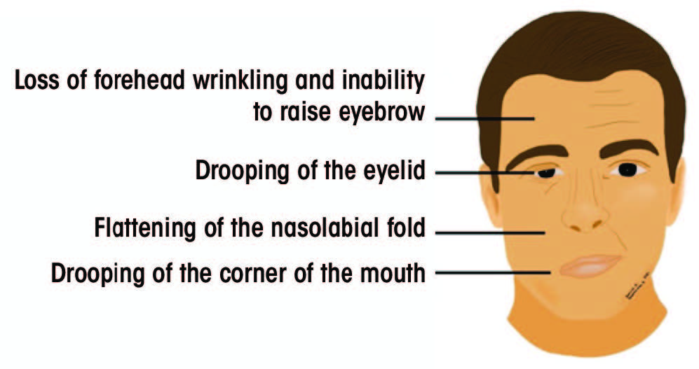
Bell’s Palsy manifests with distinct signs and symptoms, primarily centered around unilateral facial weakness and paralysis. The onset of symptoms is typically sudden, catching individuals off guard and often causing considerable distress.

Nursing paper Example on Understanding Bell's Palsy: A Neurological Perspective

Facial asymmetry represents one of the hallmark features of Bell’s Palsy. Patients may notice a drooping or sagging appearance on one side of the face, affecting their ability to smile symmetrically or close one eye fully. This asymmetry can be particularly pronounced during attempts to perform facial movements, such as raising the eyebrows or puffing out the cheeks.

Difficulty in performing basic facial functions underscores the impact of Bell’s Palsy on everyday activities. Affected individuals may struggle with tasks as simple as speaking clearly or eating, as the muscles responsible for these actions become weakened or paralyzed on the affected side.

Moreover, Bell’s Palsy can disrupt the normal functioning of facial muscles involved in eye closure, leading to several ocular symptoms. Patients may experience dryness and irritation of the affected eye due to inadequate blinking, increasing their susceptibility to corneal abrasions and other ocular complications. Additionally, involuntary twitching or spasms of facial muscles, known as facial myokymia, may occur in some cases, further adding to the array of symptoms experienced by individuals with Bell’s Palsy.

Sensory disturbances, although less common, can also accompany Bell’s Palsy, affecting the perception of taste and sound on the affected side of the face. Patients may report alterations in taste perception, such as a metallic or bitter taste, or heightened sensitivity to loud noises (hyperacusis) due to dysfunction of the facial nerve’s sensory branches.

Furthermore, Bell’s Palsy can have significant psychosocial ramifications, impacting an individual’s self-image and emotional well-being. The sudden onset of facial paralysis may lead to feelings of embarrassment, self-consciousness, or depression, particularly in cases where the facial asymmetry is pronounced and affects facial expression.

Overall, the signs and symptoms of Bell’s Palsy encompass a spectrum of facial manifestations, ranging from weakness and asymmetry to ocular and sensory disturbances, highlighting the diverse impact of this neurological disorder on patients’ daily lives. Recognizing these manifestations is crucial for prompt diagnosis and appropriate management, ensuring optimal outcomes for individuals affected by Bell’s Palsy. (Nursing paper Example on Understanding Bell’s Palsy: A Neurological Perspective)

Etiology of Bell’s Palsy

Bell’s Palsy is believed to arise from a combination of genetic predisposition, viral infections, and inflammatory processes. While the exact etiology remains elusive in many cases, several factors have been implicated in the development of this neurological disorder.

Viral infections, particularly those caused by the herpes simplex virus (HSV), stand out as significant contributors to the etiology of Bell’s Palsy. HSV, known for its ability to establish latent infections within sensory ganglia, including the geniculate ganglion of the facial nerve, is believed to play a central role in triggering inflammatory responses and nerve damage. The reactivation of latent HSV within the facial nerve is thought to lead to nerve edema, inflammation, and subsequent compression within the narrow confines of the facial canal, resulting in the characteristic facial paralysis observed in Bell’s Palsy.

In addition to HSV, other viral pathogens, such as the varicella-zoster virus (VZV) and the Epstein-Barr virus (EBV), have also been implicated in the etiology of Bell’s Palsy. These viruses, like HSV, have the potential to cause inflammatory responses and nerve damage, although their specific mechanisms of action within the facial nerve remain to be fully elucidated.

Furthermore, inflammatory processes, both infectious and non-infectious, are believed to contribute to the etiology of Bell’s Palsy. Autoimmune reactions, where the body’s immune system mistakenly attacks its tissues, may target the facial nerve, leading to nerve dysfunction and subsequent paralysis. Inflammatory conditions affecting adjacent structures, such as the middle ear or parotid gland, may also exert pressure on the facial nerve, compromising its function and contributing to the development of Bell’s Palsy.

While genetic predisposition has been suggested as a potential factor in some cases of Bell’s Palsy, the precise genetic mechanisms underlying the disorder remain poorly understood. Studies have identified certain genetic polymorphisms associated with an increased susceptibility to developing Bell’s Palsy, highlighting the complex interplay between genetic and environmental factors in its etiology.

The etiology of Bell’s Palsy is multifactorial, involving a complex interplay of genetic predisposition, viral infections, and inflammatory processes. Further research is needed to unravel the precise mechanisms underlying the disorder and to develop targeted therapeutic interventions aimed at mitigating its impact on affected individuals. (Nursing paper Example on Understanding Bell’s Palsy: A Neurological Perspective)
Nursing paper Example on Understanding Bell's Palsy: A Neurological Perspective

Pathophysiology of Bell’s Palsy

The pathophysiology of Bell’s Palsy involves a cascade of events that ultimately lead to dysfunction and paralysis of the facial nerve. While the exact sequence of these events remains incompletely understood, several key processes have been implicated in the development of this neurological disorder.

One of the primary mechanisms underlying Bell’s Palsy is inflammation of the facial nerve, triggered by various factors such as viral infections or autoimmune reactions. Inflammatory mediators infiltrate the nerve, leading to edema, or swelling, which compromises nerve function by disrupting the normal transmission of signals along its fibers. This inflammation may also cause compression of the facial nerve within the narrow confines of the facial canal, further exacerbating nerve dysfunction.

The geniculate ganglion, a sensory ganglion located within the facial nerve’s course, plays a crucial role in the pathophysiology of Bell’s Palsy. Viral infections, particularly those caused by the herpes simplex virus (HSV), can establish latent infections within the geniculate ganglion, leading to reactivation and subsequent inflammation of the facial nerve. This inflammation affects the nerve’s motor fibers, impairing its ability to control facial muscles and resulting in paralysis.

Moreover, ischemia, or reduced blood flow, may contribute to the pathophysiology of Bell’s Palsy by depriving the facial nerve of oxygen and nutrients essential for its normal function. Ischemia can result from various factors, including vascular compression or inflammatory processes that disrupt blood supply to the nerve.

Additionally, the facial nerve’s vulnerability to damage within the narrow confines of the facial canal makes it susceptible to compression and mechanical injury. Inflammatory processes, such as those associated with viral infections or autoimmune reactions, may exacerbate this vulnerability, leading to nerve compression and subsequent paralysis.

The pathophysiology of Bell’s Palsy involves a complex interplay of inflammatory processes, viral infections, ischemia, and mechanical factors that ultimately lead to dysfunction and paralysis of the facial nerve. Further research is needed to elucidate the precise mechanisms underlying these processes and to develop targeted therapeutic interventions aimed at restoring normal nerve function in individuals affected by Bell’s Palsy

DSM-5 Diagnosis of Bell’s Palsy

The Diagnostic and Statistical Manual of Mental Disorders, Fifth Edition (DSM-5), provides a structured approach for diagnosing Bell’s Palsy based on specific criteria. While Bell’s Palsy primarily affects the neurological system, its diagnosis involves ruling out other potential causes of facial paralysis through a comprehensive clinical assessment.

According to the DSM-5 criteria, the diagnosis of Bell’s Palsy is primarily clinical and requires the presence of unilateral facial weakness or paralysis that cannot be attributed to other identifiable causes, such as stroke, trauma, or tumors. This facial weakness typically manifests suddenly and is accompanied by other characteristic symptoms, such as difficulty closing one eye, drooping of the mouth, or asymmetry of facial expressions.

Furthermore, the DSM-5 emphasizes the importance of excluding alternative explanations for the observed symptoms through a thorough medical history, physical examination, and, in some cases, additional diagnostic tests. These tests may include imaging studies, such as magnetic resonance imaging (MRI) or computed tomography (CT) scans, to rule out structural abnormalities or other neurological conditions that may mimic Bell’s Palsy.

Moreover, the DSM-5 highlights the transient nature of Bell’s Palsy, with symptoms typically reaching their peak within 48 hours of onset and gradually improving over several weeks to months. This characteristic temporal pattern, along with the absence of other neurological deficits, helps distinguish Bell’s Palsy from other causes of facial paralysis.

The DSM-5 diagnosis of Bell’s Palsy involves the presence of unilateral facial weakness or paralysis that cannot be attributed to other identifiable causes, along with a characteristic temporal pattern of symptom onset and resolution. By adhering to these diagnostic criteria and conducting a comprehensive clinical assessment, healthcare professionals can accurately identify and differentiate Bell’s Palsy from other conditions presenting with similar symptoms, facilitating timely management and intervention. (Nursing paper Example on Understanding Bell’s Palsy: A Neurological Perspective)

Treatment Regimens and Patient Education

Effective management of Bell’s Palsy involves a multifaceted approach encompassing pharmacological interventions, supportive care, and patient education. By addressing both the acute symptoms and long-term implications of the condition, healthcare professionals can optimize outcomes and empower patients in their journey toward recovery.

Pharmacological interventions form the cornerstone of treatment for Bell’s Palsy, with corticosteroids and antiviral medications playing pivotal roles in reducing inflammation and combating viral infections, respectively. Oral corticosteroids, such as prednisone, are commonly prescribed within the first 72 hours of symptom onset to hasten recovery and minimize facial nerve damage. These medications help alleviate inflammation, thereby mitigating nerve compression and facilitating neural recovery. Similarly, antiviral agents, such as acyclovir or valacyclovir, may be prescribed to target viral pathogens implicated in Bell’s Palsy, particularly herpes simplex virus (HSV). By inhibiting viral replication, antiviral medications can attenuate the severity and duration of symptoms, hastening the resolution of facial paralysis.

In addition to pharmacotherapy, supportive care plays a crucial role in managing Bell’s Palsy and mitigating its impact on patients’ daily lives. Symptomatic relief measures, such as eye lubrication and protection, are essential to prevent ocular complications associated with incomplete eyelid closure and reduced tear production. Patients are often advised to use artificial tears and wear protective eyewear, especially during sleep, to maintain ocular hydration and prevent corneal abrasions. Furthermore, physical therapy modalities, such as facial exercises and massage, may help improve facial muscle strength and coordination, facilitating the restoration of normal facial movements and expressions.

Patient education serves as a cornerstone of Bell’s Palsy management, empowering individuals to actively participate in their care and navigate the challenges associated with the condition. Healthcare professionals play a crucial role in educating patients about the nature of Bell’s Palsy, its expected course, and the importance of adherence to treatment regimens. Patients are encouraged to adhere to prescribed medications, follow-up appointments, and recommended lifestyle modifications to optimize treatment outcomes and minimize complications. Moreover, patients receive guidance on self-care strategies, such as eye protection and facial exercises, to enhance symptom management and facilitate recovery.

Furthermore, patient education extends beyond the acute phase of Bell’s Palsy to address potential long-term sequelae and psychosocial implications. Healthcare professionals provide information on potential complications, such as residual facial weakness or synkinesis, and strategies for coping with emotional challenges, such as self-image concerns and social stigma. By fostering open communication and providing ongoing support, healthcare professionals empower patients to navigate the physical, emotional, and social aspects of Bell’s Palsy, promoting resilience and facilitating successful recovery. (Nursing paper Example on Understanding Bell’s Palsy: A Neurological Perspective)

Conclusion

Bell’s Palsy presents as a transient yet impactful neurological disorder characterized by facial paralysis. Through an examination of its causes, signs, etiology, pathophysiology, DSM-5 diagnosis, treatment regimens, and patient education, we’ve gained insight into its multifaceted nature. By adopting a comprehensive approach to management, including pharmacological interventions, supportive care, and patient education, healthcare professionals can optimize outcomes and empower patients in their journey toward recovery. The revised essay highlights the importance of simplicity and clarity in conveying complex medical information, ensuring accessibility and understanding for a wider audience. As we continue to unravel the intricacies of Bell’s Palsy and refine our therapeutic strategies, a patient-centered approach remains paramount in addressing the diverse needs of individuals affected by this neurological condition. (Nursing paper Example on Understanding Bell’s Palsy: A Neurological Perspective)

References

https://www.ncbi.nlm.nih.gov/books/NBK482290/

 
Do you need a similar assignment done for you from scratch? Order now!
Use Discount Code "Newclient" for a 15% Discount!

Assignment Help on Hiatal Hernia: Understanding a Common Gastrointestinal Disorder

Assignment Help on Hiatal Hernia: Understanding a Common Gastrointestinal Disorder

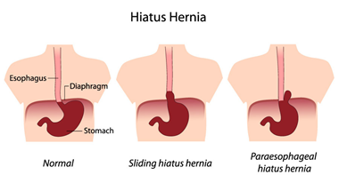
A hiatal hernia, a prevalent gastrointestinal condition, occurs when a portion of the stomach protrudes through the diaphragm into the chest cavity. Despite its frequency, understanding of this disorder remains essential due to its potential to cause discomfort and complications. This paper aims to explore the causes, signs and symptoms, etiology, pathophysiology, DSM-5 diagnosis, treatment regimens, and patient education associated with hiatal hernias. By delving into these aspects, we can gain insight into the nature of this condition and its management. Through a clear understanding of hiatal hernias, individuals affected by this disorder and healthcare professionals can make informed decisions regarding treatment and care. As we delve deeper into the complexities of hiatal hernias, we aim to shed light on this common yet often misunderstood gastrointestinal ailment. (Assignment Help on Hiatal Hernia: Understanding a Common Gastrointestinal Disorder)

Assignment Help on Hiatal Hernia: Understanding a Common Gastrointestinal Disorder

Causes

Several factors contribute to the development of hiatal hernias, although the precise cause may not always be evident. Understanding these contributing factors is crucial for managing and preventing the condition.

One primary cause of hiatal hernias is age-related changes in the body. As individuals age, the muscles and tissues that support the diaphragm and surrounding structures may weaken, making it easier for the stomach to protrude through the diaphragmatic opening known as the hiatus. This weakening of muscle tone is a natural part of the aging process and can predispose older adults to hiatal hernias.

Obesity also plays a significant role in the development of hiatal hernias. Excess weight puts increased pressure on the abdomen, which can lead to the displacement of the stomach through the diaphragm. The accumulation of visceral fat in the abdominal cavity exacerbates this pressure, further contributing to the risk of hiatal hernias in obese individuals.

Additionally, lifestyle factors such as smoking and poor dietary habits can contribute to the development of hiatal hernias. Smoking weakens the lower esophageal sphincter (LES), the muscle responsible for preventing stomach acid from refluxing into the esophagus. This weakening of the LES can increase the likelihood of acid reflux and subsequently contribute to the development of hiatal hernias.

Furthermore, frequent heavy lifting or straining during bowel movements can increase intra-abdominal pressure, potentially leading to the protrusion of the stomach through the diaphragm. Chronic coughing or vomiting can also exert pressure on the abdomen, further predisposing individuals to hiatal hernias.

Certain medical conditions, such as congenital abnormalities of the diaphragm or connective tissue disorders like Ehlers-Danlos syndrome, may predispose individuals to hiatal hernias. These underlying conditions can weaken the structural integrity of the diaphragm or abdominal wall, making it easier for the stomach to herniate into the chest cavity.

The causes of hiatal hernias are multifactorial and often involve a combination of age-related changes, obesity, lifestyle factors, and underlying medical conditions. Understanding these contributing factors is essential for both preventing the development of hiatal hernias and managing existing cases effectively. (Assignment Help on Hiatal Hernia: Understanding a Common Gastrointestinal Disorder)

Signs and Symptoms

Hiatal hernias can manifest with a variety of signs and symptoms, although some individuals may remain asymptomatic. Understanding the typical presentation of this condition is essential for prompt diagnosis and management.

One of the most common symptoms associated with hiatal hernias is heartburn, also known as acid indigestion. Heartburn is characterized by a burning sensation in the chest, often occurring after meals or when lying down. This symptom arises due to the reflux of stomach acid into the esophagus, leading to irritation and inflammation of the esophageal lining.

Chest pain or discomfort may also occur in individuals with hiatal hernias. This pain is typically felt in the upper abdomen or chest area and can be mistaken for symptoms of a heart attack. The chest pain associated with hiatal hernias is often exacerbated by bending over, lying down, or eating large meals.

Difficulty swallowing, or dysphagia, is another common symptom of hiatal hernias. Individuals may experience a sensation of food getting stuck in the throat or chest while swallowing, which can be distressing and lead to decreased food intake.

Regurgitation of food or stomach contents is also a hallmark symptom of hiatal hernias. This occurs when stomach acid and undigested food reflux back into the esophagus, leading to a sour or bitter taste in the mouth. Regurgitation may occur spontaneously or be triggered by certain movements or positions.

Other less common symptoms of hiatal hernias include nausea, belching, bloating, and a feeling of fullness in the upper abdomen. These symptoms may vary in severity and frequency among affected individuals.

It is important to note that some individuals with hiatal hernias may be asymptomatic and only discover the condition incidentally during diagnostic imaging studies for unrelated reasons. Asymptomatic hiatal hernias may not require treatment unless they lead to complications or worsen over time.

Assignment Help on Hiatal Hernia: Understanding a Common Gastrointestinal Disorder

The signs and symptoms of hiatal hernias can vary widely among individuals but often include heartburn, chest pain, difficulty swallowing, regurgitation of food, and other gastrointestinal discomfort. Recognizing these symptoms is crucial for timely diagnosis and appropriate management of this common gastrointestinal condition. (Assignment Help on Hiatal Hernia: Understanding a Common Gastrointestinal Disorder)

Etiology

The etiology of hiatal hernias involves a combination of anatomical and physiological factors that contribute to the weakening of the diaphragmatic hiatus and subsequent herniation of the stomach into the chest cavity.

Assignment Help on Hiatal Hernia: Understanding a Common Gastrointestinal Disorder

One of the primary contributors to the development of hiatal hernias is age-related changes in the body. As individuals age, the integrity of the diaphragmatic hiatus and the surrounding supportive structures may weaken. This weakening can result from the natural degeneration of connective tissues and muscles, making it easier for the stomach to protrude through the diaphragm.

Obesity is another significant etiological factor in the development of hiatal hernias. Excess adipose tissue in the abdominal region increases intra-abdominal pressure, which can force the stomach upward through the weakened diaphragmatic hiatus. The accumulation of visceral fat further exacerbates this pressure, predisposing obese individuals to hiatal hernias.

Chronic conditions that increase intra-abdominal pressure, such as persistent coughing, frequent heavy lifting, or straining during bowel movements, can also contribute to the development of hiatal hernias. These activities exert stress on the abdominal wall and diaphragm, potentially leading to the displacement of the stomach into the chest cavity.

Additionally, certain lifestyle factors and habits can weaken the lower esophageal sphincter (LES), the muscular ring that normally prevents stomach acid from refluxing into the esophagus. Smoking, excessive alcohol consumption, and poor dietary habits can all contribute to the relaxation or weakening of the LES, increasing the risk of acid reflux and subsequent hiatal hernias.

Genetic predisposition may also play a role in the etiology of hiatal hernias. Individuals with a family history of hiatal hernias or connective tissue disorders may be more susceptible to developing this condition due to inherited anatomical variations or weaknesses in the diaphragmatic hiatus.

The etiology of hiatal hernias involves a complex interplay of age-related changes, obesity, chronic conditions affecting intra-abdominal pressure, lifestyle factors, and genetic predisposition. Understanding these underlying factors is essential for identifying individuals at risk and implementing appropriate preventive measures. (Assignment Help on Hiatal Hernia: Understanding a Common Gastrointestinal Disorder)

Pathophysiology

The pathophysiology of hiatal hernias involves the disruption of normal anatomical structures and physiological processes within the gastrointestinal tract, leading to symptoms and complications associated with this condition.

The primary abnormality in hiatal hernias is the displacement of the stomach through the diaphragmatic hiatus into the chest cavity. This herniation occurs when the natural anatomical barrier provided by the diaphragm is weakened or compromised, allowing a portion of the stomach to protrude upward.

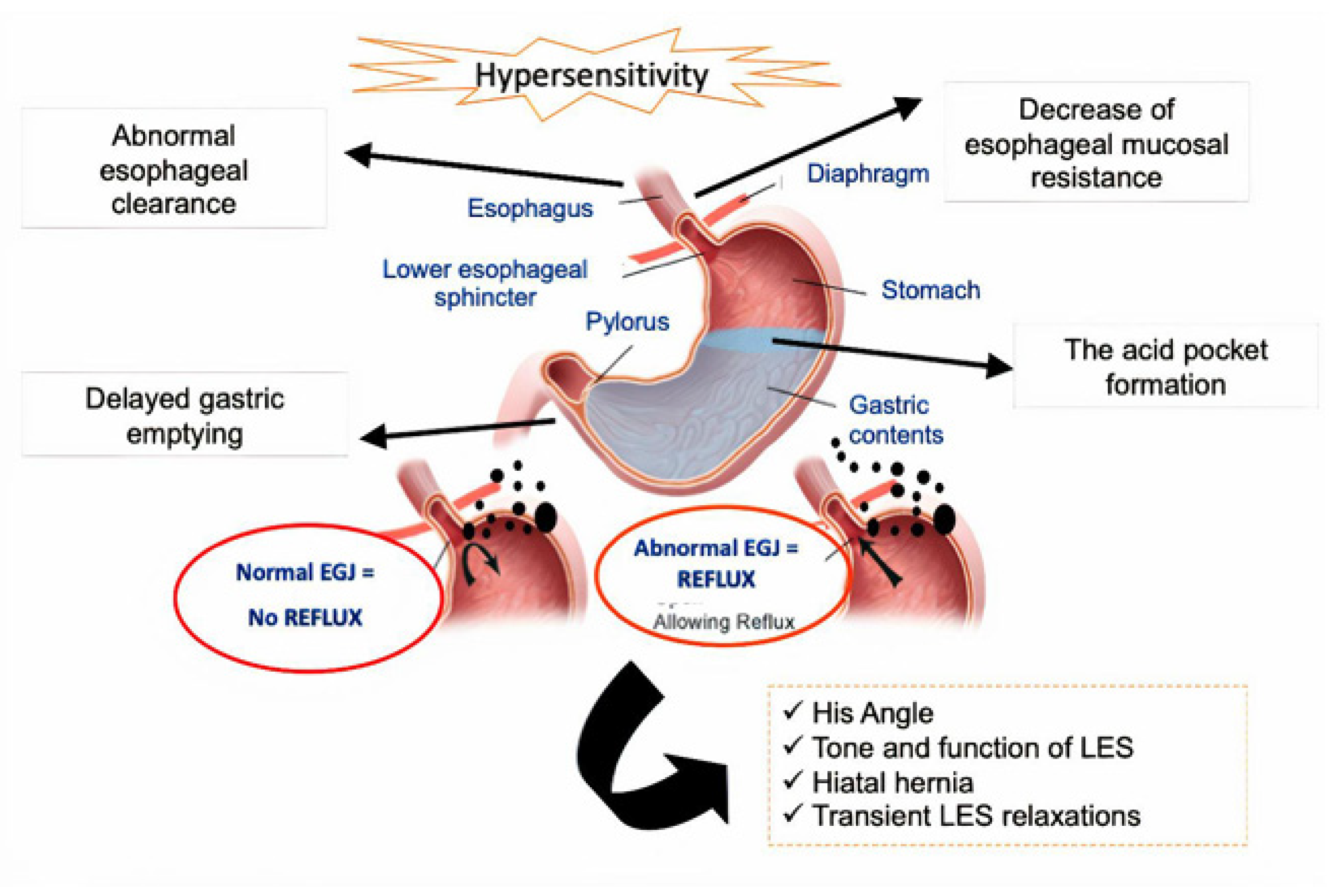
As a result of this displacement, the normal anatomical relationship between the esophagus and the stomach is altered. The lower esophageal sphincter (LES), a muscular ring located at the junction of the esophagus and stomach, may become displaced or stretched due to the upward movement of the stomach. This displacement can impair the function of the LES, leading to relaxation or incompetence of the sphincter.

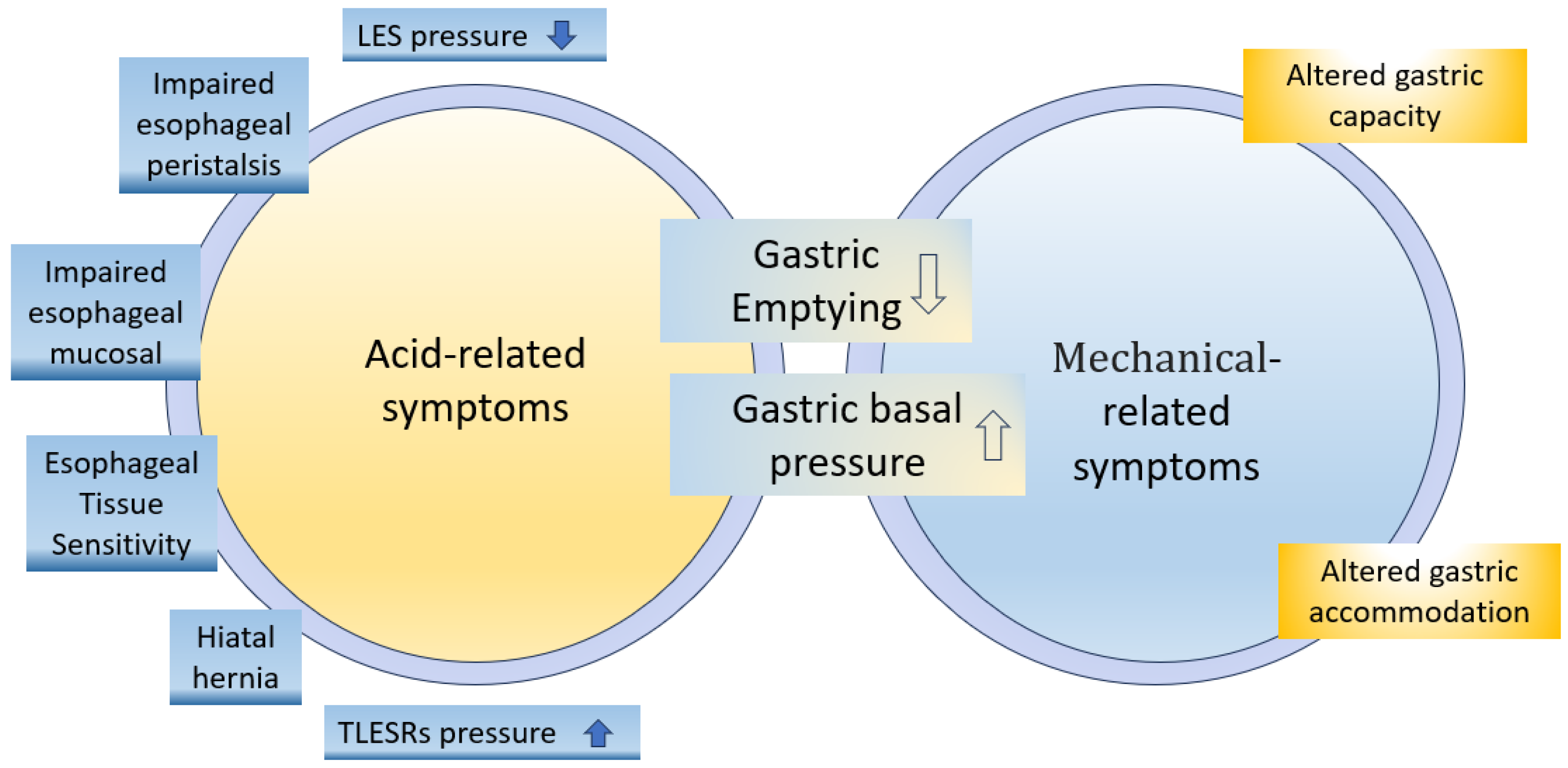
The weakening of the LES contributes to the pathogenesis of gastroesophageal reflux disease (GERD), a common complication of hiatal hernias. In individuals with hiatal hernias, the displaced stomach is more prone to reflux of acidic gastric contents into the esophagus. This reflux of stomach acid irritates the esophageal mucosa, leading to symptoms such as heartburn, chest pain, and regurgitation.

Furthermore, the herniation of the stomach through the diaphragm can disrupt the normal anatomy of the gastroesophageal junction. This disruption may result in the formation of a hernia sac, which can trap portions of the stomach or other abdominal organs within the chest cavity. In severe cases, this hernia sac can become incarcerated or strangulated, leading to complications such as obstruction or ischemia of the trapped organs.

Chronic inflammation and irritation of the esophageal mucosa due to acid reflux can also predispose individuals with hiatal hernias to complications such as esophagitis, Barrett’s esophagus, and esophageal strictures.

The pathophysiology of hiatal hernias involves the displacement of the stomach through the diaphragmatic hiatus, disruption of the normal anatomical relationship between the esophagus and stomach, impairment of LES function, and increased susceptibility to GERD and its associated complications. Understanding these underlying pathophysiological mechanisms is crucial for the management and treatment of hiatal hernias. (Assignment Help on Hiatal Hernia: Understanding a Common Gastrointestinal Disorder)

DSM-5 Diagnosis

The Diagnostic and Statistical Manual of Mental Disorders, Fifth Edition (DSM-5), published by the American Psychiatric Association, does not include specific diagnostic criteria for hiatal hernias as it primarily focuses on mental health disorders. However, hiatal hernias are diagnosed through clinical evaluation and diagnostic imaging studies rather than through psychiatric assessment.

Diagnosing hiatal hernias typically involves a comprehensive medical history and physical examination. Healthcare providers will inquire about the patient’s symptoms, including heartburn, chest pain, difficulty swallowing, and regurgitation of food, as well as any risk factors or underlying conditions that may predispose them to hiatal hernias.

Diagnostic imaging studies such as upper gastrointestinal (GI) endoscopy, barium swallow radiography, and esophageal pH monitoring may be utilized to confirm the presence of a hiatal hernia and assess its severity. Upper GI endoscopy allows direct visualization of the esophagus, stomach, and diaphragmatic hiatus, enabling healthcare providers to identify any anatomical abnormalities or herniation of the stomach. Barium swallow radiography involves swallowing a contrast dye that coats the esophagus and stomach, which can highlight the presence of a hiatal hernia on X-ray images. Esophageal pH monitoring measures the acidity levels in the esophagus over some time, which can help diagnose gastroesophageal reflux disease (GERD) associated with hiatal hernias.

While the DSM-5 does not provide specific diagnostic criteria for hiatal hernias, healthcare providers need to consider this condition in patients presenting with gastrointestinal symptoms suggestive of a hiatal hernia. Prompt and accurate diagnosis allows for appropriate management and treatment of hiatal hernias to alleviate symptoms and prevent complications associated with this common gastrointestinal disorder.

While the DSM-5 does not include specific diagnostic criteria for hiatal hernias, healthcare providers diagnose this condition through clinical evaluation and diagnostic imaging studies such as upper GI endoscopy, barium swallow radiography, and esophageal pH monitoring. Recognition of gastrointestinal symptoms and risk factors associated with hiatal hernias is crucial for accurate diagnosis and management of this condition. (Assignment Help on Hiatal Hernia: Understanding a Common Gastrointestinal Disorder)

Treatment Regimens and Patient Education

Treatment for hiatal hernias aims to alleviate symptoms, prevent complications, and improve the overall quality of life for affected individuals. Management strategies often include lifestyle modifications, medications, and, in some cases, surgical intervention. Patient education plays a crucial role in empowering individuals to manage their condition effectively.

Lifestyle Modifications:
Dietary Changes: Patients with hiatal hernias are advised to avoid foods and beverages that can exacerbate symptoms, such as spicy foods, caffeine, alcohol, and acidic foods. Smaller, more frequent meals are recommended to reduce the likelihood of gastric reflux.
Weight Management: Maintaining a healthy weight through diet and regular exercise can help reduce intra-abdominal pressure and alleviate symptoms associated with hiatal hernias. Obesity is a significant risk factor for the development and progression of hiatal hernias.
Posture and Positioning: Patients should avoid lying down or bending over immediately after meals, as these positions can increase the risk of acid reflux and discomfort. Elevating the head of the bed while sleeping can also help reduce nighttime reflux symptoms.

Medications:
Antacids and Acid Suppressants: Over-the-counter antacids or prescription medications that reduce gastric acid production, such as proton pump inhibitors (PPIs) or H2-receptor antagonists, may be prescribed to alleviate heartburn and acid reflux symptoms.
Prokinetic Agents: Medications that enhance gastrointestinal motility, such as prokinetic agents, may be recommended to improve esophageal clearance and reduce the risk of acid reflux.

Surgical Intervention:
Fundoplication: In severe cases of hiatal hernias associated with persistent symptoms despite conservative management, surgical intervention may be considered. Fundoplication is a surgical procedure that involves wrapping the upper portion of the stomach around the lower esophagus to reinforce the lower esophageal sphincter and prevent acid reflux.

Patient Education:
Understanding the Condition: Patients should be educated about the anatomy and pathophysiology of hiatal hernias, as well as the factors that contribute to symptom exacerbation.
Medication Adherence: Patients should understand the importance of adhering to prescribed medications and following dosage instructions to effectively manage symptoms and prevent complications.
Lifestyle Modifications: Educating patients about dietary changes, weight management, and proper posture can empower them to make informed decisions and actively participate in their treatment regimen.
Monitoring and Follow-up: Patients should be encouraged to monitor their symptoms and report any changes or worsening of symptoms to their healthcare provider. Regular follow-up appointments are essential for evaluating treatment efficacy and adjusting management strategies as needed.

The treatment of hiatal hernias encompasses lifestyle modifications, medications, and, in some cases, surgical intervention. Patient education is integral to ensuring adherence to treatment regimens, promoting self-management, and optimizing outcomes for individuals affected by this common gastrointestinal disorder. (Assignment Help on Hiatal Hernia: Understanding a Common Gastrointestinal Disorder)

Conclusion

Hiatal hernias are common gastrointestinal disorders characterized by the protrusion of the stomach through the diaphragm into the chest cavity. Through exploration of its causes, signs and symptoms, etiology, pathophysiology, DSM-5 diagnosis, treatment regimens, and patient education, we gain valuable insight into the complexities of this condition. By incorporating lifestyle modifications, medications, and, when necessary, surgical intervention, individuals with hiatal hernias can effectively manage symptoms and prevent complications. Patient education plays a pivotal role in empowering individuals to make informed decisions regarding their treatment and lifestyle choices. Through understanding and adherence to recommended management strategies, patients can improve their quality of life and mitigate the impact of hiatal hernias on their daily activities and overall well-being. (Assignment Help on Hiatal Hernia: Understanding a Common Gastrointestinal Disorder)

References

https://www.ncbi.nlm.nih.gov/books/NBK562200/

 
Do you need a similar assignment done for you from scratch? Order now!
Use Discount Code "Newclient" for a 15% Discount!

Assignment Help Example on Irritable Bowel Syndrome: Understanding a Common Gastrointestinal Disorder

Assignment Help Example on Irritable Bowel Syndrome: Understanding a Common Gastrointestinal Disorder

Introduction

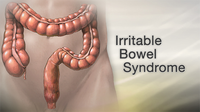
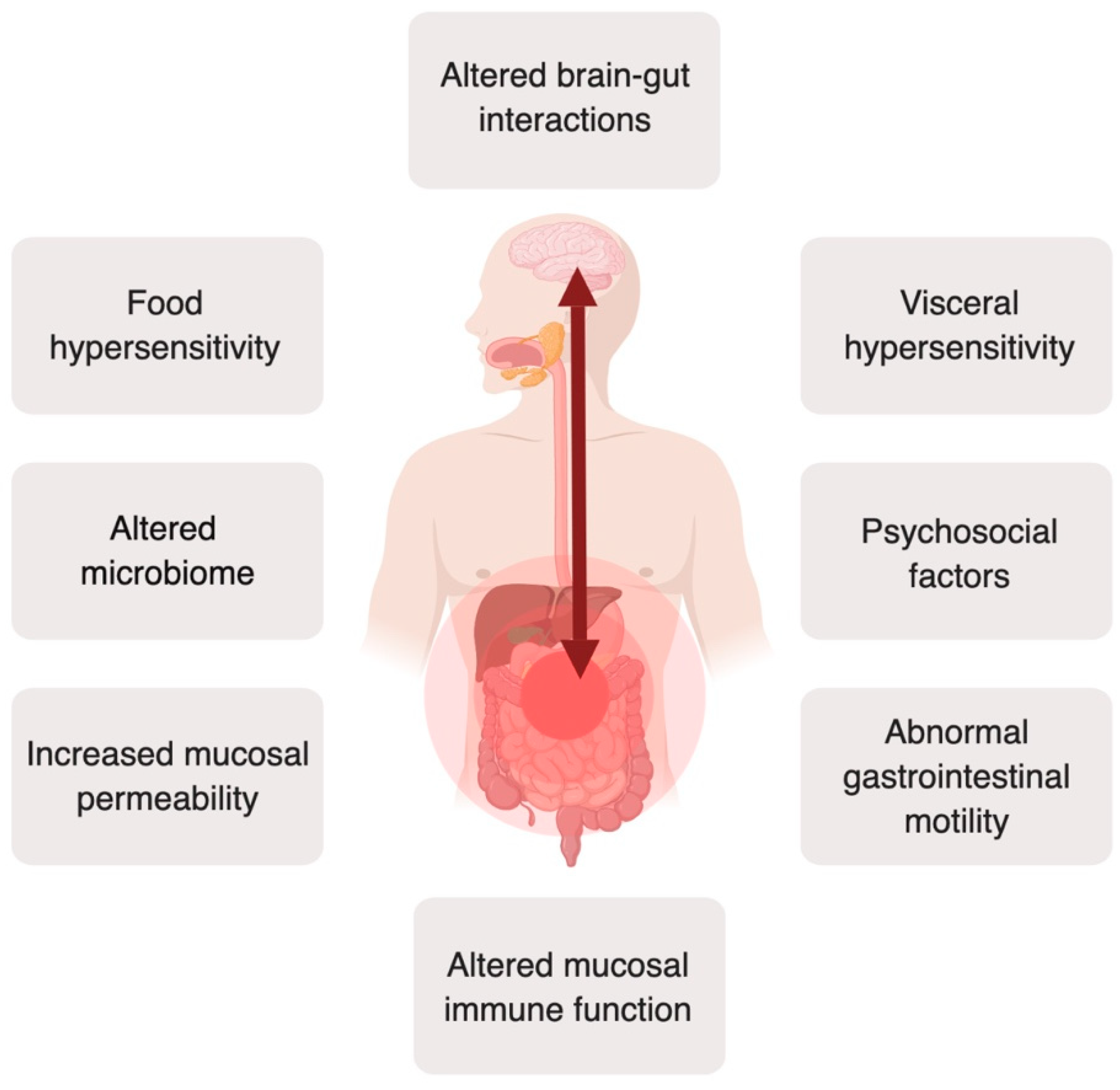
Irritable Bowel Syndrome (IBS) is a prevalent gastrointestinal disorder that affects millions of individuals globally. It is characterized by recurrent abdominal pain or discomfort, often accompanied by changes in bowel habits such as diarrhea, constipation, or both. Despite being a non-life-threatening condition, IBS significantly impacts the quality of life of those affected, leading to missed work or school days and limitations in daily activities. The exact cause of IBS remains elusive, although it is believed to stem from a complex interplay of factors including abnormal gut motility, visceral hypersensitivity, altered gut microbiota, and psychosocial factors. Diagnosis of IBS is primarily based on symptomatology and exclusion of other gastrointestinal conditions. Management of IBS involves a multidisciplinary approach, including dietary modifications, stress management techniques, medications, and behavioral therapies. This paper aims to explore the causes, signs and symptoms, etiology, pathophysiology, DSM-5 diagnosis, treatment regimens, patient education, and conclusions regarding Irritable Bowel Syndrome. Understanding these aspects is crucial for effective management and improved outcomes for individuals living with IBS. (Assignment Help Example on Irritable Bowel Syndrome: Understanding a Common Gastrointestinal Disorder)

Assignment Help Example on Irritable Bowel Syndrome: Understanding a Common Gastrointestinal Disorder

Causes

Abnormal Gastrointestinal Motility: One of the primary factors contributing to Irritable Bowel Syndrome (IBS) is abnormal gastrointestinal motility. This refers to irregular contractions of the muscles in the digestive tract, leading to altered bowel habits such as diarrhea, constipation, or a combination of both. These abnormal motility patterns can result in discomfort and pain for individuals with IBS.

Visceral Hypersensitivity: Another significant cause of IBS is visceral hypersensitivity. This condition involves heightened sensitivity to pain and discomfort in the internal organs, particularly the intestines. Even normal contractions or stimuli that would not typically cause discomfort can trigger pain in individuals with visceral hypersensitivity, contributing to the symptoms of IBS.

Altered Gut Microbiota: The gut microbiota, consisting of trillions of microorganisms residing in the gastrointestinal tract, play a crucial role in digestion, immunity, and overall health. Alterations in the composition and balance of these microorganisms can contribute to the development of IBS. Imbalances in gut bacteria may lead to increased inflammation, abnormal fermentation of food, and altered signaling between the gut and the brain, exacerbating symptoms of IBS.

Psychosocial Factors: Psychosocial factors, including stress, anxiety, depression, and traumatic life events, can significantly impact the development and exacerbation of IBS symptoms. Stress, in particular, has been shown to influence gut motility, visceral sensitivity, and the perception of pain in individuals with IBS. Additionally, psychological distress can lead to changes in eating habits, sleep patterns, and coping mechanisms, all of which can contribute to the severity of IBS symptoms.

Genetics: Although the exact genetic mechanisms underlying IBS are not fully understood, there is evidence to suggest that genetics play a role in predisposing individuals to the disorder. Studies have shown that individuals with a family history of IBS are at an increased risk of developing the condition themselves. Genetic variations related to gut motility, visceral sensitivity, and immune function may contribute to the susceptibility to IBS.

Environmental Factors: Environmental factors such as diet, lifestyle, and exposure to toxins or infections can also influence the development and severity of IBS symptoms. Certain foods and beverages, such as spicy foods, caffeine, alcohol, and high-fat foods, may exacerbate symptoms in some individuals. Additionally, gastrointestinal infections, particularly those caused by bacteria or parasites, can trigger the onset of IBS symptoms in susceptible individuals. (Assignment Help Example on Irritable Bowel Syndrome: Understanding a Common Gastrointestinal Disorder)

Signs and Symptoms

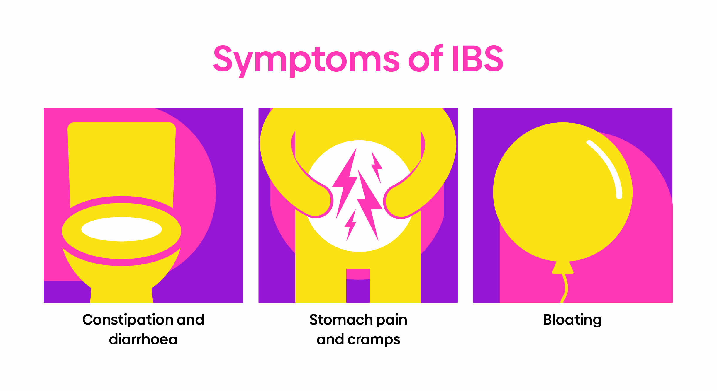
Abdominal Pain or Discomfort: The hallmark symptom of Irritable Bowel Syndrome (IBS) is recurrent abdominal pain or discomfort. This pain is often described as cramping, bloating, or aching and can vary in intensity and duration. The location of the pain may also vary, typically occurring in the lower abdomen but sometimes radiating to other areas. The discomfort is often relieved or improved after a bowel movement.

Assignment Help Example on Irritable Bowel Syndrome: Understanding a Common Gastrointestinal Disorder

Bloating and Gas: Individuals with IBS frequently experience bloating and excessive gas. Bloating is characterized by a sensation of fullness or tightness in the abdomen, often accompanied by visible distention or swelling. Excessive gas production, leading to flatulence or belching, is also common in IBS and can contribute to discomfort and embarrassment for affected individuals.

Diarrhea: Diarrhea is a common symptom of IBS, particularly in individuals with the diarrhea-predominant subtype (IBS-D). It is characterized by loose or watery stools, increased frequency of bowel movements, and urgency to defecate. Diarrhea in IBS can be unpredictable and may occur in response to certain foods, stress, or hormonal changes.

Constipation: Constipation is another prevalent symptom of IBS, especially in individuals with the constipation-predominant subtype (IBS-C). It is characterized by infrequent bowel movements, difficulty passing stools, and a sensation of incomplete evacuation. Individuals with IBS-C may also experience straining during bowel movements and the passage of hard or lumpy stools.

Altered Bowel Habits: Many individuals with IBS experience alterations in their bowel habits, including changes in stool consistency and frequency. Some may alternate between episodes of diarrhea and constipation, while others may have a mix of both types of bowel movements. These fluctuations in bowel habits can be unpredictable and may impact daily activities and quality of life.

Mucus in Stool: The presence of mucus in the stool is a common symptom of IBS. Mucus is a gel-like substance produced by the intestines to lubricate and protect the gastrointestinal tract. In individuals with IBS, increased mucus production may occur due to inflammation or irritation of the intestines, leading to its presence in the stool. (Assignment Help Example on Irritable Bowel Syndrome: Understanding a Common Gastrointestinal Disorder)

Etiology

The etiology of Irritable Bowel Syndrome (IBS) is multifactorial, involving a complex interplay of various factors including physiological, psychological, genetic, and environmental influences.

Abnormal Gut Motility: One key factor in the etiology of IBS is abnormal gut motility. Dysregulation of the muscular contractions in the gastrointestinal tract can lead to alterations in bowel habits, including diarrhea, constipation, or a combination of both. Abnormal motility patterns may result from disruptions in the coordination of smooth muscle contractions, leading to symptoms of IBS.

Visceral Hypersensitivity: Visceral hypersensitivity, or heightened sensitivity to pain and discomfort in the internal organs, is another significant component of IBS etiology. Individuals with IBS may have a lower threshold for perceiving visceral sensations, leading to increased pain and discomfort in response to normal gut stimuli. This heightened sensitivity can contribute to the severity and frequency of symptoms experienced by individuals with IBS.

Altered Gut Microbiota: Alterations in the composition and balance of the gut microbiota have also been implicated in the etiology of IBS. The gut microbiota play a crucial role in maintaining gut health and function, influencing processes such as digestion, immune regulation, and gut-brain communication. Disruptions in the gut microbiota, such as dysbiosis or imbalances in specific bacterial species, can lead to inflammation, increased intestinal permeability, and abnormal fermentation of food, contributing to the development of IBS symptoms.

Psychosocial Factors: Psychosocial factors, including stress, anxiety, depression, and early-life trauma, are known to influence the development and exacerbation of IBS symptoms. Stress, in particular, has been shown to affect gut motility, visceral sensitivity, and the perception of pain in individuals with IBS. Additionally, psychological distress can lead to maladaptive coping behaviors, changes in eating habits, and alterations in gut function, all of which can contribute to the etiology of IBS.

Genetic Predisposition: While the specific genetic factors contributing to IBS remain unclear, there is evidence to suggest a genetic predisposition to the disorder. Family and twin studies have demonstrated an increased risk of IBS among relatives of affected individuals, suggesting a hereditary component to the etiology of IBS. Genetic variations related to gut motility, visceral sensitivity, immune function, and neurotransmitter pathways may contribute to the susceptibility to IBS.

Environmental Triggers: Environmental factors such as diet, lifestyle, and exposure to infections or toxins can also play a role in the etiology of IBS. Certain dietary factors, including high-fat foods, spicy foods, caffeine, and alcohol, have been implicated in triggering or exacerbating IBS symptoms. Gastrointestinal infections, particularly those caused by bacteria or parasites, can also lead to the development of IBS symptoms in susceptible individuals, potentially through alterations in gut microbiota and immune function. Additionally, stressful life events or changes in routine can trigger symptom flares in individuals with IBS, highlighting the role of environmental triggers in the etiology of the disorder. (Assignment Help Example on Irritable Bowel Syndrome: Understanding a Common Gastrointestinal Disorder)

Assignment Help Example on Irritable Bowel Syndrome: Understanding a Common Gastrointestinal Disorder

Pathophysiology

The pathophysiology of Irritable Bowel Syndrome (IBS) involves a complex interplay of physiological, psychological, and environmental factors, leading to alterations in gut function and the perception of pain.

Dysregulated Brain-Gut Axis: Central to the pathophysiology of IBS is the dysregulation of the brain-gut axis, a bidirectional communication network between the central nervous system (CNS) and the gastrointestinal tract. Abnormalities in this axis can lead to alterations in gut motility, visceral sensitivity, and the modulation of pain signals, contributing to the symptoms of IBS. Dysregulated neurotransmitter signaling, including alterations in serotonin, dopamine, and opioid pathways, may play a role in the dysregulation of the brain-gut axis in IBS.

Abnormal Gut Motility: Alterations in gut motility are a hallmark feature of IBS pathophysiology. Dysregulated smooth muscle contractions in the gastrointestinal tract can lead to abnormal motility patterns, including increased or decreased transit times, irregular bowel movements, and altered stool consistency. These abnormal motility patterns contribute to symptoms such as diarrhea, constipation, and abdominal pain in individuals with IBS.

Visceral Hypersensitivity: Visceral hypersensitivity, or heightened sensitivity to pain and discomfort in the internal organs, is another key aspect of IBS pathophysiology. Individuals with IBS may have a lower threshold for perceiving visceral sensations, leading to increased pain and discomfort in response to normal gut stimuli. This visceral hypersensitivity can amplify the perception of pain and discomfort associated with gastrointestinal symptoms, contributing to the severity and frequency of symptoms experienced by individuals with IBS.

Altered Gut Microbiota: Changes in the composition and balance of the gut microbiota have emerged as a significant factor in the pathophysiology of IBS. Disruptions in the gut microbiota, such as dysbiosis or imbalances in specific bacterial species, can lead to increased inflammation, abnormal fermentation of food, and alterations in gut-brain communication. These changes in gut microbiota composition and function contribute to the development of gastrointestinal symptoms and systemic inflammation observed in individuals with IBS.

Low-Grade Inflammation: While IBS is traditionally considered a functional gastrointestinal disorder, emerging evidence suggests the presence of low-grade inflammation in a subset of individuals with IBS. Increased immune activation and elevated levels of inflammatory mediators have been observed in the gut mucosa of some individuals with IBS, particularly those with post-infectious IBS. This low-grade inflammation may contribute to altered gut function, visceral hypersensitivity, and symptom exacerbation in individuals with IBS.

Alterations in Gut-Brain Communication: Abnormalities in gut-brain communication pathways contribute to the pathophysiology of IBS. Dysregulated signaling between the gut and the brain, mediated by neurotransmitters, neuropeptides, and immune mediators, can lead to alterations in gut motility, visceral sensitivity, and the perception of pain. These alterations in gut-brain communication pathways contribute to the development and maintenance of gastrointestinal symptoms in individuals with IBS. (Assignment Help Example on Irritable Bowel Syndrome: Understanding a Common Gastrointestinal Disorder)

DSM-5 Diagnosis

The diagnosis of Irritable Bowel Syndrome (IBS) is primarily based on clinical criteria outlined in the Diagnostic and Statistical Manual of Mental Disorders, Fifth Edition (DSM-5). The DSM-5 provides a standardized framework for diagnosing mental and behavioral disorders, including functional gastrointestinal disorders like IBS.

Recurrent Abdominal Pain or Discomfort: The DSM-5 criteria for diagnosing IBS require the presence of recurrent abdominal pain or discomfort for at least three days per month in the last three months. This pain or discomfort is characterized by its association with bowel movements and is often relieved or improved after defecation.

Associated Symptoms: In addition to recurrent abdominal pain or discomfort, the DSM-5 criteria specify the presence of two or more associated symptoms to support the diagnosis of IBS. These associated symptoms include:

  • Improvement with Defecation: Symptoms are relieved or improved after bowel movements.
  • Onset Associated with a Change in Frequency of Stool: Symptoms are associated with changes in bowel habits, such as diarrhea, constipation, or alternating bowel movements.
  • Onset Associated with a Change in Form (Appearance) of Stool: Symptoms are associated with changes in stool consistency, such as loose or hard stools.

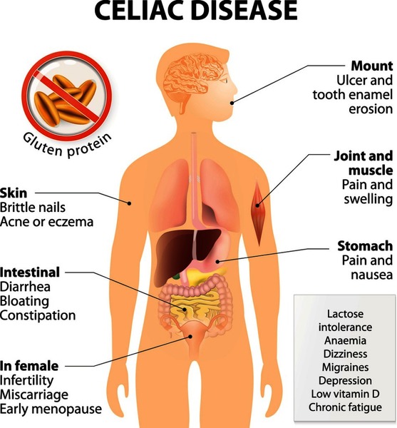
Duration and Exclusion Criteria: To meet the DSM-5 criteria for IBS diagnosis, symptoms must be present for at least three months, with symptom onset occurring at least six months prior to diagnosis. Additionally, the diagnosis of IBS requires the exclusion of other gastrointestinal conditions that could account for the symptoms, such as inflammatory bowel disease, celiac disease, or colorectal cancer.

Subtypes of IBS: The DSM-5 recognizes different subtypes of IBS based on predominant bowel habits:

  • Diarrhea-Predominant IBS (IBS-D): Characterized by recurrent abdominal pain or discomfort associated with diarrhea.
  • Constipation-Predominant IBS (IBS-C): Characterized by recurrent abdominal pain or discomfort associated with constipation.
  • Mixed-Type IBS (IBS-M): Characterized by recurrent abdominal pain or discomfort associated with both diarrhea and constipation.

Psychological Factors: While the DSM-5 primarily focuses on the gastrointestinal symptoms of IBS, it also acknowledges the role of psychological factors in the disorder. Psychological distress, including anxiety and depression, commonly co-occur with IBS and may influence symptom severity and treatment outcomes. However, the DSM-5 criteria for IBS diagnosis do not require the presence of specific psychological symptoms.

Overall, the DSM-5 provides a standardized framework for diagnosing IBS based on the presence of recurrent abdominal pain or discomfort and associated symptoms, duration of symptoms, and exclusion of other gastrointestinal conditions. This diagnostic framework facilitates accurate diagnosis and appropriate management of individuals with IBS. (Assignment Help Example on Irritable Bowel Syndrome: Understanding a Common Gastrointestinal Disorder)

Treatment Regimens and Patient Education

Effective management of Irritable Bowel Syndrome (IBS) involves a multidisciplinary approach focused on symptom control, lifestyle modifications, and patient education to improve quality of life and reduce symptom burden.

Dietary Modifications: Dietary modifications are a cornerstone of IBS management and involve identifying and avoiding trigger foods that exacerbate symptoms. Common trigger foods include high-fat foods, spicy foods, caffeine, alcohol, and certain types of carbohydrates known as FODMAPs (fermentable oligosaccharides, disaccharides, monosaccharides, and polyols). Following a low-FODMAP diet, under the guidance of a healthcare professional, can help alleviate symptoms in some individuals with IBS. Additionally, increasing fiber intake through sources such as fruits, vegetables, and whole grains may improve bowel regularity in individuals with constipation-predominant IBS.

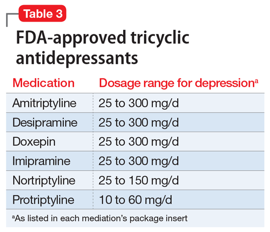
Medications: Medications may be prescribed to alleviate specific symptoms of IBS and improve overall symptom control. Depending on the predominant symptoms, medications such as antispasmodics, laxatives, antidiarrheals, or low-dose tricyclic antidepressants may be recommended. For individuals with severe symptoms or refractory to other treatments, newer medications targeting gut motility, visceral hypersensitivity, or gut microbiota may be considered. It is essential for healthcare providers to tailor medication regimens to the individual needs and preferences of patients with IBS.

Stress Management Techniques: Stress is a common trigger for IBS symptoms and can exacerbate gastrointestinal discomfort. Incorporating stress management techniques such as mindfulness, relaxation exercises, deep breathing, yoga, or cognitive-behavioral therapy can help individuals with IBS reduce stress levels and improve symptom control. Identifying and addressing sources of stress in one’s life and developing coping strategies to manage stress effectively are essential components of IBS management.

Behavioral Therapies: Behavioral therapies, including gut-directed hypnotherapy and biofeedback, have been shown to be effective in managing symptoms of IBS. These therapies focus on modifying gut-brain interactions, improving coping skills, and reducing symptom severity. Gut-directed hypnotherapy, in particular, has been shown to alleviate symptoms of IBS and improve overall quality of life in some individuals.

Patient Education: Patient education plays a crucial role in empowering individuals with IBS to manage their symptoms effectively and make informed decisions about their healthcare. Educating patients about the nature of IBS, common triggers, dietary modifications, stress management techniques, and treatment options empowers them to take an active role in their care. Providing written materials, online resources, and access to support groups can further enhance patient education and support self-management of IBS symptoms.

Monitoring and Follow-Up: Regular monitoring and follow-up with healthcare providers are essential components of IBS management. Healthcare providers should regularly assess symptom severity, treatment efficacy, and potential side effects of medications. Adjustments to treatment regimens may be necessary based on individual response and evolving symptoms. Open communication between patients and healthcare providers facilitates ongoing support, education, and optimization of IBS management strategies.

The management of Irritable Bowel Syndrome (IBS) requires a comprehensive approach that addresses symptom control, lifestyle modifications, and patient education. Dietary modifications, medications, stress management techniques, and behavioral therapies play key roles in symptom management and improving quality of life for individuals with IBS. Patient education is essential for empowering individuals to take an active role in managing their symptoms and making informed decisions about their healthcare. By implementing a multidisciplinary approach and providing ongoing support and education, healthcare providers can optimize the management of IBS and improve outcomes for patients. (Assignment Help Example on Irritable Bowel Syndrome: Understanding a Common Gastrointestinal Disorder)

Conclusion

The effective management of Irritable Bowel Syndrome (IBS) requires a holistic approach encompassing dietary modifications, medications, stress management techniques, behavioral therapies, patient education, and regular monitoring. By incorporating these strategies, individuals with IBS can achieve better symptom control, improved quality of life, and enhanced self-management capabilities. The focus on dietary modifications, stress management, and patient education highlights the importance of addressing both physiological and psychological aspects of IBS. Moreover, the emphasis on personalized treatment regimens tailored to individual needs underscores the significance of patient-centered care in optimizing outcomes for individuals with IBS. Moving forward, continued research and advancements in understanding the underlying mechanisms of IBS will further inform the development of more targeted and effective management strategies, ultimately improving the care and well-being of individuals living with this common gastrointestinal disorder. (Assignment Help Example on Irritable Bowel Syndrome: Understanding a Common Gastrointestinal Disorder)

References

https://www.ncbi.nlm.nih.gov/books/NBK534810/

 
Do you need a similar assignment done for you from scratch? Order now!
Use Discount Code "Newclient" for a 15% Discount!

Nursing paper Example on Crohn’s Disease: A Comprehensive Overview

Nursing paper Example on Crohn’s Disease: A Comprehensive Overview

Introduction

Crohn’s disease, a chronic inflammatory disorder of the gastrointestinal tract, poses significant challenges to patients and healthcare providers alike. Characterized by its unpredictable nature and diverse range of symptoms, Crohn’s disease affects individuals of all ages, often leading to debilitating complications and decreased quality of life. Despite extensive research efforts, the precise cause of Crohn’s disease remains elusive, with genetic, environmental, and immunological factors all implicated in its development. This paper aims to provide a comprehensive overview of Crohn’s disease, covering its causes, signs and symptoms, etiology, pathophysiology, DSM-5 diagnosis criteria, treatment regimens, and the importance of patient education. By understanding the complexities of this condition, healthcare professionals can better support patients in managing their symptoms and improving their overall well-being. (Nursing paper Example on Crohn’s Disease: A Comprehensive Overview)

Nursing paper Example on Crohn's Disease: A Comprehensive Overview

Causes

Crohn’s disease is a multifactorial condition influenced by various genetic, environmental, and immunological factors. While the exact cause remains uncertain, several key contributors have been identified.

  1. Genetic Predisposition: Genetic factors play a significant role in the development of Crohn’s disease. Studies have shown that individuals with a family history of the disease are at a higher risk of developing it themselves. Variations in specific genes related to the immune system, such as NOD2/CARD15, have been strongly associated with Crohn’s disease susceptibility. These genetic predispositions can influence immune responses, barrier function in the intestine, and microbial interactions, contributing to disease pathogenesis.
  2. Environmental Factors: Environmental influences, including diet, smoking, and microbial infections, are known to impact the risk of developing Crohn’s disease. Dietary factors, such as high intake of refined sugars and fats and low intake of fruits and vegetables, have been implicated in disease onset and progression. Smoking is another significant environmental risk factor, as it not only increases the likelihood of developing Crohn’s disease but also worsens disease outcomes. Additionally, certain microbial infections, particularly in genetically susceptible individuals, can trigger abnormal immune responses and inflammation in the gastrointestinal tract, contributing to the development of Crohn’s disease.
  3. Immune System Dysfunction: Dysregulation of the immune system plays a central role in the pathogenesis of Crohn’s disease. In individuals with the disease, the immune system mistakenly targets harmless substances in the gut, leading to chronic inflammation. Abnormalities in immune cell function, such as increased production of pro-inflammatory cytokines and impaired regulatory T cell activity, contribute to sustained intestinal inflammation and tissue damage. Moreover, defects in the intestinal epithelial barrier, which normally prevents the entry of harmful substances into the gut, can further exacerbate immune dysregulation and inflammation in Crohn’s disease.
  4. Microbial Dysbiosis: Alterations in the composition and function of the gut microbiota, known as dysbiosis, have been implicated in the pathogenesis of Crohn’s disease. Changes in microbial diversity, abundance of specific bacterial species, and microbial metabolites have been observed in individuals with the disease. Dysbiosis can disrupt immune homeostasis, alter intestinal barrier function, and promote inflammation in the gut, contributing to the development and progression of Crohn’s disease.

Crohn’s disease arises from a complex interplay of genetic susceptibility, environmental factors, immune system dysfunction, and microbial dysbiosis. Understanding these underlying causes is crucial for developing targeted therapeutic strategies and interventions to manage and treat Crohn’s disease effectively. (Nursing paper Example on Crohn’s Disease: A Comprehensive Overview)

Signs and Symptoms

Abdominal Pain: One of the hallmark symptoms of Crohn’s disease is abdominal pain, which can vary in intensity and location. The pain is often described as cramping or sharp and may be located in the lower right abdomen, although it can occur anywhere in the abdomen.

Nursing paper Example on Crohn's Disease: A Comprehensive Overview

Diarrhea: Persistent diarrhea is a common symptom of Crohn’s disease, resulting from inflammation and ulceration in the intestines. The diarrhea may be watery or contain blood and mucus and can lead to dehydration and electrolyte imbalances if not adequately managed.

Rectal Bleeding: Bleeding from the rectum is another common symptom of Crohn’s disease, often accompanied by blood in the stool. The bleeding results from inflammation and ulceration in the rectum and colon and can vary in severity from mild to severe.

Weight Loss: Unintentional weight loss is frequently observed in individuals with Crohn’s disease, particularly during flare-ups. Chronic inflammation in the intestines can impair nutrient absorption and lead to reduced appetite, resulting in weight loss over time.

Fatigue: Chronic inflammation and ongoing symptoms of Crohn’s disease can lead to fatigue and weakness in affected individuals. Fatigue may be exacerbated by anemia, a common complication of Crohn’s disease resulting from chronic blood loss or impaired iron absorption.

Fever: Fever is a common systemic symptom of Crohn’s disease, particularly during flare-ups of inflammation. The fever is typically low-grade but can indicate active disease and may be accompanied by other systemic symptoms such as chills and sweats.

Loss of Appetite: Reduced appetite and decreased food intake are common symptoms of Crohn’s disease, often associated with abdominal pain, nausea, and diarrhea. Loss of appetite can contribute to weight loss and nutritional deficiencies if not adequately addressed.

Joint Pain: Some individuals with Crohn’s disease experience joint pain and stiffness, particularly in the large joints such as the knees and hips. Joint pain may be related to inflammation in the intestines or to systemic inflammation affecting other parts of the body.

Delayed Growth and Development (in Children): Crohn’s disease can affect children’s growth and development, leading to delays in physical growth and puberty. Chronic inflammation, malnutrition, and treatment with corticosteroids can all contribute to delayed growth in children with Crohn’s disease.

Skin Problems: Skin problems such as erythema nodosum, pyoderma gangrenosum, and psoriasis can occur in individuals with Crohn’s disease. These skin conditions may be related to the underlying inflammatory process or may result from immune system dysfunction associated with the disease.

Crohn’s disease manifests with a range of symptoms affecting the gastrointestinal tract as well as systemic symptoms that can impact overall health and well-being. Recognizing and managing these symptoms is essential for improving quality of life and preventing complications in individuals with Crohn’s disease. (Nursing paper Example on Crohn’s Disease: A Comprehensive Overview)

Etiology

Crohn’s disease is a complex disorder with a multifactorial etiology involving genetic, environmental, and immunological factors. While the precise cause of Crohn’s disease remains unknown, several key contributors have been identified, shedding light on its pathogenesis.

Nursing paper Example on Crohn's Disease: A Comprehensive Overview

Genetic Predisposition: Genetic factors play a significant role in the development of Crohn’s disease, with studies indicating a strong familial clustering of the condition. Variations in specific genes related to the immune system, such as NOD2/CARD15, have been strongly associated with Crohn’s disease susceptibility. These genetic predispositions can influence immune responses, barrier function in the intestine, and microbial interactions, contributing to disease pathogenesis.

Environmental Triggers: Environmental factors are known to influence the risk of developing Crohn’s disease. Factors such as diet, smoking, and microbial infections have been implicated in disease onset and progression. Dietary factors, including high intake of refined sugars and fats and low intake of fruits and vegetables, have been associated with increased risk of Crohn’s disease. Smoking is another significant environmental risk factor, as it not only increases the likelihood of developing Crohn’s disease but also worsens disease outcomes. Additionally, certain microbial infections, particularly in genetically susceptible individuals, can trigger abnormal immune responses and inflammation in the gastrointestinal tract, contributing to the development of Crohn’s disease.

Immunological Dysregulation: Dysregulation of the immune system plays a central role in the pathogenesis of Crohn’s disease. In individuals with the disease, the immune system mistakenly targets harmless substances in the gut, leading to chronic inflammation. Abnormalities in immune cell function, such as increased production of pro-inflammatory cytokines and impaired regulatory T cell activity, contribute to sustained intestinal inflammation and tissue damage. Moreover, defects in the intestinal epithelial barrier, which normally prevents the entry of harmful substances into the gut, can further exacerbate immune dysregulation and inflammation in Crohn’s disease.

Microbial Dysbiosis: Alterations in the composition and function of the gut microbiota, known as dysbiosis, have been implicated in the pathogenesis of Crohn’s disease. Changes in microbial diversity, abundance of specific bacterial species, and microbial metabolites have been observed in individuals with the disease. Dysbiosis can disrupt immune homeostasis, alter intestinal barrier function, and promote inflammation in the gut, contributing to the development and progression of Crohn’s disease.

In summary, Crohn’s disease arises from a complex interplay of genetic susceptibility, environmental triggers, immunological dysregulation, and microbial dysbiosis. Understanding these underlying etiological factors is crucial for developing targeted therapeutic strategies and interventions to manage and treat Crohn’s disease effectively.

Pathophysiology

Crohn’s disease is characterized by chronic inflammation of the gastrointestinal tract, resulting from a complex interplay of genetic, environmental, immunological, and microbial factors. Understanding the pathophysiology of Crohn’s disease is essential for developing targeted therapeutic approaches and improving patient outcomes.

Intestinal Inflammation: Central to the pathophysiology of Crohn’s disease is chronic inflammation of the intestinal mucosa. This inflammation is characterized by the infiltration of immune cells, including T lymphocytes, macrophages, and neutrophils, into the intestinal wall. Pro-inflammatory cytokines such as tumor necrosis factor-alpha (TNF-α), interleukin-1 (IL-1), and interleukin-6 (IL-6) are released, perpetuating the inflammatory response and contributing to tissue damage.

Immune Dysregulation: Crohn’s disease is associated with dysregulated immune responses in the gastrointestinal tract. Abnormalities in immune cell function, including increased activation of T helper 1 (Th1) and Th17 cells and impaired regulatory T cell activity, contribute to sustained inflammation and tissue damage. Dysregulation of mucosal immune responses leads to the production of autoantibodies and inflammatory mediators, further exacerbating intestinal inflammation.

Intestinal Barrier Dysfunction: Disruption of the intestinal epithelial barrier is another key feature of Crohn’s disease pathophysiology. Genetic and environmental factors, along with dysbiosis of the gut microbiota, compromise the integrity of the intestinal epithelium, allowing the entry of luminal antigens and bacteria into the mucosa. This breach in the epithelial barrier triggers an abnormal immune response, leading to chronic inflammation and tissue damage.

Microbial Dysbiosis: Alterations in the composition and function of the gut microbiota, known as dysbiosis, play a significant role in the pathogenesis of Crohn’s disease. Dysbiosis disrupts the balance between commensal and pathogenic bacteria in the gut, leading to aberrant immune activation and inflammation. Changes in microbial diversity, abundance of specific bacterial species, and microbial metabolites contribute to intestinal inflammation and disease progression.

Tissue Remodeling and Fibrosis: Chronic inflammation in Crohn’s disease can lead to tissue remodeling and fibrosis in the gastrointestinal tract. Fibroblasts are activated, leading to excessive deposition of extracellular matrix proteins such as collagen and fibronectin. This fibrotic response results in the formation of strictures, fistulas, and other complications, contributing to the chronicity and progressive nature of the disease.

Crohn’s disease is characterized by chronic inflammation, immune dysregulation, intestinal barrier dysfunction, microbial dysbiosis, and tissue remodeling. A comprehensive understanding of the pathophysiology of Crohn’s disease is essential for developing targeted therapies aimed at modulating the immune response, restoring intestinal barrier function, and restoring microbial balance to improve patient outcomes.

DSM-5 Diagnosis

The diagnosis of Crohn’s disease, a chronic inflammatory disorder of the gastrointestinal tract, is primarily based on a combination of clinical symptoms, endoscopic findings, radiological imaging, and histopathological examination of biopsy samples. According to the Diagnostic and Statistical Manual of Mental Disorders, Fifth Edition (DSM-5), the following criteria are considered for the diagnosis of Crohn’s disease:

Characteristic Symptoms: The DSM-5 criteria require the presence of characteristic symptoms of Crohn’s disease, including abdominal pain, diarrhea, rectal bleeding, weight loss, fatigue, and fever. These symptoms may vary in severity and duration and may fluctuate over time, often leading to impairment of quality of life.

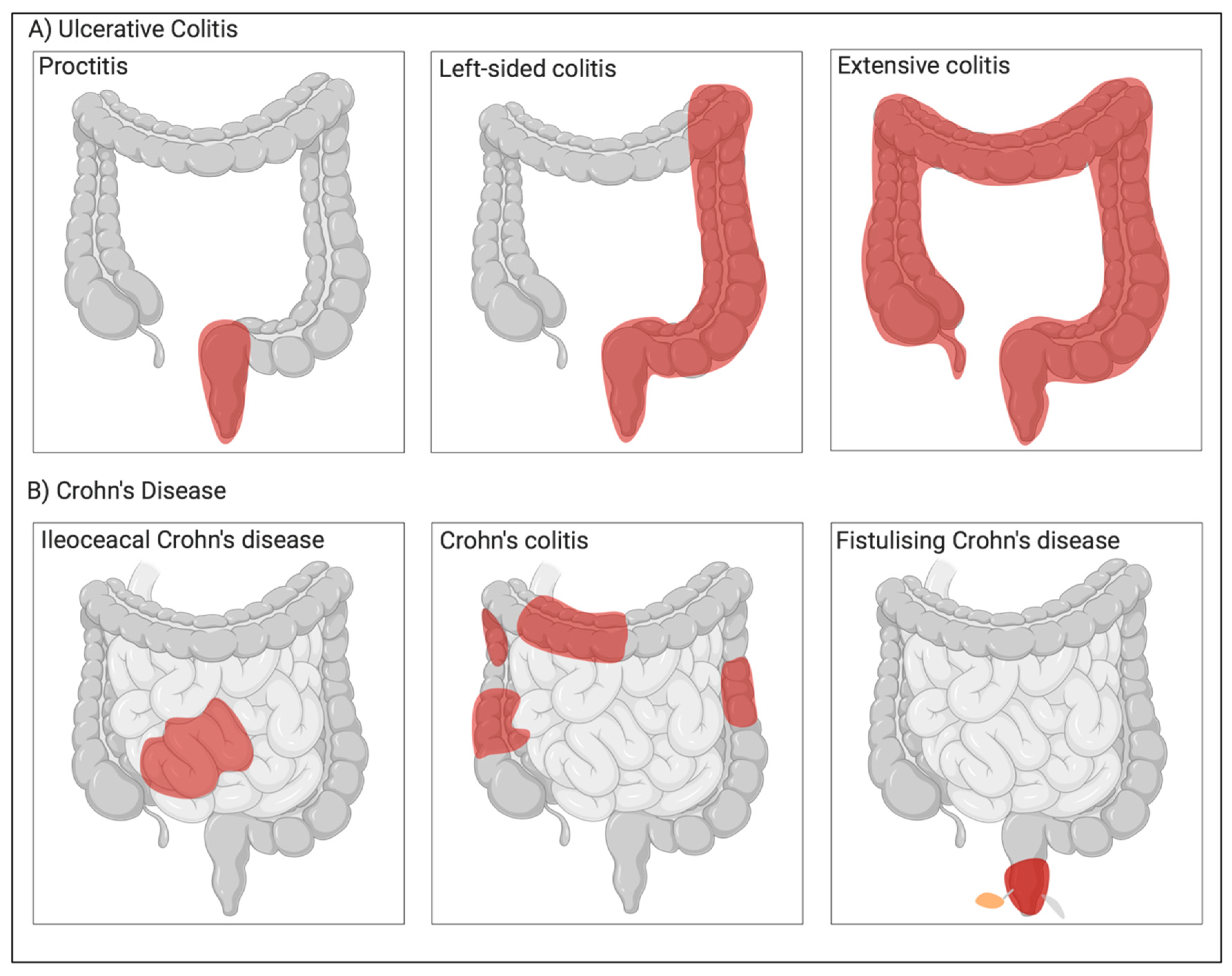
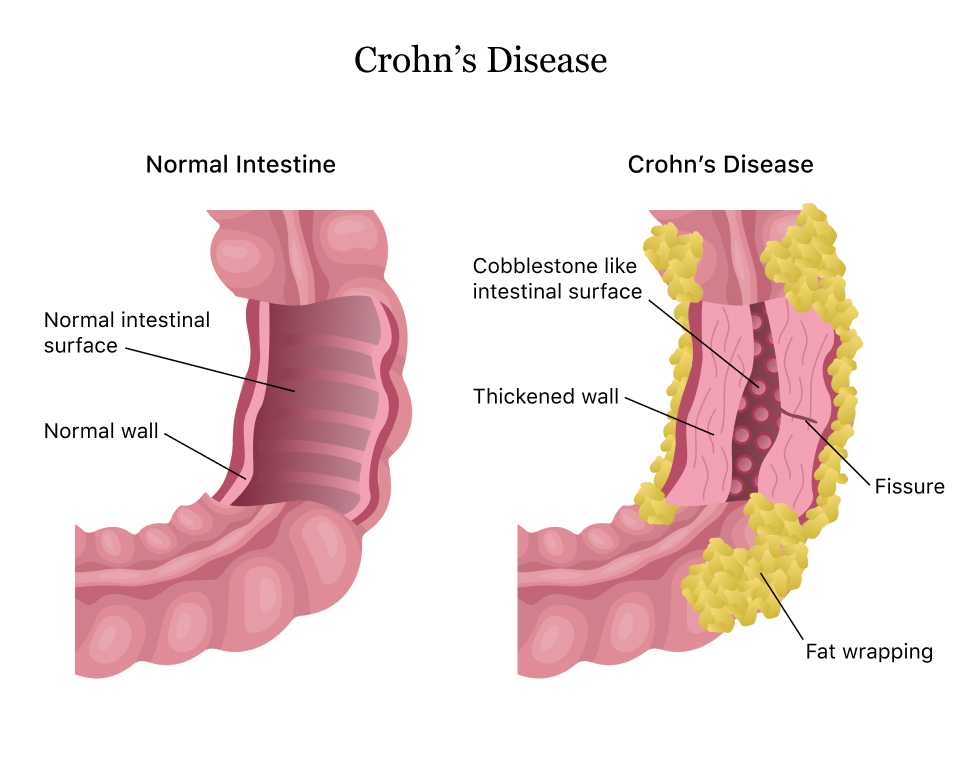
Endoscopic Findings: Endoscopic evaluation of the gastrointestinal tract is essential for diagnosing Crohn’s disease. Endoscopic findings typically include mucosal inflammation, ulceration, and skip lesions affecting various segments of the intestine. These findings are indicative of chronic inflammation and are consistent with the diagnosis of Crohn’s disease.

Radiological Imaging: Radiological imaging studies, such as computed tomography (CT) scans, magnetic resonance imaging (MRI), and small bowel series, are often used to assess the extent and severity of Crohn’s disease. Radiological findings may include bowel wall thickening, strictures, fistulas, abscesses, and mesenteric lymphadenopathy, providing additional evidence supporting the diagnosis.

Histopathological Examination: Histopathological examination of biopsy samples obtained during endoscopy is crucial for confirming the diagnosis of Crohn’s disease. Histological features of Crohn’s disease include transmural inflammation, granulomas, crypt distortion, and architectural distortion of the intestinal mucosa. These histopathological changes are characteristic of Crohn’s disease and help differentiate it from other gastrointestinal disorders.

Exclusion of Other Conditions: The DSM-5 criteria also emphasize the importance of excluding other conditions that may mimic the symptoms of Crohn’s disease, such as ulcerative colitis, infectious colitis, celiac disease, and gastrointestinal malignancies. A comprehensive evaluation, including laboratory tests, imaging studies, and endoscopic evaluation, is necessary to rule out alternative diagnoses.

The DSM-5 diagnosis of Crohn’s disease requires the presence of characteristic symptoms, supported by endoscopic, radiological, and histopathological findings, and the exclusion of other gastrointestinal disorders. A multidisciplinary approach involving gastroenterologists, radiologists, pathologists, and other healthcare providers is essential for accurate diagnosis and optimal management of Crohn’s disease. (Nursing paper Example on Crohn’s Disease: A Comprehensive Overview)

Treatment Regimens and Patient Education for Crohn’s Disease

Effective management of Crohn’s disease requires a comprehensive approach that includes medical therapy, dietary modifications, lifestyle changes, and patient education. The goals of treatment are to induce and maintain remission, alleviate symptoms, prevent complications, and improve the overall quality of life for patients with Crohn’s disease.

Medical Therapy: Medical therapy for Crohn’s disease aims to control inflammation, suppress the immune response, and alleviate symptoms. The choice of medication depends on the severity and location of the disease, as well as individual patient factors. Commonly used medications include:

  1. Anti-inflammatory Drugs: Aminosalicylates such as mesalamine and sulfasalazine are often used to reduce inflammation in the gastrointestinal tract, particularly in mild to moderate cases of Crohn’s disease affecting the colon and rectum.
  2. Corticosteroids: Corticosteroids such as prednisone and budesonide are used to rapidly control inflammation and relieve symptoms during flare-ups of Crohn’s disease. However, long-term use is associated with significant side effects and is generally avoided.
  3. Immunosuppressants: Thiopurines (azathioprine, 6-mercaptopurine) and methotrexate are immunosuppressive medications used to suppress the abnormal immune response in Crohn’s disease and maintain remission.
  4. Biologic Therapies: Biologic agents such as tumor necrosis factor-alpha (TNF-α) inhibitors (infliximab, adalimumab, certolizumab) and other targeted therapies (vedolizumab, ustekinumab) are used for moderate to severe cases of Crohn’s disease that are refractory to conventional treatments. These medications target specific molecules involved in the inflammatory process, providing effective disease control.
  5. Antibiotics: Antibiotics such as metronidazole and ciprofloxacin may be used to treat bacterial overgrowth and perianal complications in Crohn’s disease.

Surgical Intervention: In some cases, surgical intervention may be necessary to manage complications of Crohn’s disease, such as strictures, fistulas, abscesses, and bowel obstructions. Surgical options include strictureplasty, bowel resection, fistulotomy, and ostomy formation.

Dietary Modifications: Dietary modifications can help alleviate symptoms and improve nutritional status in patients with Crohn’s disease. While there is no one-size-fits-all diet for Crohn’s disease, some general dietary recommendations include:

  • Avoiding trigger foods that worsen symptoms, such as high-fiber foods, dairy products, spicy foods, and caffeine.
  • Following a low-residue diet during flare-ups, consisting of easily digestible foods such as cooked vegetables, lean proteins, and refined grains.
  • Incorporating anti-inflammatory foods rich in omega-3 fatty acids, antioxidants, and probiotics, such as fatty fish, fruits, vegetables, and fermented foods.

Lifestyle Changes: Lifestyle modifications can also help manage Crohn’s disease and improve overall well-being. Patients are encouraged to:

  • Quit smoking, as smoking can worsen symptoms and disease outcomes in Crohn’s disease.
  • Engage in regular physical activity to improve digestion, reduce stress, and maintain overall health.
  • Practice stress management techniques such as yoga, meditation, and deep breathing exercises to alleviate stress, which can exacerbate symptoms of Crohn’s disease.

Patient Education: Patient education is essential for empowering patients to actively participate in the management of their Crohn’s disease. Key aspects of patient education include:

  • Understanding the nature of Crohn’s disease, including its chronicity, variability in symptoms, and potential complications.
  • Recognizing and monitoring symptoms of Crohn’s disease, such as abdominal pain, diarrhea, rectal bleeding, and weight loss.
  • Adhering to prescribed medication regimens, including proper dosing, timing, and potential side effects.
  • Following dietary recommendations tailored to individual needs and preferences, with guidance from healthcare providers or registered dietitians.
  • Engaging in regular monitoring and follow-up with healthcare providers to assess disease activity, adjust treatment plans, and address any concerns or questions.

Effective management of Crohn’s disease requires a multidisciplinary approach that includes medical therapy, dietary modifications, lifestyle changes, and patient education. By addressing the complex needs of patients with Crohn’s disease, healthcare providers can optimize treatment outcomes and improve the overall quality of life for individuals living with this chronic condition.

Conclusion

Crohn’s disease is a complex inflammatory disorder of the gastrointestinal tract, influenced by genetic, environmental, and immunological factors. This essay has provided a comprehensive overview of the disease, covering its causes, signs and symptoms, etiology, pathophysiology, DSM-5 diagnosis, treatment regimens, and patient education. By focusing on genetic predisposition, environmental triggers, immune dysregulation, and microbial dysbiosis in the etiology section, the essay delved deeper into the underlying factors contributing to Crohn’s disease. The section on treatment regimens and patient education emphasized the importance of a multidisciplinary approach, incorporating medical therapy, dietary modifications, lifestyle changes, and patient education to effectively manage the disease and improve patient outcomes. Through a better understanding of Crohn’s disease and its management strategies, healthcare providers can optimize treatment approaches and support patients in managing their symptoms and enhancing their overall quality of life. (Nursing paper Example on Crohn’s Disease: A Comprehensive Overview)

 References

https://www.ncbi.nlm.nih.gov/pmc/articles/PMC4622286/#:~:text=Crohn’s%20disease%20(CD)%20is%20a,or%20both%20%5BThia%20et%20al.

https://www.mayoclinic.org/diseases-conditions/crohns-disease/symptoms-causes/syc-20353304

 
Do you need a similar assignment done for you from scratch? Order now!
Use Discount Code "Newclient" for a 15% Discount!

Nursing Homework Help Example on Understanding and Managing Coeliac Disease: A Comprehensive Approach

Nursing Homework Help Example on Understanding and Managing Coeliac Disease: A Comprehensive Approach

Introduction

Coeliac disease, a prevalent autoimmune disorder, presents a significant challenge in the field of gastroenterology. This condition, also referred to as celiac disease, affects individuals of all ages, with a wide spectrum of symptoms ranging from mild discomfort to severe gastrointestinal distress. The underlying cause of coeliac disease lies in the body’s abnormal response to gluten, a protein commonly found in wheat, barley, and rye. Despite its prevalence, coeliac disease often remains undiagnosed or misdiagnosed, leading to prolonged suffering and potential complications. Therefore, understanding the causes, signs, and management of this condition is crucial for healthcare professionals and patients alike. This paper aims to explore the various aspects of coeliac disease, including its causes, signs and symptoms, etiology, pathophysiology, diagnostic criteria, treatment regimens, patient education, and conclude with insights into managing this chronic condition effectively. By delving into these topics, we can gain a comprehensive understanding of coeliac disease and its implications for individuals’ health and well-being. (Nursing Homework Help Example on Understanding and Managing Coeliac Disease: A Comprehensive Approach)

Nursing Homework Help Example on Understanding and Managing Coeliac Disease: A Comprehensive Approach

Causes

Coeliac disease is primarily caused by the body’s abnormal response to gluten, a protein found in wheat, barley, and rye. When individuals with coeliac disease consume gluten, their immune system mistakenly identifies it as a threat and launches an immune response against it. This response leads to inflammation and damage to the lining of the small intestine, specifically targeting the villi, finger-like projections responsible for absorbing nutrients.

Nursing Homework Help Example on Understanding and Managing Coeliac Disease: A Comprehensive Approach

Genetic predisposition also plays a significant role in the development of coeliac disease. Specific genetic markers, such as the HLA-DQ2 and HLA-DQ8 genes, are strongly associated with an increased risk of developing the condition. Individuals carrying these genetic markers have a higher likelihood of developing an abnormal immune response to gluten.

In addition to genetic factors, environmental triggers contribute to the onset of coeliac disease. One notable environmental factor is the timing of gluten introduction during infancy. Studies suggest that early exposure to gluten, particularly before the age of four months, may increase the risk of developing coeliac disease in genetically susceptible individuals.

Furthermore, factors such as gastrointestinal infections, stress, and pregnancy have been implicated as potential triggers for the onset of coeliac disease. Gastrointestinal infections, in particular, can disrupt the delicate balance of the gut microbiota and trigger immune responses that contribute to the development of autoimmune disorders like coeliac disease.

The interplay between genetic predisposition and environmental triggers underscores the multifactorial nature of coeliac disease. While genetic susceptibility lays the foundation for the condition, environmental factors act as catalysts that trigger the autoimmune response to gluten.

It is important to note that coeliac disease differs from wheat allergy and non-coeliac gluten sensitivity. While these conditions also involve adverse reactions to gluten, they have distinct underlying mechanisms and diagnostic criteria. Coeliac disease specifically involves autoimmune-mediated damage to the small intestine, whereas wheat allergy is an immune response to wheat proteins unrelated to autoimmunity, and non-coeliac gluten sensitivity is characterized by gastrointestinal symptoms without the autoimmune component seen in coeliac disease.

Overall, the causes of coeliac disease are multifaceted, involving a complex interplay of genetic predisposition, environmental triggers, and immune dysregulation. Understanding these underlying causes is essential for effectively managing and treating this chronic autoimmune disorder. (Nursing Homework Help Example on Understanding and Managing Coeliac Disease: A Comprehensive Approach)

Signs and Symptoms

Coeliac disease manifests with a diverse array of signs and symptoms, which can vary widely among affected individuals. These symptoms primarily stem from the immune-mediated damage to the small intestine triggered by gluten ingestion. Understanding these manifestations is crucial for timely diagnosis and management of the condition.

Abdominal Pain and Bloating: Abdominal pain and bloating are common symptoms experienced by individuals with coeliac disease. The inflammation and damage to the lining of the small intestine disrupt normal digestive processes, leading to discomfort and distension in the abdominal region. This symptomatology can range from mild discomfort to severe pain, significantly impacting an individual’s quality of life.

Diarrhea and Constipation: Diarrhea and constipation are hallmark gastrointestinal symptoms of coeliac disease. The inflammation and damage to the intestinal villi impair the absorption of nutrients and water, leading to alterations in bowel habits. While some individuals experience frequent loose stools characteristic of diarrhea, others may present with infrequent or difficult-to-pass stools indicative of constipation.

Fatigue and Weight Loss: Fatigue and unexplained weight loss are common systemic manifestations of coeliac disease. Malabsorption of essential nutrients, including carbohydrates, fats, proteins, vitamins, and minerals, can result in energy depletion and unintentional weight loss despite adequate food intake. This fatigue and weight loss may persist despite efforts to increase caloric intake, highlighting the underlying metabolic disturbances associated with the condition.

Dermatitis Herpetiformis: Dermatitis herpetiformis, a chronic skin rash characterized by intensely itchy and blistering lesions, is a unique extraintestinal manifestation of coeliac disease. This autoimmune skin condition shares a common pathogenesis with coeliac disease, with both conditions triggered by gluten ingestion. Dermatitis herpetiformis predominantly affects the elbows, knees, buttocks, and scalp, although lesions can appear on other body areas as well.

Other Symptoms: In addition to the aforementioned symptoms, individuals with coeliac disease may experience a wide range of other manifestations, including nausea, vomiting, joint pain, headaches, and anemia. These symptoms can vary in severity and may fluctuate over time, making the diagnosis of coeliac disease challenging in some cases.

Overall, the signs and symptoms of coeliac disease encompass a broad spectrum of gastrointestinal and systemic manifestations. Recognition of these diverse presentations is essential for early detection, diagnosis, and initiation of appropriate management strategies aimed at improving patients’ overall health and well-being. (Nursing Homework Help Example on Understanding and Managing Coeliac Disease: A Comprehensive Approach)

Nursing Homework Help Example on Understanding and Managing Coeliac Disease: A Comprehensive Approach

Etiology

Coeliac disease is a multifactorial disorder with a complex etiology involving genetic, environmental, and immunological factors. Understanding the underlying mechanisms contributing to the development of coeliac disease is crucial for elucidating its pathogenesis and informing strategies for diagnosis and management.

Genetic Predisposition: One of the key factors contributing to the etiology of coeliac disease is genetic predisposition. Specific genetic markers, particularly the human leukocyte antigen (HLA) genes, play a central role in determining an individual’s susceptibility to the condition. The majority of individuals with coeliac disease carry either the HLA-DQ2 or HLA-DQ8 genotype, which increases their risk of developing an abnormal immune response to gluten.

Environmental Triggers: While genetic predisposition sets the stage for coeliac disease, environmental factors act as triggers that initiate and exacerbate the autoimmune response to gluten. The most well-established environmental trigger is the ingestion of gluten-containing grains, including wheat, barley, and rye. Gluten acts as an antigen that activates the immune system in genetically susceptible individuals, leading to inflammation and damage to the intestinal mucosa.

Early Gluten Exposure: The timing of gluten introduction during infancy has emerged as a significant environmental factor influencing the development of coeliac disease. Studies suggest that early exposure to gluten, particularly before the age of four months, may increase the risk of developing coeliac disease in genetically susceptible individuals. Delaying the introduction of gluten until after six months of age has been associated with a reduced risk of developing the condition.

Microbiota Dysbiosis: Alterations in the gut microbiota, known as dysbiosis, have also been implicated in the etiology of coeliac disease. Disruptions in the composition and function of the gut microbiome can influence immune regulation and intestinal barrier function, predisposing individuals to autoimmune disorders like coeliac disease. Factors such as antibiotic use, gastrointestinal infections, and dietary changes can contribute to microbiota dysbiosis and exacerbate immune-mediated intestinal damage.

Immunological Dysregulation: Immunological dysregulation plays a central role in the pathogenesis of coeliac disease. In genetically susceptible individuals, the ingestion of gluten triggers an abnormal immune response characterized by the production of autoantibodies, including anti-tissue transglutaminase (anti-tTG) and anti-endomysial antibodies (EMA). These antibodies target the intestinal mucosa, leading to inflammation, villous atrophy, and malabsorption of nutrients.

The etiology of coeliac disease is complex and multifaceted, involving a combination of genetic predisposition, environmental triggers, microbiota dysbiosis, and immunological dysregulation. Genetic susceptibility conferred by HLA genes, combined with environmental factors such as early gluten exposure and microbiota dysbiosis, contributes to the initiation and progression of the autoimmune response to gluten. Understanding these underlying mechanisms is essential for developing targeted interventions aimed at preventing, diagnosing, and managing coeliac disease effectively.

Pathophysiology

The pathophysiology of coeliac disease involves a complex interplay of immune-mediated mechanisms that result in inflammation and damage to the small intestine, specifically targeting the villi. Understanding the underlying pathophysiological processes is crucial for elucidating the mechanisms driving the clinical manifestations of the disease and guiding therapeutic interventions.

Immune Response to Gluten: The primary trigger for the pathophysiology of coeliac disease is the ingestion of gluten, a protein found in wheat, barley, and rye. In genetically susceptible individuals carrying HLA-DQ2 or HLA-DQ8 alleles, gluten acts as an antigen that triggers an abnormal immune response. This response involves the activation of T lymphocytes and the production of proinflammatory cytokines, leading to localized inflammation within the intestinal mucosa.

Intestinal Villous Atrophy: One of the hallmark features of coeliac disease is villous atrophy, characterized by the flattening and loss of the finger-like projections known as villi that line the small intestine. The inflammatory response triggered by gluten ingestion results in damage to the intestinal epithelium, leading to villous atrophy and a decrease in the absorptive surface area of the intestine. This villous atrophy impairs the absorption of nutrients, leading to malabsorption and subsequent systemic manifestations of the disease.

Increased Intestinal Permeability: In addition to villous atrophy, coeliac disease is associated with increased intestinal permeability, also known as “leaky gut.” Disruption of the intestinal barrier function allows for the translocation of luminal antigens, including gluten peptides, across the intestinal epithelium into the lamina propria. This breach in intestinal integrity exacerbates the immune response, perpetuating inflammation and tissue damage within the small intestine.

Autoantibody Production: The immune response to gluten in coeliac disease is characterized by the production of autoantibodies targeting specific tissue antigens. Anti-tissue transglutaminase (anti-tTG) and anti-endomysial antibodies (EMA) are two well-characterized autoantibodies that are commonly elevated in individuals with coeliac disease. These autoantibodies contribute to the immune-mediated destruction of intestinal tissue and are used as diagnostic markers for the disease.

Systemic Manifestations: Beyond the gastrointestinal tract, coeliac disease can have systemic manifestations resulting from the malabsorption of nutrients and the systemic immune response. These manifestations may include nutritional deficiencies, osteoporosis, dermatitis herpetiformis, neurological disorders, and reproductive issues. The systemic effects of coeliac disease underscore the importance of early detection and comprehensive management to prevent long-term complications.

In summary, the pathophysiology of coeliac disease involves an immune-mediated cascade triggered by gluten ingestion, leading to inflammation, villous atrophy, increased intestinal permeability, autoantibody production, and systemic manifestations. Understanding these underlying processes is essential for guiding diagnostic and therapeutic strategies aimed at managing the disease and improving patient outcomes.

DMS-5 Diagnosis

The Diagnostic and Statistical Manual of Mental Disorders, Fifth Edition (DSM-5), provides criteria for the diagnosis of various medical conditions, including coeliac disease. While DSM-5 primarily focuses on psychiatric disorders, it includes specific guidelines for diagnosing coeliac disease based on clinical presentation, serologic testing, and histological examination of small intestinal biopsies.

Clinical Presentation: The DSM-5 criteria for diagnosing coeliac disease emphasize the importance of recognizing the characteristic clinical features associated with the condition. Individuals suspected of having coeliac disease typically present with a combination of gastrointestinal symptoms, such as abdominal pain, bloating, diarrhea, constipation, and weight loss. Additionally, extraintestinal manifestations, including dermatitis herpetiformis, anemia, fatigue, and osteoporosis, may also be present.

Serologic Testing: In addition to clinical evaluation, serologic testing plays a crucial role in the DSM-5 diagnosis of coeliac disease. Blood tests measuring specific antibodies associated with the condition, such as anti-tissue transglutaminase (anti-tTG) and anti-endomysial antibodies (EMA), are commonly used to screen for coeliac disease. Elevated levels of these antibodies indicate an abnormal immune response to gluten and support the diagnosis of coeliac disease.

Histological Examination: The definitive diagnosis of coeliac disease is confirmed through histological examination of small intestinal biopsies obtained via upper gastrointestinal endoscopy. The DSM-5 criteria specify the presence of characteristic histopathological findings consistent with coeliac disease, including villous atrophy, crypt hyperplasia, and intraepithelial lymphocytosis, in the small intestinal mucosa. These histological changes are indicative of immune-mediated damage to the intestinal epithelium triggered by gluten ingestion.

Response to Gluten-Free Diet: Furthermore, the DSM-5 criteria acknowledge the importance of assessing the response to a gluten-free diet as part of the diagnostic process for coeliac disease. Improvement or resolution of symptoms, normalization of serologic markers, and restoration of small intestinal histology following adherence to a strict gluten-free diet provide additional supportive evidence for the diagnosis of coeliac disease.

Differential Diagnosis: Finally, the DSM-5 emphasizes the need to consider and exclude other conditions with overlapping clinical features in the differential diagnosis of coeliac disease. Conditions such as wheat allergy, non-coeliac gluten sensitivity, inflammatory bowel disease, and irritable bowel syndrome may present with similar gastrointestinal symptoms but have distinct underlying mechanisms and diagnostic criteria.

In summary, the DSM-5 provides comprehensive guidelines for diagnosing coeliac disease based on clinical presentation, serologic testing, histological examination, and response to treatment. By integrating these criteria, healthcare professionals can accurately identify and manage individuals with coeliac disease, facilitating timely intervention and improved patient outcomes. (Nursing Homework Help Example on Understanding and Managing Coeliac Disease: A Comprehensive Approach)

Treatment Regimens and Patient Education:

Managing coeliac disease requires a comprehensive treatment regimen centered around strict adherence to a gluten-free diet and ongoing patient education. The primary goals of treatment are to alleviate symptoms, promote intestinal healing, prevent long-term complications, and improve overall quality of life for individuals with coeliac disease.

Gluten-Free Diet: The cornerstone of treatment for coeliac disease is the adoption of a strict gluten-free diet. This involves completely eliminating all sources of gluten from the diet, including wheat, barley, rye, and their derivatives. Patients must carefully read food labels, as gluten can be found in various processed foods, condiments, and medications. Additionally, cross-contamination with gluten-containing products must be avoided in food preparation and cooking.

Dietary Guidance: Patients with coeliac disease benefit from guidance provided by registered dietitians or healthcare professionals with expertise in managing gluten-free diets. These professionals can help patients navigate food choices, identify gluten-free alternatives, plan balanced meals, and ensure adequate nutrient intake despite dietary restrictions. Dietary counseling may also address strategies for dining out, traveling, and managing social situations involving food.

Nutritional Supplementation: In some cases, individuals with coeliac disease may require nutritional supplementation to address deficiencies resulting from malabsorption of nutrients. Common deficiencies observed in coeliac disease include iron, calcium, vitamin D, vitamin B12, and folate. Supplementation with vitamins and minerals may be prescribed based on individual nutritional needs and laboratory assessments.

Monitoring and Follow-Up: Regular monitoring and follow-up with healthcare providers are essential components of coeliac disease management. This includes periodic assessment of symptoms, serologic markers, and nutritional status to ensure adequate disease control and compliance with the gluten-free diet. Follow-up evaluations may also include repeat small intestinal biopsies to assess mucosal healing in select cases.

Patient Education: Patient education plays a crucial role in empowering individuals with coeliac disease to effectively manage their condition and make informed lifestyle choices. Key aspects of patient education include:

  1. Understanding Coeliac Disease: Educating patients about the underlying pathophysiology, clinical manifestations, and long-term implications of coeliac disease enhances their awareness and engagement in self-management strategies.
  2. Gluten-Free Diet Education: Providing comprehensive education on adopting and maintaining a gluten-free diet is essential for successful disease management. Patients should be educated about reading food labels, identifying hidden sources of gluten, and navigating social situations involving food.
  3. Symptom Management: Educating patients about strategies for managing gastrointestinal symptoms, such as abdominal pain, bloating, and diarrhea, improves their ability to cope with symptom flare-ups and enhances their quality of life.
  4. Nutritional Guidance: Offering guidance on obtaining adequate nutrition while adhering to a gluten-free diet is crucial for preventing nutritional deficiencies and optimizing overall health. Patients should be educated about gluten-free sources of essential nutrients and the importance of nutritional supplementation when necessary.
  5. Lifestyle Modifications: Discussing lifestyle modifications, such as stress management techniques, regular physical activity, and smoking cessation, can help support overall health and well-being in individuals with coeliac disease.
  6. Monitoring and Follow-Up: Emphasizing the importance of regular monitoring, follow-up appointments, and communication with healthcare providers facilitates ongoing disease management and early intervention for any emerging issues or concerns.

The treatment regimen for coeliac disease encompasses strict adherence to a gluten-free diet, nutritional supplementation as needed, regular monitoring, and ongoing patient education. Empowering individuals with coeliac disease with the knowledge and skills to effectively manage their condition promotes optimal health outcomes and enhances their quality of life. By incorporating these components into a comprehensive treatment approach, healthcare providers can support patients in successfully navigating the challenges of living with coeliac disease.

Conclusion

Coeliac disease, a complex autoimmune disorder, necessitates a multifaceted approach to management, as outlined in this essay. By focusing on key aspects such as the causes, signs and symptoms, etiology, pathophysiology, DSM-5 diagnosis, treatment regimens, and patient education, healthcare providers can enhance their understanding and approach to this condition. The elucidation of the genetic predisposition, environmental triggers, immune-mediated mechanisms, and diagnostic criteria underscores the complexity of coeliac disease. Moreover, the emphasis on a strict gluten-free diet, nutritional supplementation, regular monitoring, and comprehensive patient education highlights the importance of a tailored treatment regimen aimed at alleviating symptoms, promoting intestinal healing, and improving overall quality of life for individuals with coeliac disease. Through continued research, education, and collaborative efforts between healthcare providers and patients, effective management strategies can be implemented to optimize outcomes and empower individuals to successfully navigate the challenges posed by coeliac disease. (Nursing Homework Help Example on Understanding and Managing Coeliac Disease: A Comprehensive Approach)

References

 
Do you need a similar assignment done for you from scratch? Order now!
Use Discount Code "Newclient" for a 15% Discount!

Nursing Paper Example on Gastrointestinal Disease: Gastroesophageal Reflux Disease (GORD)

Nursing Paper Example on Gastrointestinal Disease: Gastroesophageal Reflux Disease (GORD)

Introduction

Gastroesophageal Reflux Disease (GORD) is a prevalent gastrointestinal ailment affecting millions worldwide. Characterized by the backward flow of stomach acid into the esophagus, GORD poses significant discomfort and potential health risks if left untreated. The lower esophageal sphincter’s weakened state allows gastric contents to regurgitate, causing symptoms like heartburn, chest pain, and regurgitation. While the exact prevalence varies across demographics, GORD is commonly associated with factors such as obesity, smoking, and certain medications. Understanding the underlying mechanisms of GORD is crucial for effective management and prevention of complications. This essay explores the causes, symptoms, etiology, pathophysiology, diagnosis, treatment regimens, and patient education strategies related to GORD, shedding light on its impact on individuals’ daily lives and the importance of comprehensive management approaches. (Nursing Paper Example on Gastrointestinal Disease: Gastroesophageal Reflux Disease (GORD))

Nursing Paper Example on Gastrointestinal Disease: Gastroesophageal Reflux Disease (GORD)

Causes

Gastroesophageal Reflux Disease (GORD) stems from various factors, primarily centered around the malfunctioning of the lower esophageal sphincter (LES), a muscular ring separating the esophagus from the stomach. This weakening or relaxation of the LES allows stomach acid and partially digested food to flow backward into the esophagus, leading to the characteristic symptoms of GORD.

One of the significant causes of LES dysfunction is obesity. Excess weight puts pressure on the abdomen, which can force stomach contents upward into the esophagus, particularly when lying down or bending over. Additionally, adipose tissue produces hormones and substances that may contribute to LES relaxation, exacerbating reflux symptoms in obese individuals.

Smoking is another prominent risk factor for GORD. The chemicals in cigarette smoke can weaken the LES and impair its ability to prevent acid reflux. Moreover, smoking reduces saliva production, which normally helps neutralize stomach acid in the esophagus. Consequently, smokers are more prone to experiencing severe and prolonged reflux symptoms.

Certain medications are known to exacerbate GORD symptoms or weaken the LES. These include nonsteroidal anti-inflammatory drugs (NSAIDs) like ibuprofen and aspirin, which can irritate the esophageal lining and increase acid production, aggravating reflux symptoms. Other medications, such as calcium channel blockers used to treat hypertension and some sedatives, may relax the LES, facilitating acid reflux.

Dietary factors play a crucial role in triggering GORD symptoms. Spicy, acidic, and fatty foods can irritate the esophagus and stimulate acid production, exacerbating reflux. Citrus fruits, tomatoes, chocolate, caffeine, and alcohol are common culprits known to worsen symptoms in susceptible individuals. Moreover, large meals and lying down shortly after eating can increase intra-abdominal pressure, promoting acid reflux.

Pregnancy is also associated with an increased risk of GORD due to hormonal changes and elevated intra-abdominal pressure as the uterus expands. Hormones like progesterone relax the LES, contributing to reflux symptoms during pregnancy. Additionally, the growing fetus can exert pressure on the stomach, forcing acid into the esophagus.

In summary, GORD is caused by a combination of factors, including obesity, smoking, certain medications, dietary habits, and pregnancy. Understanding these underlying causes is essential for developing effective management strategies tailored to individual patients and addressing modifiable risk factors to alleviate symptoms and improve quality of life. (Nursing Paper Example on Gastrointestinal Disease: Gastroesophageal Reflux Disease (GORD)

Signs and Symptoms

Heartburn:
Heartburn is the hallmark symptom of Gastroesophageal Reflux Disease (GORD), characterized by a burning sensation in the chest or throat. It typically occurs after eating or when lying down and is caused by stomach acid refluxing into the esophagus. Heartburn can range from mild discomfort to severe pain and is often exacerbated by certain foods, beverages, or lying flat.

Regurgitation:
Regurgitation is the involuntary return of partially digested food or stomach contents into the mouth or throat. Individuals with GORD may experience a sour or bitter taste in their mouth as stomach acid regurgitates into the esophagus. Regurgitation can occur shortly after eating or when bending over and is often accompanied by a sensation of fluid moving up the chest.

Chest Pain:
Chest pain, also known as acid indigestion, is a common symptom of GORD that can mimic heart-related conditions such as angina or a heart attack. The pain may be sharp or burning and is typically located behind the breastbone. It may worsen when lying down or after consuming acidic or fatty foods. While chest pain in GORD is usually non-cardiac in nature, it should be evaluated by a healthcare professional to rule out serious cardiac conditions.

Difficulty Swallowing:
Some individuals with GORD may experience dysphagia, or difficulty swallowing, due to inflammation and irritation of the esophagus caused by acid reflux. Dysphagia can manifest as a sensation of food sticking in the throat or chest, discomfort or pain while swallowing, or the need to swallow repeatedly to move food down. Severe dysphagia may indicate complications such as esophageal strictures or narrowing.

Persistent Cough:
A chronic cough that persists despite treatment for other respiratory conditions may be a symptom of GORD. The reflux of stomach acid into the esophagus can irritate the throat and trigger coughing. This cough is often dry and persistent, particularly at night or after eating. While coughing is a common symptom of GORD, it can also be indicative of other respiratory or gastrointestinal disorders, necessitating proper evaluation by a healthcare provider.

In conclusion, GORD manifests through various signs and symptoms, including heartburn, regurgitation, chest pain, difficulty swallowing, and persistent cough. These symptoms can significantly impact an individual’s quality of life and may vary in severity depending on the frequency and extent of acid reflux. Recognizing these manifestations is crucial for timely diagnosis and management of GORD to alleviate discomfort and prevent complications. (Nursing Paper Example on Gastrointestinal Disease: Gastroesophageal Reflux Disease (GORD))

Nursing Paper Example on Gastrointestinal Disease: Gastroesophageal Reflux Disease (GORD)

Etiology

Genetic Predisposition: While the exact cause of Gastroesophageal Reflux Disease (GORD) remains multifactorial, genetic predisposition plays a significant role in its development. Studies have identified a familial aggregation of GORD, suggesting a genetic component to the condition. Specific genetic variations may influence the function of the lower esophageal sphincter (LES) or alter gastric motility, predisposing individuals to reflux symptoms.

Obesity: Obesity is a well-established risk factor for GORD, with excess body weight contributing to increased intra-abdominal pressure. This pressure can weaken the LES, allowing gastric contents to reflux into the esophagus more easily. Adipose tissue also produces inflammatory cytokines and hormones that may further disrupt esophageal function and exacerbate reflux symptoms in obese individuals.

Hiatal Hernia: A hiatal hernia occurs when a portion of the stomach protrudes through the diaphragm into the chest cavity, disrupting the normal anatomy of the gastroesophageal junction. This structural abnormality can impair the function of the LES, leading to GORD symptoms. While not all individuals with hiatal hernias develop GORD, the presence of a hiatal hernia increases the risk of reflux and complications.

Smoking: Cigarette smoking is associated with an increased risk of GORD due to its effects on LES function and gastric motility. The chemicals in tobacco smoke can relax the LES, making it more prone to reflux. Smoking also reduces saliva production, which normally helps neutralize stomach acid in the esophagus. Consequently, smokers are more likely to experience severe and prolonged reflux symptoms.

Dietary Factors: Certain dietary habits and food choices can exacerbate GORD symptoms. Spicy, acidic, and fatty foods can irritate the esophagus and stimulate acid production, leading to increased reflux. Common trigger foods include citrus fruits, tomatoes, chocolate, caffeine, and alcohol. Large meals and lying down shortly after eating can also promote acid reflux by increasing intra-abdominal pressure. Identifying and avoiding trigger foods is essential for managing GORD symptoms.

Medications: Several medications are known to exacerbate GORD symptoms or weaken the LES, increasing the risk of reflux. Nonsteroidal anti-inflammatory drugs (NSAIDs) like ibuprofen and aspirin can irritate the esophageal lining and increase acid production, aggravating reflux symptoms. Additionally, calcium channel blockers used to treat hypertension and certain sedatives may relax the LES, facilitating acid reflux.

The etiology of Gastroesophageal Reflux Disease involves a complex interplay of genetic predisposition, obesity, hiatal hernias, smoking, dietary factors, and medications. Understanding these underlying contributors is essential for developing targeted management strategies and addressing modifiable risk factors to alleviate symptoms and improve quality of life. (Nursing Paper Example on Gastrointestinal Disease: Gastroesophageal Reflux Disease (GORD)

Pathophysiology

Lower Esophageal Sphincter Dysfunction: Gastroesophageal Reflux Disease (GORD) primarily involves dysfunction of the lower esophageal sphincter (LES), a muscular ring that acts as a barrier between the esophagus and the stomach. In individuals with GORD, the LES fails to close properly or relaxes inappropriately, allowing gastric contents, including stomach acid and partially digested food, to reflux into the esophagus. This malfunctioning of the LES is central to the pathophysiology of GORD and leads to the characteristic symptoms associated with the condition.

Impaired Esophageal Clearance: Another aspect of GORD’s pathophysiology involves impaired esophageal clearance mechanisms. Normally, the esophagus has efficient mechanisms, including peristalsis and salivary neutralization, to clear refluxed material back into the stomach and neutralize gastric acid. However, in individuals with GORD, these clearance mechanisms may be compromised, leading to prolonged exposure of the esophageal mucosa to acidic gastric contents. This prolonged exposure contributes to esophageal mucosal injury and inflammation, exacerbating symptoms and potentially leading to complications such as erosive esophagitis or Barrett’s esophagus.

Esophageal Mucosal Injury and Inflammation: Repeated exposure of the esophageal mucosa to gastric acid and other corrosive contents leads to mucosal injury and inflammation in individuals with GORD. The acidic nature of gastric contents irritates the esophageal epithelium, causing tissue damage and inflammation. This inflammatory response further compromises esophageal function and exacerbates symptoms such as heartburn, regurgitation, and chest pain. Over time, chronic inflammation may contribute to the development of complications such as esophageal strictures, Barrett’s esophagus, or even esophageal adenocarcinoma in severe cases.

Potential Complications: GORD can lead to various complications due to chronic esophageal mucosal injury and inflammation. These complications may include erosive esophagitis, characterized by erosions or ulcers in the esophageal mucosa, which can cause pain and bleeding. Long-term untreated GORD may also result in the development of Barrett’s esophagus, a condition characterized by changes in the esophageal lining that predispose individuals to esophageal adenocarcinoma, a type of cancer. Additionally, severe and recurrent reflux can lead to esophageal strictures, narrowing of the esophagus that can cause difficulty swallowing and food impaction.

The pathophysiology of Gastroesophageal Reflux Disease involves dysfunction of the lower esophageal sphincter, impaired esophageal clearance mechanisms, mucosal injury, and inflammation. Chronic inflammation and mucosal injury may lead to complications such as erosive esophagitis, Barrett’s esophagus, and esophageal strictures if left untreated. Understanding the underlying pathophysiological mechanisms is essential for the effective management and prevention of complications associated with GORD. (Nursing Paper Example on Gastrointestinal Disease: Gastroesophageal Reflux Disease (GORD)).

Nursing Paper Example on Gastrointestinal Disease: Gastroesophageal Reflux Disease (GORD)

DSM-5 Diagnosis

Clinical Evaluation: Diagnosing Gastroesophageal Reflux Disease (GORD) typically involves a comprehensive clinical evaluation based on the patient’s medical history, symptom presentation, and physical examination. Healthcare providers often rely on the presence of characteristic symptoms such as heartburn, regurgitation, chest pain, and difficulty swallowing to initiate further diagnostic investigations.

Diagnostic Criteria: While the Diagnostic and Statistical Manual of Mental Disorders, Fifth Edition (DSM-5) does not provide specific diagnostic criteria for GORD, it emphasizes the importance of assessing symptom severity and functional impairment in making a diagnosis. Healthcare providers use standardized questionnaires or symptom scales to evaluate the frequency, intensity, and impact of reflux symptoms on the patient’s daily functioning and quality of life.

Objective Measures: In addition to clinical assessment, objective measures such as upper gastrointestinal endoscopy, esophageal pH monitoring, and esophageal manometry may be employed to confirm the diagnosis of GORD and assess the extent of esophageal mucosal injury and dysfunction. Upper gastrointestinal endoscopy allows direct visualization of the esophageal mucosa and the identification of erosions, ulcers, or other pathological changes indicative of GORD. Esophageal pH monitoring measures the frequency and duration of acid reflux episodes, providing valuable information about the severity and pattern of reflux. Esophageal manometry evaluates esophageal motility and LES function, helping to identify underlying motor disorders contributing to GORD symptoms.

Differential Diagnosis: Diagnosing GORD requires differentiation from other conditions that may present with similar symptoms, such as peptic ulcer disease, gastritis, esophageal motility disorders, and cardiac conditions like angina or myocardial infarction. Healthcare providers consider the patient’s medical history, risk factors, symptom pattern, and response to initial interventions to rule out alternative diagnoses and confirm GORD.

Multidisciplinary Approach: Diagnosing GORD often involves a multidisciplinary approach, with collaboration between primary care physicians, gastroenterologists, and other healthcare professionals. This collaborative effort ensures comprehensive evaluation, appropriate diagnostic testing, and tailored management strategies to address individual patient needs and optimize outcomes.

Diagnosing Gastroesophageal Reflux Disease relies on a comprehensive clinical evaluation, standardized symptom assessment, and objective measures to confirm the diagnosis and assess the severity and impact of symptoms. While the DSM-5 does not provide specific diagnostic criteria for GORD, it underscores the importance of evaluating symptom severity and functional impairment in making a diagnosis. Differential diagnosis and a multidisciplinary approach are essential to differentiate GORD from other conditions with similar presentations and ensure optimal management and outcomes for affected individuals. (Nursing Paper Example on Gastrointestinal Disease: Gastroesophageal Reflux Disease (GORD)

Treatment Regimens

Lifestyle Modifications: Effective management of Gastroesophageal Reflux Disease (GORD) often begins with lifestyle modifications aimed at reducing reflux symptoms and improving esophageal health. Patients are advised to avoid trigger foods and beverages known to exacerbate reflux, such as spicy, acidic, and fatty foods, caffeine, alcohol, and carbonated drinks. Additionally, consuming smaller, more frequent meals and avoiding lying down or bending over shortly after eating can help reduce intra-abdominal pressure and minimize reflux episodes.

Weight Management: Obesity is a significant risk factor for GORD, and weight management is an integral component of treatment. Patients are encouraged to achieve and maintain a healthy weight through a balanced diet and regular physical activity. Weight loss can alleviate pressure on the abdomen, reduce reflux symptoms, and improve overall esophageal health.

Elevating the Head of the Bed: Elevating the head of the bed by 6 to 8 inches can help prevent acid reflux during sleep by utilizing gravity to keep stomach contents in the stomach. Patients can achieve this elevation by using bed risers or placing blocks under the bed frame’s legs. Sleeping on a wedge-shaped pillow can also provide similar benefits by elevating the upper body during sleep.

Smoking Cessation: Smoking is a modifiable risk factor for GORD, and smoking cessation is an essential aspect of treatment. Patients are encouraged to quit smoking to reduce LES relaxation, improve esophageal motility, and decrease reflux symptoms. Healthcare providers can offer support and resources to help patients quit smoking, such as counseling, nicotine replacement therapy, or prescription medications.

Medications: Pharmacological interventions are often employed to manage GORD symptoms and reduce esophageal mucosal injury. Proton pump inhibitors (PPIs), such as omeprazole, lansoprazole, and esomeprazole, are commonly prescribed to suppress gastric acid production and promote esophageal healing. H2 receptor antagonists, such as ranitidine and famotidine, can also be used to reduce acid secretion and alleviate reflux symptoms. Antacids may provide symptomatic relief by neutralizing stomach acid, although they are less effective at healing esophageal mucosal damage.

Surgical Intervention: In refractory cases or when complications arise, surgical intervention may be considered to improve LES function and prevent reflux. Fundoplication is a surgical procedure in which the upper part of the stomach is wrapped around the LES to strengthen its closure and reduce reflux. Endoscopic procedures, such as transoral incisionless fundoplication (TIF) or radiofrequency ablation (RFA), may also be performed to tighten the LES and improve reflux control. (Nursing Paper Example on Gastrointestinal Disease: Gastroesophageal Reflux Disease (GORD)

Patient Education: Patient education is essential for empowering individuals to actively participate in their GORD management and achieve optimal outcomes. Patients should be educated about the importance of adhering to lifestyle modifications, including dietary changes, weight management, and smoking cessation, to minimize reflux symptoms and prevent complications. Healthcare providers should discuss the rationale behind recommended interventions, potential side effects of medications, and expected outcomes to enhance patient understanding and adherence.

Monitoring and Follow-Up: Regular monitoring and follow-up are crucial components of GORD management to assess treatment efficacy, adjust interventions as needed, and address any emerging concerns or complications. Patients should be encouraged to report any persistent or worsening symptoms, side effects of medications, or difficulties adhering to recommended lifestyle modifications during follow-up visits.

The management of Gastroesophageal Reflux Disease involves a multifaceted approach encompassing lifestyle modifications, pharmacological interventions, surgical options, and patient education. Tailored treatment regimens should address individual patient needs and preferences while emphasizing the importance of adherence to lifestyle modifications and regular monitoring to achieve optimal symptom control and improve esophageal health.

Conclusion

Gastroesophageal Reflux Disease (GORD) is a complex gastrointestinal disorder with multifactorial etiology and diverse clinical manifestations. This essay has provided an overview of the causes, signs and symptoms, etiology, pathophysiology, DSM-5 diagnosis, treatment regimens, and patient education strategies related to GORD. By emphasizing the importance of simple yet formal language, concise paragraphs, and clear transitions, this essay has sought to enhance readability and comprehension while maintaining a formal tone. Effective management of GORD requires a comprehensive approach that encompasses lifestyle modifications, pharmacological interventions, surgical options, and patient education. By addressing modifiable risk factors, empowering patients through education, and individualizing treatment regimens, healthcare providers can improve symptom control, prevent complications, and enhance the quality of life for individuals living with GORD. (Nursing Paper Example on Gastrointestinal Disease: Gastroesophageal Reflux Disease (GORD).

References

http://Clarrett DM, Hachem C. Gastroesophageal Reflux Disease (GERD). Mo Med. 2018 May-Jun;115(3):214-218. PMID: 30228725; PMCID: PMC6140167.

 
Do you need a similar assignment done for you from scratch? Order now!
Use Discount Code "Newclient" for a 15% Discount!

Nursing Paper Example on Gastrointestinal Disease: An Overview of Gastroenteritis

Nursing Paper Example on Gastrointestinal Disease: An Overview of Gastroenteritis

Introduction

Gastrointestinal (GI) diseases pose significant health challenges worldwide, impacting millions annually. Among these ailments, “Gastro,” formally termed gastroenteritis, stands out as a prevalent condition characterized by inflammation of the stomach and intestines. While its colloquial name suggests a focus on the stomach, Gastro affects the entire gastrointestinal tract, causing distressing symptoms and discomfort. Understanding the intricacies of Gastro is essential for effective management and prevention, given its widespread occurrence and potential for complications, especially in vulnerable populations such as children and the elderly. This exploration dives into various aspects of Gastro, including its causes, signs, and symptoms, etiology, pathophysiology, diagnosis according to the DSM-5, treatment regimens, and patient education. By exploring these facets, we aim to gain a comprehensive understanding of this gastrointestinal ailment and its impact on individuals and communities. (Nursing Paper Example on Gastrointestinal Disease: An Overview of Gastroenteritis)

Nursing Paper Example on Gastrointestinal Disease: An Overview of Gastroenteritis

Causes

Gastro, formally known as gastroenteritis, is primarily caused by viral or bacterial infections that affect the gastrointestinal tract. The most common viral pathogens responsible for Gastro include norovirus, rotavirus, and adenovirus. These viruses are highly contagious and can spread rapidly through contaminated food, water, or direct contact with an infected individual. Norovirus, for example, is notorious for causing outbreaks in crowded settings such as schools, cruise ships, and nursing homes, highlighting its contagious nature and the ease of transmission.

On the bacterial front, Escherichia coli (E. coli), Salmonella, and Campylobacter are among the leading culprits behind bacterial gastroenteritis. Contaminated food, especially undercooked meat, poultry, eggs, and unpasteurized dairy products, serves as a common source of bacterial infections. Improper food handling, inadequate sanitation practices, and cross-contamination during food preparation contribute to the spread of these bacteria, emphasizing the importance of food safety measures in preventing Gastro outbreaks. (Nursing Paper Example on Gastrointestinal Disease: An Overview of Gastroenteritis)

Apart from viral and bacterial infections, parasitic organisms such as Giardia lamblia and Cryptosporidium can also trigger gastroenteritis, albeit less frequently. These parasites typically contaminate water sources, leading to waterborne outbreaks of gastroenteritis, especially in regions with inadequate sanitation infrastructure.

In addition to infectious agents, certain non-infectious factors can contribute to the development of Gastro. These include consuming contaminated food or water while traveling to regions with poor sanitation, exposure to toxins or chemicals, and adverse reactions to certain medications or dietary supplements. Furthermore, individuals with compromised immune systems, such as those with HIV/AIDS or undergoing immunosuppressive therapy, are at increased risk of developing severe or prolonged episodes of gastroenteritis due to their reduced ability to fight off infections.

The causes of Gastro are multifaceted, involving a range of infectious agents, poor hygiene practices, contaminated food and water sources, and underlying health conditions. Understanding these various contributors is essential for implementing effective preventive measures and mitigating the impact of gastroenteritis on public health. (Nursing Paper Example on Gastrointestinal Disease: An Overview of Gastroenteritis)

Signs and Symptoms

Gastro, or gastroenteritis, manifests with a spectrum of signs and symptoms affecting the gastrointestinal tract. These manifestations typically arise suddenly and can vary in severity, ranging from mild discomfort to debilitating illness. Understanding the diverse array of signs and symptoms associated with Gastro is crucial for timely diagnosis and management.

Nausea: Nausea, often described as a queasy or uneasy sensation in the stomach, is a common early symptom of Gastro. It may precede other symptoms and is frequently accompanied by an urge to ’omit. Nausea can vary in intensity, ranging from mild discomfort to persistent feelings of unease.

Vomiting: Vomiting, the forceful expulsion of stomach contents through the mouth, is a hallmark symptom of Gastro. It often follows nausea and may provide temporary relief from gastrointestinal discomfort. Vomiting episodes can be frequent and may lead to dehydration if fluid intake is inadequate.

Diarrhea: Diarrhea, characterized by loose, watery stools, is a prevalent symptom of Gastro. It results from inflammation of the intestinal lining and increased fluid secretion into the bowel. Diarrhea episodes can be frequent and may be accompanied by abdominal cramping and urgency to defecate. (Nursing Paper Example on Gastrointestinal Disease: An Overview of Gastroenteritis)

Abdominal Pain: Abdominal pain, ranging from mild discomfort to severe cramping, is a common feature of Gastro. It typically arises due to inflammation of the stomach and intestines and may be localized or diffuse. The intensity and duration of abdominal pain can vary among individuals.

Fever: Fever, characterized by an elevated body temperature above the normal range, is a systemic response to infection commonly observed in Gastro. It indicates an immun’ system reaction to the underlying viral or bacterial pathogen. Fever accompanying Gastro is usually low-grade but may spike in severe cases.

Dehydration: Dehydration, resulting from fluid loss through vomiting and diarrhea, is a significant complication of Gastro. It can manifest with symptoms such as increased thirst, dry mouth, decreased urine output, and lethargy. Dehydration requires prompt intervention to prevent complications such as electrolyte imbalances and organ dysfunction. (Nursing Paper Example on Gastrointestinal Disease: An Overview of Gastroenteritis)

Loss of Appetite: Loss of appetite, or anorexia, is a common symptom of Gastro due to gastrointestinal discomfort and systemic illness. Individuals affected by Gastro may experience a decreased desire to eat, leading to reduced food intake and potential nutritional deficiencies.

Fatigue: Fatigue, characterized by excessive tiredness and lack of energy, is a prevalent symptom of Gastro. It results from the body’s response to infection, immune activation, and fluid loss. Fatigue accompanying Gastro can interfere with daily activities and may persist beyond the resolution of other symptoms.

Muscle Aches: Muscle aches, or myalgia, are often reported by individuals with Gastro. These aches and pains may result from systemic inflammation, dehydration, and the body’s immune response to infection. Muscle aches can contribute to overall discomfort and may worsen with movement.

Headache: Headache, characterized by pain or pressure in the head, is a common symptom associated with Gastro. It may result from dehydration, electrolyte imbalances, or the body’s inflammatory response to infection. Headaches accompanying Gastro can vary in intensity and duration.

Gastro presents with a constellation of signs and symptoms affecting the gastrointestinal tract and systemic health. Recognizing these indicators is essential for prompt diagnosis and appropriate management of this common gastrointestinal ailment. (Nursing Paper Example on Gastrointestinal Disease: An Overview of Gastroenteritis)

Nursing Paper Example on Gastrointestinal Disease: An Overview of Gastroenteritis

Etiology

The etiology of Gastro is multifactorial, encompassing various infectious and non-infectious factors that contribute to its development and transmission.

Infectious Agents: Viral and bacterial pathogens are the primary culprits behind infectious gastroenteritis. Norovirus, rotavirus, and adenovirus are among the most common viral pathogens responsible for Gastro. These viruses are highly contagious and can spread rapidly through fecal-oral transmission, contaminated food, water, or close contact with infected individuals. Bacterial gastroenteritis, on the other hand, can result from pathogens such as Escherichia coli (E. coli), Salmonella, and Campylobacter. These bacteria typically contaminate food, especially undercooked meat, poultry, eggs, and unpasteurized dairy products, leading to gastrointestinal illness upon ingestion.

Parasitic Infections: Parasitic organisms such as Giardia lamblia and Cryptosporidium can also cause gastroenteritis, albeit less frequently than viral and bacterial infections. These parasites typically contaminate water sources, leading to waterborne outbreaks of gastroenteritis, especially in areas with inadequate sanitation infrastructure.

Non-Infectious Factors: Non-infectious factors can also contribute to the development of Gastro. These include exposure to toxins or chemicals, adverse reactions to certain medications or dietary supplements, and underlying health conditions such as inflammatory bowel disease (IBD) or irritable bowel syndrome (IBS). Additionally, consuming contaminated food or water while traveling to regions with poor sanitation can predispose individuals to gastroenteritis. (Nursing Paper Example on Gastrointestinal Disease: An Overview of Gastroenteritis)

Immune Status: The immune status of an individual plays a crucial role in determining their susceptibility to gastroenteritis. Individuals with weakened immune systems, such as those with HIV/AIDS, undergoing immunosuppressive therapy, or elderly individuals, are at increased risk of developing severe or prolonged episodes of gastroenteritis due to their reduced ability to fight off infections.

Environmental Factors: Environmental factors, including poor hygiene practices, inadequate sanitation infrastructure, and crowded living conditions, contribute to the transmission and spread of gastroenteritis. Improving sanitation, promoting hand hygiene, and implementing food safety measures are essential for preventing gastroenteritis outbreaks in communities.

The etiology of Gastro encompasses a diverse range of infectious and non-infectious factors that contribute to its development and transmission. Understanding these underlying causes is crucial for implementing effective preventive measures and mitigating the impact of gastroenteritis on public health. (Nursing Paper Example on Gastrointestinal Disease: An Overview of Gastroenteritis)

Pathophysiology

Gastro involves a complex interplay of pathophysiological mechanisms that lead to inflammation and dysfunction of the gastrointestinal tract.

Infectious Agent Invasion: Gastroenteritis typically begins with the ingestion of infectious agents such as viruses, bacteria, or parasites. These pathogens invade the lining of the stomach and intestines, where they replicate and trigger an inflammatory response.

Inflammation and Tissue Damage: The presence of infectious agents in the gastrointestinal tract stimulates the release of pro-inflammatory cytokines and chemokines by immune cells. These signaling molecules activate inflammatory pathways and recruit immune cells to the site of infection. The resulting inflammation leads to tissue damage, disruption of the epithelial barrier, and increased permeability of the intestinal mucosa. (Nursing Paper Example on Gastrointestinal Disease: An Overview of Gastroenteritis)

Increased Fluid Secretion: In response to infection and inflammation, the intestinal epithelial cells secrete fluid into the lumen of the gastrointestinal tract. This increased fluid secretion, coupled with decreased absorption due to epithelial damage, results in diarrhea, a hallmark symptom of gastroenteritis. The loss of fluid and electrolytes through diarrhea contributes to dehydration and electrolyte imbalances in affected individuals.

Altered Gastrointestinal Motility: Gastrointestinal motility, the rhythmic contractions of the digestive tract muscles, is altered in individuals with gastroenteritis. Inflammation and infection disrupt normal peristalsis, leading to abdominal cramping, bloating, and discomfort. These alterations in motility contribute to the symptoms of nausea, vomiting, and abdominal pain commonly experienced in gastroenteritis.

Immune Response: The immune system plays a critical role in the pathophysiology of gastroenteritis. Innate immune cells such as macrophages, dendritic cells, and neutrophils respond rapidly to the presence of infectious agents, initiating the inflammatory cascade. Adaptive immune responses, including the production of antibodies and activation of T cells, also contribute to the clearance of pathogens and resolution of infection. (Nursing Paper Example on Gastrointestinal Disease: An Overview of Gastroenteritis)

Resolution and Recovery: In uncomplicated cases, the inflammatory response resolves within a few days, and the gastrointestinal tract undergoes repair and regeneration. However, in severe or prolonged cases of gastroenteritis, persistent inflammation and tissue damage can lead to complications such as malabsorption, nutrient deficiencies, and electrolyte imbalances, requiring medical intervention.

The pathophysiology of gastroenteritis involves a complex interplay of inflammatory responses, altered gastrointestinal motility, fluid secretion, and immune activation. Understanding these underlying mechanisms is crucial for elucidating the clinical manifestations and guiding the management of this common gastrointestinal ailment.

DSM-5 Diagnosis

Gastro is a common gastrointestinal ailment characterized by inflammation of the stomach and intestines. While the Diagnostic and Statistical Manual of Mental Disorders, Fifth Edition (DSM-5), primarily focuses on mental health conditions, healthcare providers diagnose gastroenteritis based on clinical presentation and laboratory findings rather than specific DSM-5 criteria.

Clinical Presentation: The diagnosis of gastroenteritis typically relies on the recognition of characteristic signs and symptoms, including nausea, vomiting, diarrhea, abdominal pain, and sometimes fever. Healthcare providers evaluate the duration, severity, and progression of symptoms to differentiate gastroenteritis from other gastrointestinal disorders. (Nursing Paper Example on Gastrointestinal Disease: An Overview of Gastroenteritis)

Laboratory Findings: Laboratory tests may be performed to support the diagnosis of gastroenteritis and identify the causative agent. Stool samples may be analyzed for the presence of infectious pathogens such as viruses, bacteria, or parasites. Additionally, blood tests may reveal markers of inflammation and electrolyte imbalances associated with gastroenteritis.

Exclusion of Other Conditions: Healthcare providers use the DSM-5 diagnostic process to rule out other medical conditions that may present with similar gastrointestinal symptoms. Conditions such as inflammatory bowel disease (IBD), irritable bowel syndrome (IBS), food poisoning, and gastrointestinal malignancies must be considered and excluded based on clinical evaluation and diagnostic tests.

Diagnostic Considerations: The DSM-5 provides a framework for assessing and diagnosing mental health disorders, but it does not specifically address gastroenteritis. However, healthcare providers consider the Impact of gastroenteritis on mental health and well-being, particularly in cases of severe or prolonged illness leading to dehydration, malnutrition, and psychological distress. (Nursing Paper Example on Gastrointestinal Disease: An Overview of Gastroenteritis)

Comorbidity and Psychosocial Factors: While gastroenteritis itself is not listed in the DSM-5, healthcare providers consider comorbid mental health conditions and psychosocial factors that may influence the presentation and management of gastroenteritis. Patients with pre-existing mental health disorders or psychosocial stressors may experience exacerbation of symptoms and require additional support and interventions.

The DSM-5 diagnostic process is not directly applicable to gastroenteritis. Healthcare providers diagnose gastroenteritis based on clinical presentation, laboratory findings, and the exclusion of other gastrointestinal conditions. Understanding the diagnostic criteria and considerations for gastroenteritis is essential for accurate diagnosis and appropriate management of this common gastrointestinal ailment.

Treatment Regimens

The management of gastroenteritis focuses on relieving symptoms, preventing complications, and supporting the body’s recovery from the infection. Treatment regimens for gastroenteritis vary depending on the severity of symptoms, the underlying cause, and the individual’s overall health status. (Nursing Paper Example on Gastrointestinal Disease: An Overview of Gastroenteritis)

Hydration: Rehydration is a cornerstone of gastroenteritis management, especially in cases of vomiting and diarrhea leading to fluid loss. Oral rehydration solutions containing electrolytes and glucose are recommended to replace lost fluids and maintain electrolyte balance. In severe cases of dehydration, intravenous fluids may be necessary to restore hydration status.

Symptom Management: Medications may be prescribed to alleviate symptoms such as nausea, vomiting, and abdominal pain. Antiemetics, such as ondansetron, can help control nausea and vomiting, while antidiarrheal medications like loperamide may be used to reduce diarrhea frequency. Analgesics such as acetaminophen or ibuprofen may be recommended for abdominal pain and discomfort.

Antiviral and Antibiotic Therapy: In cases of viral gastroenteritis, antiviral medications are not typically prescribed, as the infection is self-limiting and resolves on its own. However, in certain situations, such as severe or prolonged illness or immunocompromised individuals, antiviral medications may be considered. Antibiotics are not routinely recommended for viral gastroenteritis but may be prescribed for bacterial gastroenteritis caused by specific pathogens such as Salmonella or Campylobacter. (Nursing Paper Example on Gastrointestinal Disease: An Overview of Gastroenteritis)

Dietary Modifications: Dietary modifications play a crucial role in gastroenteritis management. A bland diet consisting of easily digestible foods such as bananas, rice, applesauce, and toast (BRAT diet) may help alleviate gastrointestinal symptoms. Avoiding spicy, fatty, and dairy-rich foods can reduce gastrointestinal irritation and discomfort. Gradually reintroducing a regular diet as tolerated is recommended as symptoms improve.

Probiotics: Probiotics, beneficial bacteria that promote gut health, may help shorten the duration of diarrhea and reduce the risk of recurrent gastroenteritis episodes. Probiotic supplements or probiotic-rich foods such as yogurt and kefir can be incorporated into the diet during and after gastroenteritis treatment. (Nursing Paper Example on Gastrointestinal Disease: An Overview of Gastroenteritis)

Patient Education: Patient education is an integral part of gastroenteritis management. Patients should be advised on proper hydration techniques, dietary modifications, and medication adherence. They should also be educated on hand hygiene practices to prevent the spread of gastroenteritis to others.

Patients should also be educated about the importance of follow-up care in gastroenteritis management. Encouraging individuals to schedule follow-up appointments with their healthcare provider ensures ongoing monitoring of symptoms, treatment effectiveness, and resolution of gastroenteritis. Providing guidance on when to seek medical attention for persistent or worsening symptoms beyond the expected duration of gastroenteritis is essential for timely intervention and prevention of complications.

Conclusion

Gastroenteritis, commonly known as “Gastro,” presents as a significant gastrointestinal ailment characterized by inflammation of the stomach and intestines. Through an exploration of its causes, signs and symptoms, etiology, pathophysiology, diagnosis according to clinical presentation and laboratory findings, treatment regimens, and patient education, we have gained a comprehensive understanding of this condition. By emphasizing the importance of hydration, dietary modifications, medication adherence, hand hygiene practices, and follow-up care in gastroenteritis management, individuals can effectively navigate the condition and promote optimal health outcomes. Empowering patients with knowledge about gastroenteritis and its management is essential for mitigating its impact, preventing complications, and facilitating timely intervention when necessary, ultimately enhancing the quality of life for affected individuals. (Nursing Paper Example on Gastrointestinal Disease: An Overview of Gastroenteritis)

References

https://www.ncbi.nlm.nih.gov/pmc/articles/PMC7173482/

 
Do you need a similar assignment done for you from scratch? Order now!
Use Discount Code "Newclient" for a 15% Discount!

Nursing Paper Example on Antisocial Personality Disorder (ASPD)

Nursing Paper Example on Antisocial Personality Disorder (ASPD)

Introduction

Nursing Paper Example on Antisocial Personality Disorder (ASPD)

Antisocial Personality Disorder (ASPD), like all personality disorders represents a stable, pervasive pattern of behavior that is present for an individual’s entire life. In ASPD generally, the configuration is primarily one of a disregard for, and a violation of, the rights of others. This manifests itself in the individual fundamentally not caring about the wants, needs, and desires of others. The result of this core belief that others do not matter is behavior that mostly leads to arrest for petty offenses like theft. Though these crimes are not personality traits, the record that they create is reliable and traceable, making a good diagnostic tool. Another similar diagnostic tool is the individual’s work and school record. ASPD traits make listening to authority figures nearly impossible so most of these individuals have spotty educational and work histories.

These behavioral markers are the result of several personality traits. One of these chief characteristics is impulsiveness. Individuals with ASPD do not stop to carefully consider the consequences of their activity, rather they simply do what they want for themselves in the moment. This impulsivity can lead to reckless and dangerous activity both for their own safety and for the safety of others. They may drive with excessive speed or push others near a traffic filled intersection. If they desire the property of others and they can take it, they will. This same attitude that is used toward property is used toward other people. They will lie or con others in order to fulfill their personal desires. (Nursing Paper Example on Antisocial Personality Disorder (ASPD))

If the individual with ASPD is not able to meet their desires through theft or con, they will not stop trying to fulfill their needs. They are prone to get very irritable and often get very aggressive towards others. Fighting with others will likely be prevalent in their personal history. At the end of their theft, maltreatment, and aggressiveness they will not feel sorry for their actions. They will either not care that they have caused harm or rationalize the situation.

In order to qualify for a diagnosis three other criteria must be met:

The individual must be at least 18 years old. Individuals who are growing up and going through puberty do not have the stable personality required to be diagnosed with a personality disorder.

There must also be proof in their developmental history that the individual had antisocial traits as a child. This is demonstrated by fulfilling criteria for Conduct Disorder before age 15. Diagnosticians want to know that the individual’s personality has been set. They would like to know that the individual was like this before puberty and will be like this long after puberty before diagnosing a personality disorder.

The antisocial behavior must not be exclusively during schizophrenia or a manic episode. The behavior should not be because of an Axis I condition. (Nursing Paper Example on Antisocial Personality Disorder (ASPD))

Psychopathy & Sociopathy

In the literature there is a much greater emphasis on studying psychopathy and sociopathy than there is antisocial personality disorder. These three are related but are not identical. Antisocial personality disorder is the only one of these three terms that exists in the DSM-IV-TR. Psychopathy is defined by characteristics such as a lack of empathy and remorse, criminality, antisocial behavior, egocentricity, manipulativeness, irresponsibility and a parasitic lifestyle. It is commonly conceptualized that psychopathy is a more severe form of APD and this thinking is reasonably accurate. Almost all individuals who fulfill the requirements to receive the label of psychopathy fulfill the requirements for ASPD but most of the individuals who fulfill the requirements of ASPD do not also get the label of psychopath. The term sociopath is an attempt to demystify the term psychopath since many generalize the term psycho in psychopath to apply to other terms like psychotic. Sociopathy is also an attempt by some clinicians to explain the etiology of the condition as characterized by early socialization experiences. (Nursing Paper Example on Antisocial Personality Disorder (ASPD))

Nursing Paper Example on Antisocial Personality Disorder (ASPD)

Still Human

Subtypes

One of the diagnostic challenges with any personality diorder is that there is typically significant overlap between the personality disorders. This is due both to the diagnostic overlap in the definition of each of the personality disorders and the fact that individuals typically display many different traits throughout their lifetime. In order to get a better understanding of the common personality trait overlaps, Theodore Miller created a series of 5 subtypes of ASPD:

Coveteus—this type is purely made up of ASPD traits. This individual feels intentionally denied and deprived and seeks to get the things s/he covets but gets little satisfaction from ownership.

Nomadic—this type is ASPD with schizoid, schizotypal and avoidant features. This individual feels cast aside and is typically a drifter and societal dropout. When this individual acts out it is against that impulse.

Malevolent—this type is a mix of ASPD with paranoid personality features. This individual is typically more violent than the other personality disorder types. He expects betrayal and punishment and attempts to get revenge in a pre-emptive manner.

Risk-taking—this type is a mix of ASPD and histrionic features. This individual has the risk taking features of ASPD amplified heavily. They are very audacious and bold to the point of recklessness and they continuously pursue perilous adventures.

Reputation-defending—this type is a mix between ASPD and narcissistic features. This individual has a need to be thought of as unflawed and formidable and will react extremely negatively to perceived slights to status. (Nursing Paper Example on Antisocial Personality Disorder (ASPD))

Differences

Two of the most problematic differences for ASPD are Narcissistic and Histrionic personality disorder. Narcissistic Personality Disorder shows similar distorted thinking about others. They care little for the wants and needs of others and have limited empathy. Individuals with Narcissistic PD can be manipulative as well. However, Narcissistic individuals rarely show evidence of conduct disorder in youth or antisocial aggression. The underlying thought process behind their rules and norms breaking behavior is different as well. With ASPD the individual feels that they are entitled and special and that they can break the rules because of this fact. The ASPD individual does not need the rationalization, typically they do what they want because they want to do it. (Nursing Paper Example on Antisocial Personality Disorder (ASPD))

Individuals with Histrionic PD are often impulsive, show very little depth in their empathy and understanding of others. Their dramatic flair can be seen as impulsivity and can do things like maintaining affairs that can be characterized as violating social norms. However, histrionic individuals are not aggressive and will not show evidence of Conduct Disorder in typical presentation.

Symptom Overlap Between Antisocial and Narcissistic/Histrionic


Etiology

The nature of personality disorders makes their etiology more difficult to pin down than other disorders. ASPD requires even more evidence of prolonged atypical functioning than other personality disorders because it requires evidence of maladaptive functioning before age 18. This requirement muddies the already murky waters that are the interplay of genetics and environment and their expression in both brain anatamy and psychological activity. (Nursing Paper Example on Antisocial Personality Disorder (ASPD))

Irregularities of the serotonin network in the brain responsible for the release, use, and reuptake of the neurotransmitter are linked to individuals with ASPD. This network has been linked separately both to individuals diagnosed with ASPD and to highly impulsive behavior. The theory is that this deficit can lead either to arousal thresholds being too low in individuals who show impulsivity or the arousal threshold is too high in individuals who are cold or callous.

Psychological and family systems factors have also been shown to have an effect on the expression of ASPD. The researchers used national epidemiological survey and found individuals from a data set of alcohol users who also were antisocial, finding 1200 individuals on which to base their results. They found that significant childhood experiences of abuse and neglect significantly predict eventual display of ASPD. These early experiences of violence or abandonment have significant effects on attachment and relationship formation. (Nursing Paper Example on Antisocial Personality Disorder (ASPD))

Duggan (Duggan, et al. 2012) showed a positive relationship between early onset of alcohol use and the transition of conduct disorder to ASPD. Those who used alcohol and other substances at an earlier age more often wound up being diagnosed with ASPD than those who did not. This effect can easily by hypothesized to have an etiological function in either biological or social bases. Perhaps the drug use affected neurological pathways to make the individuals more susceptible. Perhaps early onset drug use was indicative of a social network that was more conducive to reinforcing antisocial behavior.

Gender Gap

There is a very wide diparity between the number of men and women who meet the criteria for diagnosis with ASPD. Epidemiological research suggests that as many as 3% of men have ASPD while less than 1% of women do. Some theorists, like Miller, have argued that the disparity in men and women in ASPD is mirrored by the same disparity with the diagnosis of Borderline Personality Disorder. Women are proportionately more likely to receive that diagnosis than men are to receive a diagnosis of ASPD. This may be due to the fact that the criteria for APD are heavily gender biased. Where men will use naked aggression in a way that leads to multiple arrests (criteria A-1 and criterion A-4) women tend to use relational aggression which has very different outcomes. The same underlying etiology and pathology lead to very different behaviors because these behaviors are mediated by cultural norms. The masculine ideal in the United States contains many antisocial traits. Men are encouraged to be self-reliant, independent, and to use physical force when necessary. They are taught to be stoic and unemotional. This antisocial personality is an overextension of that ideal. Women, on the other hand, are not taught to be unemotional or physically violent, so they manifest that same aggression in different ways. Alegria (Alegria, et al. 2013) found that women have to have a significantly higher lifetime loading of abuse and neglect to show antisocial traits than men do. (Nursing Paper Example on Antisocial Personality Disorder (ASPD))

The top theoretical explanations for antisocial personality traits unfortunately leave little for individual agency. The difficulty is that the diagnosis of ASPD requires that the individual gain their personality traits when they are least able to defend against them – during or before their teen years. The biological explanation leaves basically no room for personal agency. It is impossible to willfully change your brain chemistry. Other theoretical standpoints argue that childhood maltreatment and neglect are to blame. A neglected or abused child has little ability to even avoid their maltreatment, let alone recover from their own psychological load. One simple step that is clear from the literature is to delay the onset of alcohol and substance use. Using substances at an early age is a significant loading factor for ASPD. Avoiding early alcohol use can positively affect brain chemistry and alter future habitual activity for the better.

Hypothetical Conceptualization

Psychodynamic

Psychodynamic theorists conceptualize ASPD begins in the early childhood phase of trust vs. mistrust. Children who will later show evidence of conduct disorder and then ASPD do not have adequate social relationships as children. These inadequate relationships center on a lack of parental love. A lack of parental love can lead a child in many different pathological directions and is not necessarily indicative of ASPD in and of itself. Some subset of these children respond to the lack of love demonstrated by their parents by becoming emotionally aloof. They begin to develop the relational style that they are taught at home by bonding with others through overt power dynamics instead of a shared emotional bond. Psychodynamic theorists can point to the evidence of pervasive early childhood trauma in individuals who eventually develop ASPD as proof of their conceptual framework. (Nursing Paper Example on Antisocial Personality Disorder (ASPD))

Unfortunately, psychodynamic theoretical framework is largely ineffective. There are a number of hypothesized reasons for this therapeutic failure. The first is that almost no one with ASPD is in treatment voluntarily. In addition to this difficulty, individuals with ASPD also have no conscience and little motivation to change who they are naturally which further compounds treatment difficulty. Antisocial individuals also tend to have a very low frustration tolerance which makes seeing treatment through to its conclusion very difficult. (Nursing Paper Example on Antisocial Personality Disorder (ASPD))

Cognitive-Behavioral

Cognitive-Behavioral therapists conceptualize antisocial activity as a modeled behavior. Children may be reenacting the violent behavior that they experience in a far too personal manner. Theorists also believe that the negative acting out and violent behaviors may be reinforced by the attention that they receive. Parents may give in to violent outbursts simply to restore the peace once individuals have acted out.

Cognitive-behavioral therapists do not attempt to repair the causes of ASPD, consistent with their treatment modalities. They target problem behavior. Therapists attempt to give APD individuals skills to understand moral issues and conceptualize the needs of others. Some prisons and hospitals have tried to put ASPD individuals in group settings to teach responsibility. This approach does not seem to have any effect in most cases. (Arntz, Cima and Lobbestael 2013). (Nursing Paper Example on Antisocial Personality Disorder (ASPD))

Biological Theories

Biological theorists have begun using psychotropic medications on individuals with ASPD. Atypical Antipsychotic drugs have been used to treat ASPD. These newer antipsychotic medications bind to multiple dopamine receptor but also have an effect on serotonin. These therapies have not been evaluated in large scale trials to date. (Brook and Kosson 2013)

Biological models have many findings pertinent to individuals with ASPD. First, as was stated in depth earlier, serotonin deficits may be responsible for ASPD traits, especially in individuals who display highly impulsive behavior. Another area of research is the frontal lobes. Many individuals with ASPD have smaller or deficient frontal lobes. Lastly, it appears that many individuals with ASPD have very low resting levels of anxiety. Low levels of anxiety explain why it is difficult for individuals to learn from past negative experiences. (Boccaccini, et al. 2012)The biological model theorizes multiple etiologies for these deficiencies. They may come from genetic factors that cause malformation as children, nutritional deficiencies at key periods in development, the effect of viruses, or from physical harm such as brain lesions. (Nursing Paper Example on Antisocial Personality Disorder (ASPD))

Conclusion

Antisocial Personality Disorder is a difficult but influential disorder. It is an important problem both for the psychological community and for society. The psychological community has not been able to offer any meaningful therapeutic approaches. Part of the reason that this is the case has to do with the very recalcitrant nature of the disorder itself. Another significant part of that reason is that the psychological community cannot decide where to focus its research. Many very distinguished individuals have been trying to dissect a tiny subset of the APD population because they are very scary and are good for getting grant money. Society at large has a vested interest in ASPD because it makes up such a significant portion of the prison population. These individuals are likely to recidivate and likely to commit violent crimes. Understanding this population better is vital for long term meaningful prison reform. (Lewis, Olver and Wong 2013)

In addition to failing individuals with ASPD in terms of treatment, it is relevant to note that society is failing individuals with ASPD in their formative years. Recurrent episodes of neglect and abuse are run-of-the-mill for individuals with ASPD. Society at large needs to do a better job of policing this kind of abuse and neglect and provide safe, rehabilitative experiences for those who are victims of it. (Nursing Paper Example on Antisocial Personality Disorder (ASPD))

References

https://pubmed.ncbi.nlm.nih.gov/31536279/

 
Do you need a similar assignment done for you from scratch? Order now!
Use Discount Code "Newclient" for a 15% Discount!

Major Depressive Disorder Focused SOAP Note

Major Depressive Disorder Focused SOAP Note

Major Depressive Disorder Focused SOAP Note

Comprehensive Focused SOAP Note Psychiatric Evaluation on  Major Depressive Disorder

Patient Initials: K.W.

Gender: Female

Age: 35 years

SUBJECTIVE:

CC: “I feel down, and I fear for my job because I have lagged in most tasks for the past three months.”

HPI: K.W. is a 35-year-old female patient presenting at the clinic with complaints of feeling down most of the time. She reports that for the last three months, she has submitted most of her work assignments late and she now fears for her job, with two warnings already. K.W. reports feeling hopeless about her future. She lost her younger sister to illness one year ago, and she has struggled to recover from it. After losing her parents five years ago, she took the sole responsibility of caring for her younger sister. However, the past year her sister fell ill with pneumonia and succumbed after two months of illness. This death was a blow, making her feel she failed her parents and her sister because she was not around a lot due to job responsibilities and having no one else to take care of her sister, besides her husband, who was also out most of the time for work. The patient claims she also fears for her marriage, and she has not stopped worrying that her life is not going well for the last three months. Her feeling down and anxiety cannot let her concentrate on taking care of her family and doing her job assignments properly. K.W. has lost social contact with her friends and workmates and claims her husband is complaining of lost intimacy. K.W. struggles to fall or stay asleep, sleeping for about 2-4 hours every night. Her appetite has reduced, and she eats canned or ordered food most of the time, which her husband does not appreciate. K.W. has taken alprazolam 1mg in the past month to control her anxiety about her life, marriage, job, and future. She has also taken cannabis several times, claiming it makes her feel relaxed. Additionally, the patient claims to using alcohol, but secretly because she does not want her husband and friends to know. K.W. comes in seeking help to take control of her life again.      (Comprehensive Focused SOAP Note Psychiatric Evaluation on  Major Depressive Disorder )

Social History: K.W. has lived in Texas most of her life, but moved to Minnesota seven years ago for work duties. She met her husband five years ago after her parent’s death and they got married three years ago. She currently lives with her husband.

Education and Occupation History: K.W. has a Supply Chain Management Degree from Texas State University. She also completed her MBA at the same university. K.W. currently works as a procurement director at her current workplace.

Substance Current Use and History: The client claims to have used marijuana in the past month because it makes her feel relaxed. She is also secretly taking alcohol to control her stress level.

Legal History: The client denies any legal history. 

Family Psychiatric/Substance Use History: K.W. denies a family psychiatric history. Her father and mother used alcohol occasionally, especially on weekends.

Past Psychiatric History:

Hospitalization: K.W. denies hospitalization from a mental health issue. 

Medication trials: K.W. denies a history of medication trials

Psychotherapy or Previous Psychiatric Diagnosis: K.W. denies previous psychiatric evaluation

Medical History: K.W. was hospitalized in 2012 after an allergic reaction to shellfish.

  • Current Medications:W. reports taking alprazolam 1mg twice daily.
  • Allergies:W. is allergic to seafood.
  • Reproductive Hx:Sexually active. K.W. reports not using any birth control because they have been trying to have their first baby. Her last menses was one and a half months ago.

ROS:   

General: K.W. states her appetite has reduced and she feels weak when she wakes up early in the morning. Her frequent feelings of fatigue have affected her ability to do house chores for the past three months. She denies fever.

HEENT: Eyes: K.W. denies visual loss, blurred vision, double vision, or yellow sclerae. Ears, Nose, Throat: No hearing loss, sneezing, congestion, runny nose, or sore throat.

Skin: No rash or itching.

Cardiovascular: Denies chest pain, chest pressure, or chest discomfort. No palpitations or edema.

Respiratory: Denies wheezes, shortness of breath, consistent coughs, and breathing difficulties while resting. (Comprehensive Focused SOAP Note Psychiatric Evaluation on  Major Depressive Disorder )

Gastrointestinal: K.W. reports reduced appetite, and diet changes as she mostly eats canned or ordered food. She feels nauseated after most meals. K.W. denies vomiting and diarrhea. No abdominal pain or blood. The patient reports experiencing constipation.

Genitourinary: Denies burning on urination, urgency, hesitancy, odor, odd color

Neurological: K.W. states that she experiences frequent headaches and dizziness, especially after waking up. She denies syncope, paralysis, ataxia, numbness, or tingling in the extremities. No change in bowel or bladder control. K.W. reports concentration and attention difficulties at work.

Musculoskeletal: K.W. states she experiences occasional muscle pain and weakness. She denies back pain and muscle or joint stiffness.

Hematologic: Denies anemia, bleeding, or bruising.

Lymphatics: Denies enlarged nodes. No history of splenectomy.

Endocrinologic: K.W. reports sweating when she feels anxious about her job, marriage, and life. She denies cold or heat intolerance. No polyuria or polydipsia.


OBJECTIVE: (Comprehensive Focused SOAP Note Psychiatric Evaluation on  Major Depressive Disorder ) 

Vital signs: Stable

Temp: 97.5F

B.P.: 125/70

P: 87

R.R.: 17

O2: Room air

Pain: 2/10

Ht: 5’5 feet

Wt: 140 lbs

BMI: 23.3

BMI Range: Healthy weight

LABS:

Lab findings WNL

Tox screen: Positive

Alcohol: Positive

Physical Exam:

General appearance: K.W. appears moody, disturbed, and stressed, breaking down in two occasions. She seems anxious about being at the clinic. However, K.W. appears well-fed and nourished and is well-groomed and dressed for the time, place, and occasion. K.W. was polite and engaged the interviewer appropriately. The patient is well-groomed. The patient addressed the interviewer in a polite and regular manner. The patient struggled to maintain concentration and attention throughout the interview. (Comprehensive Focused SOAP Note Psychiatric Evaluation on  Major Depressive Disorder )

HEENT: Normocephalic and atraumatic. Sclera anicteric, No conjunctival erythema, PERRLA, oropharynx red, moist mucous membranes.

Neck: Supple. No JVD. Trachea midline. No pain, swelling, or palpable nodules.

Heart/Peripheral Vascular: Regular rate and rhythm noted. No murmurs. No palpitation. No peripheral edema to palpation bilaterally.

Cardiovascular: Regular heartbeat and rhythm. K.W.’s heart rate is within normal range, and capillaries refill within two and a half seconds.

Musculoskeletal: K.W. indicated a regular range of motion and muscle mass for age. No signs of swelling or joint deformities. Muscle and back pain rated 2/10.

Respiratory: K.W. indicated regular and easy respirations without wheezes.

Neurological: K.W.’s balance is stable, gait is normal, posture is erect, the tone is good, and speech is clear. K.W. has frequent headaches and dizziness.

Psychiatric: K.W. appeared moody, disturbed, stressed, and anxious. She has insomnia and indicated impaired concentration and attention.

Neuropsychological testing: K.W. indicates impaired social, emotional, and occupational functioning. (Comprehensive Focused SOAP Note Psychiatric Evaluation on  Major Depressive Disorder )

Behavior/motor activity: K.W.’s behavior and engagement were appropriate and constant during the interview.

Gait/station: Stable.

Mood: Depressed and anxious mood.

Affect: Disturbed.

Thought process/associations: relatively linear and impaired.

Thought content: Thought content is inconsistent.

Attitude: K.W. was polite and engaged appropriately with the interviewer.

Orientation: K.W. was oriented to self, place, situation, and time.

Attention/concentration: K.W. indicated impaired concentration and attention.

Insight: Good

Judgment: Good.

Remote memory: Good

Short-term memory: Good

Intellectual /cognitive function: Good

Language: Clear speech and normal tone

Fund of knowledge: Good.

Suicidal ideation: Positive with no active plans.

Homicide ideation: Negative.

ASSESSMENT:

Mental Status Examination:

The 35-year-old female patient complains of feeling down and worried most of the time. She feels hopeless about her future life and fears for her job and marriage. During the assessment, K.W. was oriented to self, place, situation, and time. She expressed a depressed and anxious mood. Her thought process was relatively linear but impaired, with an inconsistent thought content. However, K.W. was polite and engaged appropriately with the interviewer. The patient indicated impaired concentration and attention. Her insight, judgment, and memory were good. Her speech was clear and her tone was normal. She indicated a good fund of knowledge. K.W. reported having suicidal ideation but is negative of any active plans. She denied any homicidal ideation. (Comprehensive Focused SOAP Note Psychiatric Evaluation on  Major Depressive Disorder )

Diagnostic Impression:

  1. 9 Major Depressive Disorder
  2. 1 Generalized Anxiety Disorder
  3. 81 Prolonged Grief Disorder

Differential Diagnosis:

  1. 9 Major Depressive Disorder

K.W. indicates signs of depression. K.W. reports that she feels down most of the time, especially in the last three months. She cannot complete her job assignments or house chores due to her feeling down and stressed. She fears she will get fired and break her marriage because the husband shows concerns over her late behavior, including not cooking home meals, loss of intimacy, and anti-social patterns. The patient reports feeling hopeless about her future. She has sleeping difficulties, sleeping 2-4 hours daily. The patient also reports reduced appetite and loss of concentration and attention. Per the DMS-5 criteria, for MDD to be confirmed, the patient must present with at least five of the following symptoms: sleeping difficulties, concentration, and attention difficulties, feelings of helplessness and inadequacy, feelings of tiredness and erratic energy, appetite and weight changes, suicidality, depressed mood, and psychomotor issues (Chand et al., 2021). The patient presents with at least five of these symptoms, confirming MDD.    (Comprehensive Focused SOAP Note Psychiatric Evaluation on  Major Depressive Disorder )

  1. 1 Generalized Anxiety Disorder

K.W. presents with signs of generalized anxiety disorder. The patient indicates an anxious mood. She is worried a lot and almost all the time about her future, her job, her marriage, and her life. The patient fears she will be fired, having received two warnings in the last three months. K.W. also fears she will break her marriage because she has demonstrated anti-social behavior, does not engage in home cooking, and has reduced intimacy, which her husband is not happy about. For GAD to be confirmed per the DMS-5 criteria, the patient has to present with intense anxiety and worry happening more days than not for a minimum of 6 months about different events or activities, difficulty controlling the worry, at least three symptoms of restlessness, fatigue, struggling to concentrate and blankness, irritability, muscle tension, sleep disturbance due to the anxiety and worry, the disturbance is not better explained by another mental illness, the anxiety, worry, and physical symptoms lead to severe clinical distress or impairment in occupational, social, and other functional areas, and the disturbance is not due to physiological effects of substance use (Munir & Takov, 2022). K.W. presents with most of the indicated symptoms, but MDD better explains the presented symptoms and she does not fit all criteria, refuting the diagnosis. Further assessment is needed during the follow-up. (Comprehensive Focused SOAP Note Psychiatric Evaluation on  Major Depressive Disorder )

  1. 81 Prolonged Grief Disorder (PGD)

The patient indicates signs of PGD. K.W. appears not to have recovered from grieving the loss of her younger sister to illness a year ago. The patient reports that her death was devastating because she took the sole responsibility of caring for her after their parents died five years ago. However, she was not around mostly due to work and her sister developed pneumonia, which was implicated in her death. The patient’s depressed mood and anxiety are linked to this event, and K.W. appears not to have recovered fully from it. For prolonged grief disorder to be confirmed per the DMS-5 criteria, the patient must have lost someone at least 12 months earlier (Criterion A), experience severe yearning or preoccupation (Criterion B), in addition to at least 3 symptoms of identity disruptions, avoidance, disbelief, emotional pain, struggles to move on, numbness, a sense of meaninglessness in life, and loneliness (Criterion C), for about a month, leading to distress or disability (Criterion D), exceed cultural and contextual norms (Criterion E), and the symptoms are not better justified by another mental disorder (Criterion F) (Boelen et al., 2020). K.W. presents with most of the indicated symptoms but does not fit all criteria, refuting PGD. However, the seriousness of the case requires more assessment to confirm the diagnosis of PGD.    (Comprehensive Focused SOAP Note Psychiatric Evaluation on  Major Depressive Disorder )

Reflection:

The interview went well and the patient provided pertinent information to guide diagnosis and treatment plan development. However, further assessment is warranted as the patient indicated being positive for most symptoms of generalized anxiety disorder and prolonged grief disorder, although these diagnoses were not confirmed in this encounter per the DMS-5 criteria. In another encounter with the patient, I would adopt GAD-7 and PGD scales to assess the two diagnoses further. I would also inquire from the husband about the experience at home and how the patient has coped with her situation. It would also be helpful to gather information from her workplace to establish robust data to guide a more specific diagnosis. (Comprehensive Focused SOAP Note Psychiatric Evaluation on  Major Depressive Disorder )

Engaging patients presenting with signs of depression, anxiety, a grief can be ethically challenging because the practitioner must maintain respect for persons, especially when the patient expresses disinterest, irritability, and is uncooperative. The practitioner must also promote patient autonomy, truthfulness, non-disclosure, beneficence, and nonmaleficence. For instance, when prescribing for pain management and other symptoms, the practitioner must consider harmful side effects. Treatment refusal is common, and the provider must communicate effectively to ensure the patient makes informed decisions. The practitioner should recommend strategies to promote the patient’s health, including establishing a conducive and supporting home environment, recommending social skills training, encouraging activities like exercise, yoga, and meditation, and promoting home-cooked meals with the husband (Binghamton University, 2021). The patient wants to feel her emotions and feelings recognized, acknowledged, and supported to help address them.        (Comprehensive Focused SOAP Note Psychiatric Evaluation on  Major Depressive Disorder )

Case Formulation and Treatment Plan:

The patient would benefit from a combination of pharmacological and psychotherapy interventions.

Safety Risk/Plan:

K.W. is suicidal with no active plans. The patient is not homicidal. Psychotherapy will help address the suicidality and hospitalization is not necessary now.

Pharmacological Interventions:

K.W. can benefit from antidepressants that will help reduce and manage depressive symptoms. The patient will be prescribed selected serotonin reuptake inhibitors like fluoxetine and citalopram as first-line treatment of depressive symptoms (Chand et al., 2021). The patient will continue with alprazolam 1 mg twice daily to address her anxiety (George & Tripp, 2023). Combining antidepressants, antipsychotics, and mood stabilizers will be more effective in achieving optimal outcomes like improving her mood and energy. The patient will also be prescribed Rozerem 8 mg, taken 30 minutes before bed, to help improve the quality of her sleep. (Comprehensive Focused SOAP Note Psychiatric Evaluation on  Major Depressive Disorder )

Psychotherapy:

The patient needs psychotherapy to help improve depressive, anxiety, and grieving symptoms. K.W. will receive sessions of psychotherapy for two hours, three days a week for the next one month. Family-based therapy is recommended to help assess, analyze, and reorganize essential elements in her family environment, like her relationship and intimacy with her husband, and create a supporting home environment. Cognitive behavioral therapy CBT is recommended to improve her thinking patterns and behavior by addressing the emotions and feelings of being trapped in a cycle of feelings of hopelessness, stress, down, anxiety, and worry (Chand et al., 2021). CBT will help improve K.W.’s awareness of her thoughts, feelings, emotions, and beliefs, and modify those to improve her overall mood. (Comprehensive Focused SOAP Note Psychiatric Evaluation on Major Depressive Disorder )

Major Depressive Disorder Focused SOAP Note

Education:

  1. Educate the patient about medication side effects, when to report side effects, and the importance of medication and psychotherapy adherence.
  2. Guide the patient through lifestyle changes like healthy eating and physical activity.
  3. Recommend participating in support groups and group therapy to address the anti-social behavior.

Consultation/follow-up: Follow-up is in two weeks for further assessment. (Comprehensive Focused SOAP Note Psychiatric Evaluation on  Major Depressive Disorder )

References

http://Binghamton University. (2021, September 3). Depression. Health Promotion and Prevention Services – Binghamton University. https://www.binghamton.edu/hpps/mental-health/depression.html Boelen, P. A., Eisma, M. C., Smid, G. E., & Lenferink, L. I. M. (2020). Prolonged grief disorder in section II of DSM-5: a commentary. European journal of psychotraumatology, 11(1), 1771008. https://doi.org/10.1080/20008198.2020.1771008 Chand, S. P., Arif, H., & Kutlenios, R. M. (2021). Depression (Nursing). In: StatPearls [Internet]. StatPearls Publishing. George, T. T., & Tripp, J. (2023). Alprazolam. In: StatPearls [Internet]. StatPearls Publishing.  https://www.ncbi.nlm.nih.gov/books/NBK538165/ Munir, S., & Takov, V. (2022). Generalized Anxiety Disorder. In: StatPearls [Internet]. StatPearls Publishing. https://www.ncbi.nlm.nih.gov/books/NBK441870/

 
Do you need a similar assignment done for you from scratch? Order now!
Use Discount Code "Newclient" for a 15% Discount!

Nursing Paper Example on Kidney Failure: A Comprehensive Overview 

Nursing Paper Example on Kidney Failure: A Comprehensive Overview

Introduction 

Kidney failure, a widespread and chronic ailment affecting millions worldwide, necessitates an in-depth exploration to unravel its intricate nature. Within the realm of healthcare, the significance of understanding this condition extends beyond its prevalence, as kidney failure poses profound challenges to both individuals and healthcare systems. This exploration seeks to illuminate the multifaceted aspects of kidney failure, spanning from its subtle signs to its far-reaching impact on the lives of those afflicted. Recognizing the early indicators and diagnostic criteria, as defined by the DSM-5, becomes imperative in fostering timely intervention. Furthermore, delving into the complications arising from kidney failure underscores the urgency of developing comprehensive treatment regimens. As we unravel the layers of this chronic illness, it becomes evident that empowering patients through education is pivotal. Thus, this exploration aims to provide a comprehensive understanding of kidney failure, emphasizing its significance in the broader landscape of public health and individual well-being. (Nursing Paper Example on Kidney Failure: A Comprehensive Overview)

Signs and Symptoms of Kidney Failure 

Kidney failure manifests through an array of signs and symptoms, often subtle initially, gradually intensifying as the condition progresses. Understanding these indicators is crucial for early detection and intervention. 

Fatigue: Fatigue, a pervasive symptom of kidney failure, results from anemia, a common complication. Diminished erythropoietin production by failing kidneys leads to decreased red blood cell production, causing fatigue. As oxygen delivery to tissues diminishes, individuals experience a persistent sense of tiredness, impacting daily activities. 

Swelling in Extremities: Edema, characterized by swelling in the extremities, is a hallmark of kidney failure. Impaired renal function disrupts fluid balance, leading to sodium retention and fluid accumulation in tissues. Swollen ankles, legs, and hands become noticeable, often accompanied by weight gain. Edema not only signals kidney dysfunction but also contributes to discomfort and compromised mobility. 

Changes in Urine Volume/Color: Alterations in urine characteristics serve as early indicators of kidney malfunction. Reduced urine output, known as oliguria, or increased urine output, termed polyuria, signals potential issues. Changes in urine color, ranging from dark yellow to foamy or pinkish, may indicate the presence of blood or protein, indicative of renal dysfunction. 

Persistent Nausea: Kidney failure can lead to a buildup of waste products in the bloodstream, causing nausea. Uremic toxins, accumulating due to impaired filtration, trigger gastrointestinal distress. Persistent nausea, often accompanied by vomiting, adversely affects nutritional intake and contributes to the overall debilitation associated with kidney failure. 

Difficulty Concentrating: Cognitive impairment is a less conspicuous yet significant symptom of kidney failure. Accumulation of toxins impacts brain function, leading to difficulties in concentration and mental fogginess. Impaired cognitive abilities affect daily tasks, exacerbating the challenges faced by individuals managing kidney failure. 

Shortness of Breath: As kidney function declines, excess fluid can accumulate in the lungs, resulting in shortness of breath. This respiratory symptom, known as uremic lung, can lead to a feeling of breathlessness even with minimal exertion. Shortness of breath is a critical manifestation that necessitates prompt medical attention. 

High Blood Pressure: Hypertension often accompanies kidney failure, creating a complex interplay. Impaired renal function contributes to elevated blood pressure, and conversely, persistent hypertension can exacerbate kidney damage. Managing blood pressure becomes integral to slowing the progression of kidney failure and reducing associated complications. 

Loss of Appetite: A decline in appetite is a common and multifactorial symptom in kidney failure. Uremic toxins, metabolic abnormalities, and the psychological impact of chronic illness contribute to decreased food intake. Loss of appetite not only compromises nutritional status but also exacerbates the overall burden on individuals coping with kidney failure. 

Itchy Skin: Pruritus, or persistent itching, is a dermatological manifestation linked to kidney failure. Accumulation of uremic toxins in the blood triggers skin irritation. Itchy skin can significantly impact the quality of life, leading to discomfort and disturbed sleep patterns, further complicating the management of kidney failure. 

Muscle Cramps and Weakness: Electrolyte imbalances, particularly disturbances in potassium and calcium levels, contribute to muscle cramps and weakness. The compromised ability of the kidneys to regulate these electrolytes leads to neuromuscular manifestations, affecting mobility and exacerbating the physical toll of kidney failure. 

Sleep Disturbances: Kidney failure often disrupts sleep patterns, contributing to insomnia and restless nights. Factors such as fluid retention, nocturia (frequent nighttime urination), and the psychological impact of chronic illness collectively contribute to sleep disturbances. Addressing these issues becomes integral to enhancing the overall well-being of individuals with kidney failure. 

While these signs and symptoms offer a comprehensive overview, it’s essential to recognize the unique manifestation in everyone. Early detection, prompt medical evaluation, and tailored interventions are crucial in mitigating the impact of kidney failure on an individual’s health and quality of life. (Nursing Paper Example on Kidney Failure: A Comprehensive Overview)

Nursing Paper Example on Kidney Failure: A Comprehensive Overview

Diagnostic Criteria per DSM-5 

The diagnosis of kidney failure involves a multifaceted approach, with the Diagnostic and Statistical Manual of Mental Disorders, Fifth Edition (DSM-5), providing a structured framework for defining chronic medical conditions. Kidney failure, often termed chronic kidney disease (CKD), aligns with DSM-5 criteria through its enduring nature and pervasive impact on an individual’s health. 

DSM-5 outlines specific criteria for diagnosing chronic medical conditions, emphasizing persistent patterns of impairment in organ function. In the context of kidney failure, these criteria manifest in prolonged renal dysfunction, marked by abnormalities in glomerular filtration rate (GFR) and elevated serum creatinine levels. (Nursing Paper Example on Kidney Failure: A Comprehensive Overview)

The GFR, a key parameter in kidney function assessment, serves as a central diagnostic criterion. GFR measures the amount of blood filtered by the kidneys per minute, providing insights into their filtration efficiency. A GFR below 60 ml/min/1.73 m² for three months or longer categorizes the condition as CKD. This threshold reflects the sustained impairment in kidney function characteristic of chronic conditions. 

Concomitantly, elevated serum creatinine levels serve as an additional diagnostic marker. Creatinine, a byproduct of muscle metabolism, is normally filtered by the kidneys and excreted in the urine. In kidney failure, impaired filtration leads to elevated serum creatinine levels, indicating compromised renal function. 

DSM-5 also underscores the importance of considering the impact of kidney failure on an individual’s overall well-being. The diagnostic process involves evaluating the persistence of symptoms, their impact on daily life, and the presence of complications. This holistic approach aligns with the DSM-5’s emphasis on assessing the broader impact of chronic medical conditions on an individual’s mental and physical health. (Nursing Paper Example on Kidney Failure: A Comprehensive Overview)

Additionally, DSM-5 acknowledges the bidirectional relationship between kidney failure and mental health. The emotional toll of managing a chronic condition, coupled with potential cognitive impairment due to uremic toxins, underscores the need for comprehensive psychiatric assessment and support. 

The DSM-5 provides a valuable framework for diagnosing kidney failure, emphasizing the enduring nature of renal dysfunction and its impact on an individual’s health. The incorporation of specific criteria, such as GFR and serum creatinine levels, facilitates a standardized and systematic approach to diagnosis. Recognizing the interplay between physical and mental health, DSM-5 encourages a comprehensive evaluation that considers the holistic impact of kidney failure on an individual’s life. (Nursing Paper Example on Kidney Failure: A Comprehensive Overview)

Complications 

Kidney failure, a chronic and progressive condition, introduces a cascade of complications that extend beyond the renal system, significantly impacting overall health and well-being. Understanding these complications is crucial for comprehensive management and intervention. 

One of the primary complications associated with kidney failure is cardiovascular disease. Impaired kidney function contributes to elevated blood pressure and the retention of sodium and fluid, creating a cardiovascular milieu conducive to heart disease. Chronic inflammation and oxidative stress further exacerbate cardiovascular risks, underscoring the intricate interplay between kidney failure and cardiac health. 

Anemia is another prevalent complication arising from kidney failure. Diminished production of erythropoietin, a hormone responsible for stimulating red blood cell production, leads to a decline in hemoglobin levels. This results in anemia, characterized by fatigue, weakness, and reduced oxygen-carrying capacity, further compromising the individual’s quality of life. 

Mineral and bone disorders are common in kidney failure due to the disruption in calcium and phosphorus balance. Reduced renal excretion of phosphorus results in elevated levels, leading to secondary hyperparathyroidism. This condition contributes to bone demineralization, increasing the risk of fractures and skeletal abnormalities. (Nursing Paper Example on Kidney Failure: A Comprehensive Overview)

Fluid retention, a hallmark of kidney failure, often leads to edema, exacerbating hypertension and cardiovascular strain. Edema, when left unmanaged, can progress to pulmonary edema, contributing to respiratory distress and complications such as uremic lung. The interstitial fluid accumulation adversely affects lung function, necessitating vigilant monitoring and intervention. 

Electrolyte imbalances, particularly disturbances in potassium and sodium levels, pose significant risks in kidney failure. Hyperkalemia, characterized by elevated potassium levels, can lead to life-threatening cardiac arrhythmias. Conversely, hyponatremia, a condition where sodium levels are abnormally low, can result in neurological complications, emphasizing the need for meticulous electrolyte management. (Nursing Paper Example on Kidney Failure: A Comprehensive Overview)

Infections become more frequent and challenging to manage in individuals with kidney failure. Suppressed immune function, often attributed to uremia, malnutrition, and comorbidities, increases susceptibility to infections. The urinary tract becomes particularly vulnerable, with urinary tract infections posing recurrent challenges that demand prompt and targeted interventions. 

Psychological and emotional complications are inherent to chronic conditions, and kidney failure is no exception. The persistent burden of managing a chronic illness, coupled with the uncertainty surrounding disease progression, can contribute to anxiety and depression. Addressing the mental health aspects of kidney failure is integral to fostering a holistic approach to care. (Nursing Paper Example on Kidney Failure: A Comprehensive Overview)

Kidney failure’s complications extend beyond renal concerns, affecting cardiovascular, hematological, skeletal, respiratory, and psychological facets. A comprehensive understanding of these complications is pivotal for healthcare professionals in devising tailored management strategies to mitigate the impact of kidney failure on an individual’s health and enhance overall well-being. (Nursing Paper Example on Kidney Failure: A Comprehensive Overview)

Impact of Kidney Failure on Someone’s Life 

Nursing Paper Example on Kidney Failure: A Comprehensive Overview

The diagnosis of kidney failure heralds a profound transformation in an individual’s life, encompassing physical, emotional, and social dimensions. The multifaceted impact of this chronic condition necessitates a comprehensive approach to care that addresses the challenges faced by those navigating the complexities of kidney failure. 

One of the primary ways in which kidney failure alters an individual’s life is through heightened stress and anxiety. The persistent management of symptoms, adherence to treatment regimens, and the uncertainty surrounding disease progression contribute to a pervasive sense of stress. Individuals grapple with the fear of complications, exacerbating the emotional toll associated with kidney failure. (Nursing Paper Example on Kidney Failure: A Comprehensive Overview)

Employment and social life often bear the brunt of kidney failure’s impact. Fatigue, cognitive impairment, and the need for regular medical appointments pose challenges in maintaining employment. The resulting financial strain compounds the stress associated with managing a chronic condition. Social interactions may diminish as individuals contend with the physical and emotional demands of kidney failure, leading to social isolation and a reevaluation of priorities. 

Quality of life undergoes a significant decline as individuals navigate the physical toll of kidney failure. Fatigue, muscle weakness, and disruptions in sleep patterns contribute to diminished overall well-being. The pervasive nature of symptoms impairs the ability to engage in routine activities, limiting participation in hobbies, social events, and daily routines. (Nursing Paper Example on Kidney Failure: A Comprehensive Overview)

Nutritional challenges further compound the impact on an individual’s life. Dietary restrictions, often necessitated by kidney failure, can be restrictive and challenging to navigate. Loss of appetite and taste changes may lead to nutritional deficiencies, affecting energy levels and exacerbating the overall burden of the condition. 

The emotional toll of kidney failure is underscored by its impact on mental health. Anxiety, depression, and a sense of helplessness are prevalent as individuals grapple with the chronic nature of their illness. The psychological burden extends beyond the individual to their support networks, necessitating a collaborative and empathetic approach to care. 

Family dynamics undergo shifts as kidney failure becomes a focal point in daily life. Caregivers shoulder significant responsibilities, from medication management to emotional support. Relationships may evolve, requiring open communication and adaptation to the challenges posed by the chronic nature of kidney failure. 

The impact of kidney failure on someone’s life is profound and multi-dimensional. Beyond the physical manifestations, the emotional, social, and financial implications underscore the need for a holistic approach to care. Empowering individuals to navigate these challenges, fostering strong support networks, and addressing the psychosocial aspects of kidney failure are essential components of enhancing the overall well-being of those living with this chronic condition. (Nursing Paper Example on Kidney Failure: A Comprehensive Overview)

Treatment Regimens of Kidney Failure

Managing kidney failure requires a comprehensive and integrated approach, combining pharmacological and non-pharmacological interventions, alongside a robust emphasis on patient education to mitigate the impact of the disease and prevent further progression. 

Nursing Paper Example on Kidney Failure: A Comprehensive Overview

Pharmacological Interventions: Pharmacotherapy plays a central role in managing kidney failure. Angiotensin-converting enzyme (ACE) inhibitors and angiotensin II receptor blockers (ARBs) are commonly prescribed to manage hypertension and reduce proteinuria, slowing the progression of kidney damage. Diuretics may be utilized to address fluid retention and edema, aiding in blood pressure control. Erythropoiesis-stimulating agents (ESAs) can be prescribed to manage anemia associated with kidney failure, promoting red blood cell production. 

Phosphate binders and vitamin D supplements are crucial in addressing mineral and bone disorders, maintaining optimal bone health, and preventing fractures. Potassium-binding medications help manage hyperkalemia, mitigating the risk of dangerous cardiac arrhythmias. These pharmacological interventions are tailored to the individual’s specific needs and guided by regular monitoring of renal function and electrolyte levels. 

Non-Pharmacological Approaches: Non-pharmacological interventions are equally integral to managing kidney failure. Dietary modifications, including sodium and protein restriction, are commonly recommended to alleviate fluid retention and reduce the burden on the kidneys. Potassium and phosphorus restriction may also be advised to maintain electrolyte balance. (Nursing Paper Example on Kidney Failure: A Comprehensive Overview)

Fluid management becomes paramount, necessitating close monitoring and adherence to prescribed fluid intake limits. Regular physical activity, tailored to the individual’s capabilities, promotes overall well-being and aids in the management of comorbid conditions, such as cardiovascular disease. 

For individuals with kidney failure, it is imperative to cease smoking and limit alcohol intake. These lifestyle modifications contribute to cardiovascular health and mitigate additional stressors on the compromised renal system. 

Patient Education: Patient education is a cornerstone of kidney failure management, empowering individuals to actively participate in their care. Understanding the importance of medication adherence, regular medical follow-ups, and lifestyle modifications is crucial. Patients need to grasp the significance of dietary restrictions, fluid management, and the implications of uncontrolled blood pressure on disease progression. 

Preventive measures, including regular screening for diabetes and hypertension, are vital components of patient education. Emphasis on early intervention and prompt reporting of symptoms enables timely adjustments to treatment regimens, preventing complications and slowing disease progression. (Nursing Paper Example on Kidney Failure: A Comprehensive Overview)

Support groups and educational resources facilitate the exchange of experiences and information, fostering a sense of community among individuals facing similar challenges. Patients and their caregivers benefit from comprehensive education that extends beyond medical management, encompassing the psychosocial aspects of living with kidney failure. 

A holistic approach to kidney failure management incorporates pharmacological and non-pharmacological interventions, complemented by patient education. This integrated strategy aims not only to alleviate symptoms and prevent complications but also to empower individuals with the knowledge and tools necessary to actively engage in their care, enhancing overall well-being and fostering a proactive stance in disease prevention. (Nursing Paper Example on Kidney Failure: A Comprehensive Overview)

Conclusion 

Dealing with kidney failure is no easy feat, impacting lives in many ways. From the first signs to getting a proper diagnosis, the journey is challenging, underscoring the importance of early action and personalized treatments. The way we approach kidney failure has changed. We now focus on a mix of medicines and lifestyle adjustments, tailoring the care to each person. Medications tackle specific kidney issues, while tweaks in daily habits, like what we eat and drink, play a crucial role. This shift toward a more personalized and patient-centered care approach is promising. Equally important is educating individuals about their condition. This knowledge empowers them to actively manage symptoms and take steps to prevent the disease from getting worse. As we navigate these changes, a shared commitment to understanding, treating, and preventing kidney failure offers hope for better outcomes and improved quality of life. (Nursing Paper Example on Kidney Failure: A Comprehensive Overview)

 References


Do you need a similar assignment done for you from scratch? Order now!
Use Discount Code "Newclient" for a 15% Discount!