Posts

Nursing Paper Examples on Behcet’s Disease: Understanding a Complex Disorder

Nursing Paper Examples on Behcet’s Disease: Understanding a Complex Disorder

Behcet’s Disease, also known as Behcet’s Syndrome, is a rare autoimmune disorder characterized by recurrent oral and genital ulcers, skin lesions, and inflammation of the eyes. First identified by the Turkish dermatologist Hulusi Behcet in 1937, this condition primarily affects individuals in the Mediterranean, Middle Eastern, and Asian regions. Despite decades of research, the exact cause of Behcet’s Disease remains elusive, with genetic predisposition and environmental triggers believed to play key roles. The disease’s pathophysiology involves systemic inflammation and vasculitis, leading to various manifestations across multiple organs and tissues. Diagnosis is based on clinical criteria, and treatment aims to manage symptoms, reduce inflammation, and prevent complications. Patient education is crucial for empowering individuals with Behcet’s Disease to understand their condition and effectively manage their health. This paper aims to explore the causes, signs and symptoms, etiology, pathophysiology, DSM-5 diagnosis, treatment regimens, and patient education strategies related to Behcet’s Disease. (Nursing Paper Examples on Behcet’s Disease: Understanding a Complex Disorder)

Nursing Paper Examples on Behcet's Disease: Understanding a Complex Disorder

Causes of Behcet’s Disease

Behcet’s Disease is a multifactorial condition with complex underlying causes. While the precise etiology remains uncertain, several factors are believed to contribute to the development and progression of the disease.

Genetic Predisposition: Genetic susceptibility plays a significant role in Behcet’s Disease, with certain genetic markers associated with increased susceptibility to the condition. Notably, the HLA-B51 gene has been identified as a potential genetic risk factor, particularly in populations with a higher prevalence of the disease. However, it is essential to recognize that genetic predisposition alone is not sufficient to cause Behcet’s Disease, indicating the involvement of additional factors in disease pathogenesis.

Environmental Triggers: Environmental factors are thought to trigger and exacerbate Behcet’s Disease in genetically susceptible individuals. Infections, particularly viral and bacterial pathogens, have been proposed as potential triggers due to their ability to stimulate the immune system and initiate inflammatory responses. Additionally, environmental factors such as exposure to certain chemicals, dietary components, and climatic conditions may influence disease susceptibility and severity.

Immune System Dysregulation: Behcet’s Disease is characterized by dysregulation of the immune system, leading to abnormal immune responses and inflammation. Dysfunction in immune pathways, including aberrant activation of T cells and cytokine imbalances, contributes to the chronic inflammatory process observed in the disease. Dysregulation of innate and adaptive immune responses further perpetuates the inflammatory cascade, resulting in tissue damage and organ involvement.

Abnormal Responses to Microorganisms: Some evidence suggests that Behcet’s Disease may result from abnormal immune responses to specific microorganisms. Molecular mimicry, where microbial antigens resemble self-antigens, may trigger autoimmune reactions, leading to tissue damage and inflammation. Furthermore, alterations in the microbiome composition and dysbiosis in the gut microbiota have been implicated in Behcet’s Disease pathogenesis, highlighting the potential role of microbial factors in disease development.

Overall, Behcet’s Disease is a complex disorder influenced by a combination of genetic predisposition, environmental triggers, immune system dysregulation, and abnormal responses to microorganisms. Further research is needed to elucidate the precise mechanisms underlying disease pathogenesis and identify targeted therapeutic approaches. (Nursing Paper Examples on Behcet’s Disease: Understanding a Complex Disorder)

Nursing Paper Examples on Behcet's Disease: Understanding a Complex Disorder

Signs and Symptoms

Behcet’s Disease is characterized by a wide range of signs and symptoms affecting various organs and tissues throughout the body. The severity and frequency of symptoms can vary among affected individuals and may fluctuate over time.

Recurrent Oral and Genital Ulcers: One of the hallmark features of Behcet’s Disease is the presence of recurrent oral ulcers, which are often painful and can affect the lips, tongue, and oral mucosa. Genital ulcers, occurring on the vulva or scrotum, are also common and may recur frequently, leading to discomfort and impaired quality of life.

Skin Lesions: Behcet’s Disease can cause a variety of skin lesions, including erythema nodosum-like lesions, papulopustular lesions resembling acne, and pathergy, which is an exaggerated skin reaction to minor trauma. These skin manifestations can vary in appearance and distribution but are typically inflammatory and may leave scars upon healing.

Eye Inflammation (Uveitis): Inflammation of the eyes, specifically uveitis, is a significant complication of Behcet’s Disease and can lead to vision impairment or blindness if left untreated. Uveitis may present with symptoms such as eye pain, redness, blurred vision, sensitivity to light (photophobia), and floaters.

Arthritis: Joint involvement is common in Behcet’s Disease and can manifest as arthritis, causing joint pain, swelling, and stiffness. The arthritis associated with Behcet’s Disease often affects large joints such as the knees, ankles, and wrists, but can also involve smaller joints.

Gastrointestinal Involvement: Behcet’s Disease can affect the gastrointestinal tract, leading to symptoms such as abdominal pain, diarrhea, and gastrointestinal bleeding. Inflammation of the intestines, known as intestinal Behcet’s Disease, can mimic inflammatory bowel diseases like Crohn’s disease or ulcerative colitis.

Neurological Symptoms: In rare cases, Behcet’s Disease can involve the nervous system, leading to neurological symptoms such as headaches, cognitive dysfunction, seizures, and movement disorders. Neurological involvement typically occurs as a result of inflammation of the brain or spinal cord.

Vascular Complications: Behcet’s Disease can affect blood vessels, leading to various vascular complications such as deep vein thrombosis (DVT), arterial thrombosis, and arterial aneurysms. These vascular manifestations can pose significant risks and may require urgent medical intervention to prevent complications such as stroke or pulmonary embolism.

Overall, Behcet’s Disease is characterized by a diverse array of signs and symptoms affecting multiple organ systems, highlighting the systemic nature of the condition. Early recognition and appropriate management of these manifestations are essential for improving patient outcomes and preventing long-term complications. (Nursing Paper Examples on Behcet’s Disease: Understanding a Complex Disorder)

Nursing Paper Examples on Behcet's Disease: Understanding a Complex Disorder

Etiology

Behcet’s Disease is a complex disorder with an intricate etiology involving a combination of genetic, environmental, and immunological factors. While the precise cause of Behcet’s Disease remains unclear, several hypotheses have been proposed to elucidate its underlying etiology.

Genetic Predisposition: Genetic factors play a significant role in Behcet’s Disease, with evidence suggesting a genetic predisposition to the condition. Certain genetic markers, particularly variations in the HLA-B51 gene, have been associated with increased susceptibility to Behcet’s Disease, particularly in populations with a higher prevalence of the disorder. However, the inheritance pattern of Behcet’s Disease is complex and likely involves multiple genetic factors interacting with environmental triggers.

Environmental Triggers: Environmental factors are thought to contribute to the development and progression of Behcet’s Disease by triggering immune dysregulation and inflammatory responses in genetically susceptible individuals. Infections, particularly viral and bacterial pathogens, have been proposed as potential environmental triggers due to their ability to stimulate the immune system and initiate inflammatory cascades. Additionally, environmental factors such as dietary components, smoking, and climatic conditions may influence disease susceptibility and severity.

Immune Dysregulation: Behcet’s Disease is characterized by dysregulation of the immune system, leading to abnormal immune responses and chronic inflammation. Dysfunction in immune pathways, including aberrant activation of T cells, dysregulated cytokine production, and impaired regulation of inflammatory responses, contributes to the pathogenesis of the disease. These immunological abnormalities result in systemic inflammation and tissue damage, leading to the characteristic manifestations of Behcet’s Disease across multiple organ systems.

Microbial Factors: Some evidence suggests that Behcet’s Disease may result from abnormal immune responses to specific microbial antigens. Molecular mimicry, where microbial antigens resemble self-antigens, may trigger autoimmune reactions, leading to chronic inflammation and tissue damage. Furthermore, alterations in the composition of the microbiome and dysbiosis in the gut microbiota have been implicated in Behcet’s Disease pathogenesis, suggesting a potential role for microbial factors in disease development.

Behcet’s Disease is a complex disorder with a multifactorial etiology involving genetic predisposition, environmental triggers, immune dysregulation, and abnormal responses to microbial factors. Further research is needed to unravel the intricate interplay between these factors and identify targeted therapeutic approaches for Behcet’s Disease. (Nursing Paper Examples on Behcet’s Disease: Understanding a Complex Disorder)

Pathophysiology

Behcet’s Disease is characterized by systemic inflammation and vasculitis, leading to various manifestations across multiple organs and tissues. The pathophysiology of Behcet’s Disease involves a complex interplay of immune dysregulation, endothelial dysfunction, and inflammatory mediators.

Immune Dysregulation: Dysregulation of the immune system plays a central role in the pathogenesis of Behcet’s Disease. Abnormal activation of T cells, particularly CD4+ T cells, and dysregulated cytokine production contribute to the chronic inflammatory response observed in the disease. Elevated levels of pro-inflammatory cytokines, such as tumor necrosis factor-alpha (TNF-α), interleukin-1 (IL-1), and interleukin-6 (IL-6), further perpetuate the inflammatory cascade, leading to tissue damage and organ involvement.

Endothelial Dysfunction: Endothelial dysfunction, characterized by impaired endothelial cell function and integrity, is a key feature of Behcet’s Disease. Endothelial cells play a crucial role in maintaining vascular homeostasis and regulating inflammatory responses. In Behcet’s Disease, endothelial dysfunction leads to aberrant expression of adhesion molecules, increased vascular permeability, and enhanced leukocyte adhesion and migration into tissues. These alterations contribute to the development of vasculitis and tissue inflammation observed in Behcet’s Disease.

Vasculitis: Vasculitis, inflammation of blood vessels, is a hallmark feature of Behcet’s Disease and underlies many of its clinical manifestations. Vasculitis in Behcet’s Disease can affect blood vessels of all sizes, including arteries, veins, and capillaries, leading to a wide range of vascular complications such as thrombosis, aneurysms, and vessel occlusion. The inflammatory infiltrates in vessel walls, consisting of T cells, macrophages, and neutrophils, contribute to vascular damage and tissue injury, further perpetuating the inflammatory process.

Overall, Behcet’s Disease is characterized by immune dysregulation, endothelial dysfunction, and vasculitis, leading to systemic inflammation and tissue damage across multiple organ systems. Understanding the underlying pathophysiological mechanisms of Behcet’s Disease is crucial for developing targeted therapeutic strategies aimed at modulating the immune response and reducing inflammation to improve patient outcomes. (Nursing Paper Examples on Behcet’s Disease: Understanding a Complex Disorder)

DSM-5 Diagnosis

Behcet’s Disease is primarily diagnosed based on clinical criteria established by the International Study Group for Behcet’s Disease. According to the Diagnostic and Statistical Manual of Mental Disorders, Fifth Edition (DSM-5), the diagnosis of Behcet’s Disease requires the presence of recurrent oral ulcers plus any two of the following:

  1. Recurrent Genital Ulcers: The presence of recurrent genital ulcers, typically observed on the vulva or scrotum, is a common manifestation of Behcet’s Disease and is considered a diagnostic criterion.
  2. Eye Inflammation (Uveitis): Uveitis, characterized by inflammation of the uvea (middle layer of the eye), is a significant complication of Behcet’s Disease. Eye involvement, presenting as symptoms such as eye pain, redness, blurred vision, photophobia, or floaters, fulfills the diagnostic criteria.
  3. Skin Lesions: Various skin lesions, including erythema nodosum-like lesions, papulopustular lesions resembling acne, and pathergy (an exaggerated skin reaction to minor trauma), are characteristic of Behcet’s Disease and contribute to the diagnostic criteria.
  4. Positive Pathergy Test: The pathergy test is a diagnostic procedure in which a small needle prick is made on the skin, typically on the forearm, and the reaction is observed. A positive pathergy test, defined as the development of a papule or pustule at the site of the needle prick within 24 to 48 hours, is considered indicative of Behcet’s Disease.

In addition to these clinical criteria, other diagnostic tests such as laboratory investigations (e.g., inflammatory markers, HLA-B51 genetic testing) and imaging studies (e.g., ocular examinations, MRI) may be performed to rule out other conditions and assess for complications associated with Behcet’s Disease. (Nursing Paper Examples on Behcet’s Disease: Understanding a Complex Disorder)

Treatment Regimens

Treatment for Behcet’s Disease aims to alleviate symptoms, reduce inflammation, prevent complications, and improve the quality of life for affected individuals. The choice of treatment depends on the severity and specific manifestations of the disease in each individual.

Nonsteroidal Anti-Inflammatory Drugs (NSAIDs): NSAIDs such as ibuprofen or naproxen may be used to manage pain, reduce inflammation, and relieve symptoms associated with Behcet’s Disease, particularly during mild flares.

Corticosteroids: Corticosteroids, such as prednisone or methylprednisolone, are often prescribed to suppress immune-mediated inflammation during acute flares of Behcet’s Disease. These medications can help alleviate symptoms and reduce the severity of inflammatory manifestations, but long-term use may be associated with significant side effects.

Immunomodulatory Agents: Immunomodulatory agents such as colchicine, azathioprine, methotrexate, cyclosporine, and mycophenolate mofetil may be used to control disease activity, prevent relapses, and reduce the need for long-term corticosteroid therapy. Biologic therapies targeting specific immune pathways, such as tumor necrosis factor-alpha (TNF-α) inhibitors or interleukin-1 (IL-1) inhibitors, may also be considered for refractory cases or severe manifestations of Behcet’s Disease.

Topical Treatments: Topical treatments such as corticosteroid creams or ointments may be used to manage oral and genital ulcers and skin lesions associated with Behcet’s Disease. These topical therapies can help reduce pain, promote healing, and improve local symptoms. (Nursing Paper Examples on Behcet’s Disease: Understanding a Complex Disorder)

Patient Education and Self-Management

Patient education is essential for empowering individuals with Behcet’s Disease to understand their condition, manage symptoms, and make informed decisions about their health. Key components of patient education and self-management include:

  1. Understanding the Disease: Educating patients about the nature of Behcet’s Disease, its chronicity, and the potential impact on various organ systems helps individuals develop realistic expectations and cope with the challenges associated with the condition.
  2. Medication Adherence: Emphasizing the importance of adhering to prescribed medications as directed by healthcare providers helps optimize treatment outcomes and reduce the risk of disease flares and complications.
  3. Lifestyle Modifications: Encouraging patients to adopt healthy lifestyle habits such as regular exercise, balanced nutrition, adequate sleep, stress management, and smoking cessation can help improve overall well-being and potentially reduce disease activity.
  4. Monitoring and Self-Assessment: Teaching patients how to monitor disease symptoms, recognize signs of flares or complications, and seek prompt medical attention when necessary empowers individuals to actively participate in their care and collaborate with healthcare providers to optimize treatment outcomes.
  5. Disease-Specific Education: Providing tailored education about specific manifestations of Behcet’s Disease, such as oral and genital ulcer management, eye care, skin lesion care, and joint protection strategies, helps individuals manage symptoms and minimize the impact of the disease on their daily lives.

By providing comprehensive education and support, healthcare providers can empower individuals with Behcet’s Disease to effectively manage their condition, improve their quality of life, and achieve better long-term outcomes. (Nursing Paper Examples on Behcet’s Disease: Understanding a Complex Disorder)

Conclusion

Behcet’s Disease is a complex autoimmune disorder characterized by recurrent oral and genital ulcers, skin lesions, and inflammation of the eyes. While the exact cause remains unknown, a multifactorial etiology involving genetic predisposition, environmental triggers, immune dysregulation, and abnormal responses to microorganisms is implicated. The pathophysiology of Behcet’s Disease is characterized by systemic inflammation, vasculitis, and endothelial dysfunction. Diagnosis is based on clinical criteria, and treatment aims to alleviate symptoms, reduce inflammation, and prevent complications through a combination of NSAIDs, corticosteroids, immunomodulatory agents, and biological therapies. Patient education is crucial for empowering individuals to understand their condition, adhere to treatment regimens, and adopt self-management strategies to improve their quality of life. By providing comprehensive education and support, healthcare providers can help individuals with Behcet’s Disease effectively manage their condition and achieve better long-term outcomes. (Nursing Paper Examples on Behcet’s Disease: Understanding a Complex Disorder)

References

https://www.ncbi.nlm.nih.gov/books/NBK470257/

 
Do you need a similar assignment done for you from scratch? Order now!
Use Discount Code "Newclient" for a 15% Discount!

Nursing Paper Example on Alpers Disease: Understanding a Rare Neurological Disorder

Nursing Paper Example on Alpers Disease: Understanding a Rare Neurological Disorder

Alpers Disease, also recognized as Alpers-Huttenlocher syndrome, is an uncommon neurological disorder that profoundly impacts infants and young children, often presenting within the initial years of life. This condition, characterized by a trio of symptoms encompassing seizures, liver dysfunction, and progressive neurological degeneration, was first delineated by Bernard Jacob Alpers in 1931. Despite its rarity, Alpers Disease poses significant challenges to affected individuals and their families due to its severe and debilitating nature. Understanding the underlying causes, clinical manifestations, and management strategies for Alpers Disease is crucial for healthcare professionals and caregivers alike. In this paper, we will delve into the causes, signs and symptoms, etiology, pathophysiology, DMS-5 diagnosis criteria, treatment regimens and patient education, and conclude by highlighting the importance of comprehensive care for individuals grappling with this challenging condition. (Nursing Paper Example on Alpers Disease: Understanding a Rare Neurological Disorder)

Nursing Paper Example on Alpers Disease: Understanding a Rare Neurological Disorder

Causes

The exact cause of Alpers Disease remains enigmatic, yet it is predominantly attributed to genetic mutations affecting mitochondrial DNA (mtDNA). Mitochondria, revered as the powerhouse of the cell, play a pivotal role in energy production. The mutations implicated in Alpers Disease primarily target genes responsible for mitochondrial function, particularly those crucial for DNA replication and maintenance within mitochondria.

These genetic mutations engender mitochondrial dysfunction, thereby impeding energy metabolism, particularly in tissues with elevated energy requisites like the brain and liver. Consequently, affected individuals endure a cascade of physiological disturbances, culminating in the characteristic triad of symptoms associated with Alpers Disease.

Mitochondrial DNA mutations are typically inherited in an autosomal recessive pattern, signifying that both parents must carry a mutated gene for their offspring to manifest the disease. Occasionally, Alpers Disease can also result from spontaneous mutations occurring in the mtDNA during embryonic development.

It is essential to recognize that while genetic mutations serve as the primary precipitant for Alpers Disease, environmental factors, and additional genetic modifiers may influence the severity and clinical presentation of the disorder. Furthermore, the variable penetrance and expressivity of mitochondrial DNA mutations contribute to the heterogeneous nature of Alpers Disease, wherein affected individuals may exhibit a spectrum of symptoms ranging from mild to severe.

Given the intricate interplay between genetic predisposition and environmental factors in the pathogenesis of Alpers Disease, further research endeavors are imperative to elucidate the precise mechanisms underlying this debilitating disorder. Such insights hold the potential to inform the development of novel therapeutic interventions aimed at ameliorating the clinical course and enhancing the quality of life for individuals grappling with Alpers Disease. (Nursing Paper Example on Alpers Disease: Understanding a Rare Neurological Disorder)

Signs and Symptoms

Alpers Disease presents a constellation of symptoms that progressively worsen over time, severely impairing affected individuals’ quality of life. The clinical manifestations of this disorder are diverse, encompassing neurological, hepatic, and systemic abnormalities.

Nursing Paper Example on Alpers Disease: Understanding a Rare Neurological Disorder

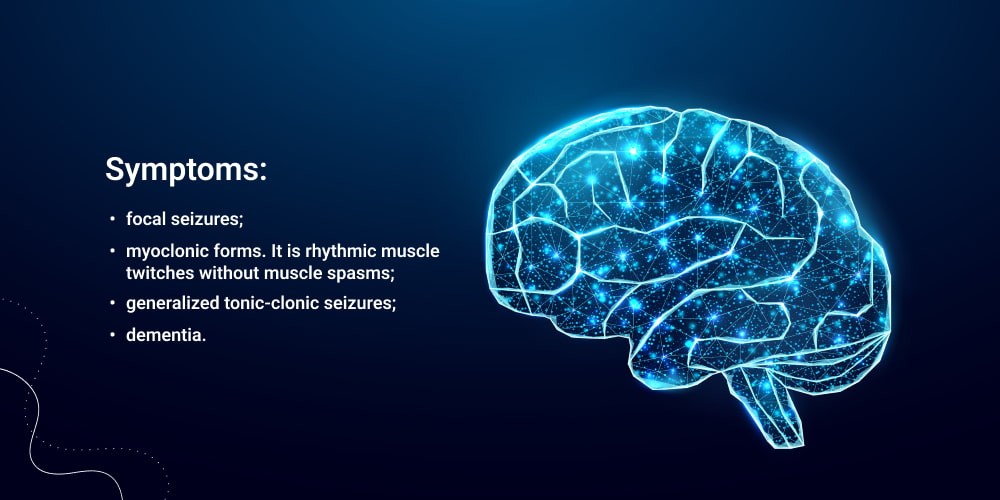
Neurological Symptoms: Seizures represent a hallmark feature of Alpers Disease and often serve as the initial presenting symptom. These seizures typically commence in infancy or early childhood and tend to be refractory to conventional antiepileptic medications. As the disease progresses, affected individuals may experience various types of seizures, including focal, generalized, or myoclonic seizures.

Developmental Regression: Progressive developmental regression is a prominent feature of Alpers Disease, characterized by a loss of previously acquired developmental milestones. Affected individuals may exhibit a decline in motor skills, speech, and cognitive abilities, ultimately leading to profound intellectual disability.

Neurological Decline: Over time, individuals with Alpers Disease experience a relentless deterioration of neurological function, marked by a decline in motor coordination, muscle weakness, and spasticity. The progressive neurodegeneration may also manifest as movement disorders, including dystonia and ataxia.

Hepatic Dysfunction: Liver involvement is another cardinal feature of Alpers Disease, presenting as hepatomegaly, jaundice, and signs of hepatic failure. Liver dysfunction may manifest early in infancy or later in the disease course and can significantly impact overall prognosis.

Systemic Symptoms: In addition to neurological and hepatic manifestations, individuals with Alpers Disease may exhibit systemic symptoms, including failure to thrive, feeding difficulties, and metabolic disturbances.

Ophthalmological Abnormalities: Ophthalmological abnormalities, such as optic atrophy and retinitis pigmentosa, have also been reported in some individuals with Alpers Disease, further contributing to the multisystemic nature of the disorder.

The progressive nature and multisystemic involvement of Alpers Disease underscore the complex pathophysiology underlying this devastating disorder, necessitating a multidisciplinary approach to management and care. (Nursing Paper Example on Alpers Disease: Understanding a Rare Neurological Disorder)

Etiology

Alpers Disease is primarily rooted in genetic aberrations that disrupt mitochondrial function, leading to the characteristic clinical features observed in affected individuals. The genetic etiology of Alpers Disease predominantly revolves around mutations in genes encoding proteins essential for mitochondrial DNA (mtDNA) replication and maintenance.

Mitochondria play a pivotal role in cellular energy production through oxidative phosphorylation, a process that generates adenosine triphosphate (ATP). Mutations in genes encoding components of the mitochondrial DNA polymerase gamma (POLG) enzyme complex, such as the POLG gene itself, have been identified as major contributors to Alpers Disease. POLG is crucial for mtDNA replication and repair, and mutations in this gene impair mitochondrial DNA synthesis, leading to mitochondrial dysfunction.

Furthermore, mutations in other genes involved in mitochondrial DNA replication and maintenance, including TWNK (Twinkle) and DGUOK (Deoxyguanosine Kinase), have also been implicated in the pathogenesis of Alpers Disease. These genetic mutations disrupt mitochondrial DNA replication and repair processes, culminating in mitochondrial dysfunction and cellular energy depletion.

The inheritance pattern of Alpers Disease is typically autosomal recessive, necessitating the presence of mutations in both alleles of the causative gene for disease manifestation. However, in some cases, Alpers Disease can result from de novo mutations occurring sporadically during embryonic development.

Although the genetic etiology of Alpers Disease predominantly involves mutations in nuclear genes encoding mitochondrial proteins, mitochondrial DNA mutations can also contribute to disease pathogenesis. Mitochondrial DNA mutations can arise spontaneously or be maternally inherited and can further exacerbate mitochondrial dysfunction, amplifying the severity of clinical manifestations in affected individuals.

Understanding the underlying genetic basis of Alpers Disease is critical for accurate diagnosis, genetic counseling, and potential targeted therapeutic interventions aimed at mitigating the devastating consequences of this debilitating disorder. (Nursing Paper Example on Alpers Disease: Understanding a Rare Neurological Disorder)

Pathophysiology

The pathophysiology of Alpers Disease revolves around mitochondrial dysfunction, which disrupts cellular energy metabolism and leads to widespread neuronal and hepatic damage. Mitochondria, organelles responsible for generating cellular energy in the form of adenosine triphosphate (ATP), play a central role in the pathogenesis of this disorder.

Mutations in genes encoding proteins involved in mitochondrial DNA (mtDNA) replication and maintenance, such as POLG, TWNK, and DGUOK, impair mitochondrial DNA synthesis and repair processes. Consequently, mitochondrial dysfunction ensues, characterized by reduced ATP production and increased production of reactive oxygen species (ROS).

The impaired energy metabolism resulting from mitochondrial dysfunction particularly affects tissues with high energy demands, such as the brain and liver. Neurons are highly susceptible to mitochondrial dysfunction due to their reliance on ATP for synaptic transmission and neuronal signaling. The progressive neuronal degeneration observed in Alpers Disease is attributed to energy depletion, oxidative stress, and impaired mitochondrial function, leading to neuronal cell death.

Hepatic dysfunction in Alpers Disease stems from mitochondrial impairment and the resultant disruption of liver metabolism. Mitochondria play a crucial role in hepatic energy metabolism, and their dysfunction leads to hepatocyte injury, hepatomegaly, and liver failure.

The accumulation of reactive oxygen species (ROS) resulting from mitochondrial dysfunction exacerbates cellular damage, leading to oxidative stress and further amplifying tissue injury. Additionally, impaired mitochondrial function disrupts calcium homeostasis and alters cellular signaling pathways, contributing to the pathogenesis of Alpers Disease.

Overall, the pathophysiology of Alpers Disease is characterized by mitochondrial dysfunction, energy depletion, oxidative stress, and widespread cellular damage, particularly affecting the brain and liver. Understanding these underlying pathogenic mechanisms is crucial for developing targeted therapeutic interventions aimed at ameliorating mitochondrial dysfunction and mitigating the devastating consequences of this debilitating disorder. (Nursing Paper Example on Alpers Disease: Understanding a Rare Neurological Disorder)

DMS-5 Diagnosis

Diagnosing Alpers Disease typically involves a comprehensive clinical evaluation, neuroimaging studies, and genetic testing to ascertain the presence of characteristic symptoms and identify underlying genetic mutations. The Diagnostic and Statistical Manual of Mental Disorders, Fifth Edition (DSM-5), does not delineate specific diagnostic criteria for Alpers Disease but provides guidelines for assessing the clinical presentation and determining the appropriate diagnostic approach.

The DSM-5 criteria for epilepsy, a common feature of Alpers Disease, encompass recurrent, unprovoked seizures, which align with the clinical manifestation of seizures observed in affected individuals. Additionally, developmental regression, cognitive decline, and neurological abnormalities evident in Alpers Disease may fulfill the criteria for intellectual disability or other neurodevelopmental disorders outlined in the DSM-5.

Neuroimaging studies, particularly magnetic resonance imaging (MRI), play a crucial role in the diagnostic workup of Alpers Disease. MRI findings may reveal characteristic abnormalities, including cerebral atrophy, white matter changes, and signal abnormalities in the basal ganglia, which are consistent with the neurodegenerative nature of the disorder.

Genetic testing is paramount for confirming the diagnosis of Alpers Disease, as it enables the identification of mutations in genes associated with mitochondrial dysfunction, such as POLG, TWNK, and DGUOK. Targeted sequencing or whole-exome sequencing techniques are utilized to detect pathogenic variants in these genes, providing definitive confirmation of the diagnosis.

The diagnostic evaluation of Alpers Disease necessitates a multidisciplinary approach involving neurologists, geneticists, and other healthcare professionals. By integrating clinical assessment, neuroimaging studies, and genetic testing, healthcare providers can establish an accurate diagnosis and initiate appropriate management strategies tailored to the individual needs of patients with Alpers Disease. (Nursing Paper Example on Alpers Disease: Understanding a Rare Neurological Disorder)

Treatment Regimens and Patient Education

As of now, there is no cure for Alpers Disease, and treatment primarily focuses on managing symptoms and providing supportive care to improve the quality of life for affected individuals. Given the complex nature of this disorder, treatment regimens typically entail a multidisciplinary approach involving neurologists, hepatologists, geneticists, and other healthcare professionals.

Symptom Management: Antiepileptic drugs (AEDs) are commonly prescribed to manage seizures in individuals with Alpers Disease. However, seizures associated with this disorder are often refractory to conventional AEDs, necessitating a tailored approach to medication management. Adjunctive therapies, such as ketogenic diet therapy or other dietary interventions, may be considered in some cases to help control seizures.

Supportive Care: Supportive care plays a pivotal role in managing the diverse array of symptoms associated with Alpers Disease. Physical and occupational therapies are integral components of supportive care, aiming to optimize motor function, enhance mobility, and improve the overall quality of life for affected individuals. Speech therapy may also be beneficial in addressing communication difficulties and speech impairments.

Nutritional Support: Given the hepatic involvement observed in Alpers Disease, ensuring adequate nutritional support is essential to support overall health and well-being. Nutritional interventions may include specialized formulas, dietary modifications, or gastrostomy tube placement to facilitate adequate nutrient intake, particularly in individuals with feeding difficulties or failure to thrive.

Liver Transplantation: In cases of severe liver dysfunction or liver failure secondary to Alpers Disease, liver transplantation may be considered as a therapeutic option. Liver transplantation can provide a life-saving intervention by replacing the diseased liver with a healthy donor organ, thereby improving the overall prognosis and quality of life for affected individuals.

Patient Education: Patient education is integral to the management of Alpers Disease, as it empowers patients and their families to understand the nature of the disorder and actively participate in treatment decisions. Educating patients and caregivers about the signs and symptoms of Alpers Disease, available treatment options, and strategies for symptom management can help alleviate anxiety and improve coping mechanisms.

Additionally, providing information about available support services, such as patient advocacy groups and community resources, can facilitate access to additional support and assistance for affected individuals and their families. By fostering open communication and providing comprehensive education, healthcare providers can empower patients and caregivers to navigate the challenges associated with Alpers Disease effectively.

While there is currently no cure for Alpers Disease, a multidisciplinary approach to treatment focusing on symptom management, supportive care, and patient education can significantly improve the quality of life for affected individuals and their families. By addressing the diverse array of symptoms and providing comprehensive support, healthcare providers can optimize outcomes and enhance the overall well-being of individuals grappling with this rare and debilitating disorder. (Nursing Paper Example on Alpers Disease: Understanding a Rare Neurological Disorder)

Conclusion

Alpers Disease presents a significant challenge due to its rare occurrence and severe clinical manifestations affecting infants and young children. Through an exploration of its causes, signs and symptoms, etiology, pathophysiology, DSM-5 diagnosis, treatment regimens, and patient education, we have gained a deeper understanding of this debilitating disorder. While the exact cause remains elusive, genetic mutations disrupting mitochondrial function play a central role in its pathogenesis. Symptom management, supportive care, and patient education are paramount in addressing the diverse array of symptoms and optimizing outcomes for affected individuals and their families. Despite the absence of a cure, a multidisciplinary approach to treatment, including antiepileptic drugs, supportive therapies, and nutritional support, can significantly enhance the quality of life for those grappling with Alpers Disease. By fostering open communication and providing comprehensive education, healthcare providers can empower patients and caregivers to navigate the challenges associated with this rare and devastating disorder effectively. (Nursing Paper Example on Alpers Disease: Understanding a Rare Neurological Disorder)

References

https://www.ncbi.nlm.nih.gov/books/NBK540966/

 
Do you need a similar assignment done for you from scratch? Order now!
Use Discount Code "Newclient" for a 15% Discount!

Nursing Paper Example on The Absence of the Septum Pellucidum: Understanding a Rare Disorder

Nursing Paper Example on The Absence of the Septum Pellucidum: Understanding a Rare Disorder

The absence of the septum pellucidum is a rare condition where the structure that typically separates the two lateral ventricles of the brain is either partially or entirely missing. This disorder, although infrequent, presents significant challenges due to its association with various neurological and developmental abnormalities. Understanding the intricacies of this condition is essential for healthcare professionals to provide appropriate care and support to affected individuals and their families. Despite its rarity, the absence of the septum pellucidum underscores the complexity of brain development and highlights the need for further research to elucidate its underlying causes and improve diagnostic and therapeutic strategies. This paper explores the causes, signs, and symptoms, etiology, pathophysiology, DSM-5 diagnosis, treatment regimens, and patient education, and concludes with insights into the management and impact of this unique neurological disorder. (Nursing Paper Example on The Absence of the Septum Pellucidum: Understanding a Rare Disorder)

Nursing Paper Example on The Absence of the Septum Pellucidum: Understanding a Rare Disorder

Causes

The causes of the absence of the septum pellucidum are not yet fully understood, but researchers have identified several potential factors contributing to its development. One significant factor is believed to be abnormalities during fetal brain development. During the early stages of pregnancy, intricate processes guide the formation and organization of brain structures, including the septum pellucidum. Any disruption or deviation from these processes can lead to structural anomalies in the developing brain.

Genetic factors also play a role in predisposing individuals to this condition. Studies have suggested that certain genetic mutations or variations may increase the likelihood of developing the absence of the septum pellucidum. However, the specific genes involved and their precise contributions to the disorder remain areas of ongoing investigation.

Furthermore, environmental influences during pregnancy may contribute to the development of this condition. Factors such as maternal infections, exposure to toxins, or maternal health conditions can impact fetal brain development and increase the risk of structural abnormalities like the absence of the septum pellucidum.

Additionally, disruptions in the signaling pathways or molecular mechanisms involved in brain development may also contribute to the pathogenesis of this disorder. Research in this area aims to elucidate the intricate cellular and molecular processes underlying normal and abnormal brain development, providing valuable insights into the causes of conditions like the absence of the septum pellucidum.

Overall, the causes of the absence of the septum pellucidum are likely multifactorial, involving complex interactions between genetic predisposition and environmental influences during critical periods of fetal brain development. Further research is needed to unravel the precise mechanisms contributing to this condition and to develop targeted interventions for affected individuals. (Nursing Paper Example on The Absence of the Septum Pellucidum: Understanding a Rare Disorder)

Signs and Symptoms

The absence of the septum pellucidum presents a range of signs and symptoms that can vary in severity and presentation among affected individuals. Common manifestations of this disorder often become apparent during infancy, childhood, or adolescence.

Nursing Paper Example on The Absence of the Septum Pellucidum: Understanding a Rare Disorder

One of the primary symptoms associated with the absence of the septum pellucidum is cognitive deficits. Individuals affected by this condition may experience difficulties with learning, memory, attention, and problem-solving skills. These cognitive impairments can significantly impact academic performance and daily functioning.

Developmental delays are also commonly observed in individuals with the absence of the septum pellucidum. Motor skills, language development, and social-emotional milestones may be delayed compared to their peers. This can result in challenges with coordination, speech and language abilities, and social interactions.

Behavioral issues are another hallmark of the absence of the septum pellucidum. Individuals may exhibit behavioral problems such as impulsivity, hyperactivity, aggression, or emotional lability. These behavioral challenges can pose significant challenges for caregivers and may require behavioral interventions or management strategies.

Neurological abnormalities are frequently associated with the absence of the septum pellucidum. Seizures are a common neurological symptom, occurring in a significant portion of affected individuals. Visual impairments, including abnormalities in visual perception or processing, may also be present.

Additionally, some individuals with the absence of the septum pellucidum may experience other neurological symptoms such as headaches, balance problems, or sensory issues. These symptoms can further contribute to the overall impact of the disorder on an individual’s quality of life and daily functioning.

Overall, the signs and symptoms of the absence of the septum pellucidum encompass a wide range of cognitive, developmental, behavioral, and neurological manifestations that require comprehensive evaluation and management by healthcare professionals. Early recognition and intervention are essential for optimizing outcomes and supporting affected individuals in reaching their full potential. (Nursing Paper Example on The Absence of the Septum Pellucidum: Understanding a Rare Disorder)

Etiology

The etiology of the absence of the septum pellucidum is complex and involves multiple factors contributing to its development. While the precise cause remains unclear, researchers have identified several potential etiological factors that may play a role in the pathogenesis of this disorder.

Genetic factors are thought to contribute significantly to the development of the absence of the septum pellucidum. Studies have suggested a genetic predisposition, with certain gene mutations or variations increasing the susceptibility to structural abnormalities in the brain, including the absence of the septum pellucidum. However, the specific genes involved and their exact contributions to the disorder require further investigation.

Environmental influences during critical periods of fetal brain development are also believed to play a role in the etiology of the absence of the septum pellucidum. Factors such as maternal infections, exposure to toxins, nutritional deficiencies, or maternal health conditions can disrupt normal brain development and increase the risk of structural anomalies.

Furthermore, interactions between genetic and environmental factors may contribute to the pathogenesis of this disorder. Studies suggest that gene-environment interactions during prenatal development may influence the susceptibility to structural brain abnormalities like the absence of the septum pellucidum. These interactions may involve complex molecular mechanisms that impact neural development and organization.

Overall, the etiology of the absence of the septum pellucidum is likely multifactorial, involving a combination of genetic predisposition and environmental influences during critical periods of fetal brain development. Further research is needed to elucidate the specific genetic and environmental factors contributing to this disorder and to improve our understanding of its underlying mechanisms. Such insights are essential for the development of targeted interventions and management strategies for affected individuals. (Nursing Paper Example on The Absence of the Septum Pellucidum: Understanding a Rare Disorder)

Pathophysiology

The pathophysiology of the absence of the septum pellucidum involves disruptions in the normal development and organization of the brain, leading to structural abnormalities and functional deficits in affected individuals.

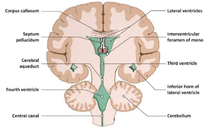
During fetal brain development, the septum pellucidum forms a thin membrane that separates the two lateral ventricles of the brain. In individuals with the absence of the septum pellucidum, this membrane fails to develop properly or is completely absent, resulting in a lack of separation between the ventricles.

This structural anomaly disrupts the normal architecture of the brain and affects the connectivity between different regions, leading to functional impairments. The absence of the septum pellucidum may disrupt neural pathways involved in cognitive processing, sensory perception, motor coordination, and emotional regulation.

Furthermore, the absence of the septum pellucidum can impact the flow of cerebrospinal fluid (CSF) within the brain. Normally, the septum pellucidum helps regulate the flow of CSF between the lateral ventricles. In its absence, CSF circulation may be altered, leading to changes in intracranial pressure and potentially contributing to symptoms such as headaches or neurological deficits.

The absence of the septum pellucidum is often associated with other structural brain abnormalities, such as agenesis of the corpus callosum or abnormalities in the development of the hippocampus. These additional anomalies further exacerbate the neurological and cognitive impairments observed in affected individuals.

Overall, the pathophysiology of the absence of the septum pellucidum involves disruptions in brain development, structural abnormalities, altered neural connectivity, and potential changes in CSF dynamics. These pathophysiological changes contribute to the wide range of symptoms and functional deficits observed in individuals with this disorder. Further research is needed to elucidate the underlying mechanisms and identify potential targets for therapeutic interventions. (Nursing Paper Example on The Absence of the Septum Pellucidum: Understanding a Rare Disorder)

DSM-5 Diagnosis

According to the Diagnostic and Statistical Manual of Mental Disorders, Fifth Edition (DSM-5), the absence of the septum pellucidum is not listed as a specific diagnostic entity. However, this condition may be recognized within the context of related disorders and associated symptoms.

Structural brain abnormalities, including the absence of the septum pellucidum, may be considered in the diagnosis of intellectual disabilities or developmental disorders. These disorders are characterized by significant limitations in intellectual functioning and adaptive behavior, affecting various domains of daily life.

In cases where the absence of the septum pellucidum is associated with neurological symptoms such as seizures, visual impairments, or other neurological deficits, additional diagnostic considerations may be warranted. Depending on the specific clinical presentation, the absence of the septum pellucidum may be identified as a contributing factor in the diagnosis of neurological conditions or disorders.

Furthermore, if the absence of the septum pellucidum is associated with behavioral disturbances or psychiatric symptoms, it may be considered within the diagnostic criteria for psychiatric disorders such as attention-deficit/hyperactivity disorder (ADHD), autism spectrum disorder (ASD), or mood disorders.

Overall, while the absence of the septum pellucidum is not specifically listed as a standalone diagnosis in the DSM-5, it may be recognized and considered within the context of associated symptoms and related disorders. Accurate diagnosis and comprehensive evaluation by healthcare professionals are essential for identifying and addressing the complex clinical presentations associated with this structural brain abnormality. (Nursing Paper Example on The Absence of the Septum Pellucidum: Understanding a Rare Disorder)

Treatment Regimens and Patient Education

Management of the absence of the septum pellucidum focuses on addressing symptoms, providing supportive care, and optimizing the overall well-being of affected individuals. Treatment regimens are tailored to address the specific needs and challenges associated with this rare neurological disorder.

1. Symptom Management:

  • Cognitive deficits: Educational interventions and specialized programs can help address learning difficulties and promote cognitive development. Individualized educational plans (IEPs) and accommodations in academic settings may be implemented.
  • Behavioral issues: Behavioral therapy, counseling, and psychosocial interventions can help manage behavioral challenges such as impulsivity, aggression, and emotional lability. Parent training programs may also be beneficial.
  • Neurological symptoms: Medications may be prescribed to manage seizures or other neurological symptoms. Close monitoring and adjustment of medication regimens are essential for optimal symptom control.

2. Supportive Care:

  • Speech and language therapy: For individuals with speech and language delays or difficulties, speech therapy can improve communication skills and enhance social interactions.
  • Occupational therapy: Occupational therapy focuses on improving motor skills, coordination, and activities of daily living to promote independence and functional abilities.
  • Physical therapy: Physical therapy may be beneficial for individuals with motor impairments, helping to improve mobility, strength, and coordination.

3. Patient Education:

  • Understanding the disorder: Educating patients and their families about the nature of the absence of the septum pellucidum, its potential impact on various aspects of life, and available resources for support is crucial.
  • Treatment options: Providing information about treatment options, including therapies, medications, and supportive services, empowers patients and families to make informed decisions about their care.
  • Monitoring and follow-up: Emphasizing the importance of regular monitoring, follow-up appointments, and collaboration with healthcare providers ensures ongoing management and adjustments to treatment regimens as needed.
  • Community resources: Connecting patients and families with community resources, support groups, and advocacy organizations can provide additional support and opportunities for networking and sharing experiences.

Patient education plays a vital role in promoting self-management, enhancing coping skills, and improving the overall quality of life for individuals with the absence of the septum pellucidum. By providing comprehensive information and support, healthcare professionals empower patients and families to navigate the challenges associated with this rare neurological disorder effectively. (Nursing Paper Example on The Absence of the Septum Pellucidum: Understanding a Rare Disorder)

Conclusion

The absence of the septum pellucidum presents a rare yet significant challenge in neurological and developmental disorders. Through a comprehensive understanding of its causes, signs, and symptoms, along with its complex etiology and pathophysiology, healthcare professionals can better diagnose and manage this condition. While not specifically listed in the DSM-5, recognition within related disorders facilitates tailored treatment approaches focused on symptom management and supportive care. Patient education plays a pivotal role in empowering individuals and families to navigate the challenges associated with the absence of the septum pellucidum, facilitating informed decision-making, and accessing appropriate resources. By implementing treatment regimens addressing cognitive, behavioral, and neurological symptoms, along with providing supportive services and community resources, healthcare professionals can enhance the overall well-being and quality of life of individuals affected by this disorder. Ongoing research and collaboration aim to advance our understanding and management of this unique neurological condition, ensuring optimal outcomes for affected individuals. (Nursing Paper Example on The Absence of the Septum Pellucidum: Understanding a Rare Disorder)

References

https://www.ncbi.nlm.nih.gov/pmc/articles/PMC7069873/

 
Do you need a similar assignment done for you from scratch? Order now!
Use Discount Code "Newclient" for a 15% Discount!

Abnormal Behavior – Case Study-Car Salesman Assignment Help

Abnormal Behavior – Case Study-Car Salesman Assignment Help

I need this back ASAP! Please list at least two references diagnosis code and v codes.

Diagnosis is one of the following


1.Physical Disorders and Health Psychology

2. Anxiety, Trauma- and Stressor-Related 

and Obsessive-Compulsive and Related Disorders

3. Somatic Symptom and Related Disorders and Dissociative Disorders

4. Eating and Sleep-Wake Disorders

5. Neurodevelopmental Disorders and Conduct Disorder

6. Mood Disorders and Suicide

Case

Car Salesman
A 29-year-old car salesman was referred by his current girlfriend, a psychiatric nurse, who suspected he had a mood disorder, even though the patient was reluctant to admit that he might be a moody person. According to him, since the age of 14, he has experienced repeated alternating cycles that he terms “good times and bad times.” During a bad period, usually lasting four to seven days, he oversleeps 10-14 hours daily, lacks energy, confidence, and motivation—“just vegetating,” as he puts it. Often he abruptly shifts, characteristically upon waking up in the morning, to a three-to-four day stretch of overconfidence, heightened social awareness, promiscuity, and sharpened thinking—“Things would flash in my mind.” At such times he indulges in alcohol to enhance the experience but also to help him sleep. Occasionally the good periods last seven to ten days, but culminate in irritable and hostile outbursts, which often herald the transition back to another period of bad days. He admits to frequent use of marijuana, which he claims helps him adjust to daily routines.

In school, As and Bs alternated with Cs and Ds, with the result that the patient was considered a bright student whose performance was mediocre overall because of unstable motivation. As a car salesman his performance has also been uneven, with good days canceling out the bad days; yet even during his good days, he is sometimes perilously argumentative with customers and loses sales that appeared sure. Although considered a charming man in many social circles, he alienates friends when he is hostile and irritable. He typically accumulates social obligations during the bad days and takes care of them all at once on the first day of a good period.


Case study

The Case of Ellen Waters Ellen Waters’ counselor referred her for a medication consultation because of her continuing depressed mood and panic attacks. She is a 37-year-old, part-time graduate student who lives alone and supports herself by working as a home health aide. She completed the course work for a Ph.D. in sociology 3 years ago, but has not yet begun her dissertation.

Ellen is indeed an unhappy-looking woman, and describes being unhappy through much of her life, with no long periods of feeling really good. Her father had a history of alcohol problems, and there was always a great deal of strife in her parents’ marriage. She denies sexual or physical abuse, but feels that her parents were “emotionally abusive” to her. She was first referred for treatment after she made a suicide attempt at age 14, and there have been many times over the years during which her usual low-level depression has become considerably worse, but she has not sought treatment.

Two years ago, when she had been seeing her current boyfriend for about 4 years, it finally became clear that he was unwilling to marry her or live with her. She began to get more depressed and to experience acute panic attacks, and it was at that time that she entered counseling.

In the month before the consultation, she says she was depressed most of the time. She had gained about 10 pounds because she was constantly nibbling on chips or cookies or making herself peanut butter sandwiches. She often awakened in the middle of the night, was unable to go back to sleep for hours, and then overslept the following day, often sleeping up to 18 hours. She says she feels like dead weight, her legs and arms are heavy, and she is always tired, she ruminates about her own failures and cannot concentrate on any serious reading. Although she often wished to be dead, she has not made any recent suicide attempts.

Ellen’s mood is clearly reactive to favorable events. Small attentions from her therapist of her boyfriend can cause her to feel really good for hours at a time. She has an equally extreme reaction to any sort of rejection. If a friend does not return a call, or if someone appears romantically interested then withdraws, she feels devastated.

Although Ellen reports chronic depression, when she is asked about “high” periods, she describes many episodes of abnormally elevated mood that have lasted for several months. During these times she would function on 4 or 5 hours of sleep a night, run up huge telephone bills, and feel that her thoughts were speeded up. She was able to get a lot done, but her friends were obviously concerned about the change in her behavior, urging her to “slow down” and “calm down.” She has never gotten into any real trouble during these episodes.


EXAMPLE WORK

Ellen meets the Diagnostic and Statistical Manual of Mental Disorders Fifth Edition  (DSM-5)(2013) criteria for hypomanic episode, A-F. Criteria: A– She experiences many episodes of abnormally elevated mood that have lasted for several months; B– She meets four of the seven symptoms (2- would function on 4 or 5 hours of sleep a night; 4- felt that her thoughts were speeded up; 6- was able to get a lot done; and 7- ran up huge telephone bills) … “which represent a noticeable change from usual behavior, and have been present to a significant degree” (APA, 2013, p. 132); C and D– friends were obviously concerned about the change in her behavior, urging her to “slow down” and “calm down”; E– has never gotten into any real trouble during these episodes; and F– There are no reports of any medications or other substances that might cause the episode.

Ellen meets the criteria for major depressive episode (DSM-5), A-C. Criteria: A– She meets six of the nine symptoms (1- was depressed most of the time for a month; 3- gained about 10 pounds; 6- always tired; 7- ruminates about her failures; 8- cannot concentrate on any serious reading; 9- often wished to be dead without any specific plans to commit suicide); B– She sought counseling for the continuing depressed mood; C– There are no reports of medications or other substances that might cause the episode. Medical tests should be conducted to eliminate any physiological condition that might cause this episode.

Ellen meets the criteria for Bipolar II Disorder A-D. Criteria: A– She has experienced a hypomanic episode; B– She has never had a manic episode; and C– Her hypomanic and depressive episodes are not better explained by: schizoaffective disorder, schizophrenia, schizophreniform disorder, delusional disorder or other specified or unspecified schizophrenia spectrum and other psychotic disorders; D– The symptoms of depression cause clinically significant distress.

Ellen’s current episode is depressed with moderate severity. Her symptoms fall between the mild and severe levels of severity. She has one more than the minimum number of symptoms for both hypomanic episode and major depressive episode so, the symptoms are not excessive.

Specifier- with atypical features, Criteria A-C (DSM-5, pp. 151-152). Criteria: A– Her mood is clearly reactive to favorable events; B– She meets three of the four symptoms (1- gained about 10 pounds, constantly nibbling on chips or cookies or making herself peanut butter sandwiches, 3- feels like dead weight, her legs and arms are heavy; and 4- She has an extreme reaction to any sort of rejection. If a friend does not return a call, or if someone appears romantically interested then withdraws, she feels devastated.); C– Criteria are not met for with melancholic features or with catatonia. In addition, a condition worth noting is Ellen’s report of parental emotional abuse so the V- code V15.42, personal history of psychological abuse in childhood, is included with the diagnosis of bipolar II disorder (American Psychiatric Association, 2013). As noted in the DSM-5 on page 138 (American Psychiatric Association, 2013) about 1/3 of individuals have a history of suicide attempts which Ellen has admitted accordingly a safety plan would need to be discussed. Since Ellen reported panic attacks in the past and anxiety disorders are often comorbid with bipolar II disorders, I considered panic disorder but subsequently discarded that idea as Ellen is not reporting current panic attacks. Ellen does not meet the criteria for bipolar I disorder as she did not meet the criteria for manic episode because she doesn’t meet criterion C under manic episode.


Does not met the criteria specifier (criterion B, hypomania episode or depressive episode) with mixed features because her symptoms are mostly subjective in nature.

Reference

American Psychiatric Association. (2013). Diagnostic and statistical manual of mental disorders (5th ed.), pp. 132-139, 151. Arlington, VA: American Psychiatric Association Publishing.

 
Do you need a similar assignment done for you from scratch? Order now!
Use Discount Code "Newclient" for a 15% Discount!