Posts

Nursing Paper Example on Barrett’s Esophagus: Understanding, Diagnosis, and Treatment

Nursing Paper Example on Barrett’s Esophagus: Understanding, Diagnosis, and Treatment

Barrett’s esophagus is a condition in which the tissue lining the esophagus changes due to prolonged exposure to stomach acid. This condition is significant because it serves as a precursor to esophageal adenocarcinoma, a type of cancer with a poor prognosis. Understanding Barrett’s esophagus is essential for early detection and intervention to prevent the progression of cancer. Despite its importance, many individuals may not be familiar with this condition and its implications. Therefore, this essay aims to provide a comprehensive overview of Barrett’s esophagus, covering its causes, signs and symptoms, etiology, pathophysiology, DSM-5 diagnosis, treatment regimens, patient education, and concluding with the importance of awareness and proactive management. By shedding light on this condition, individuals can better recognize its symptoms, seek appropriate medical attention, and take proactive steps to manage their health effectively. (Nursing Paper Example on Barrett’s Esophagus: Understanding, Diagnosis, and Treatment)

Nursing Paper Example on Barrett's Esophagus: Understanding, Diagnosis, and Treatment

Causes

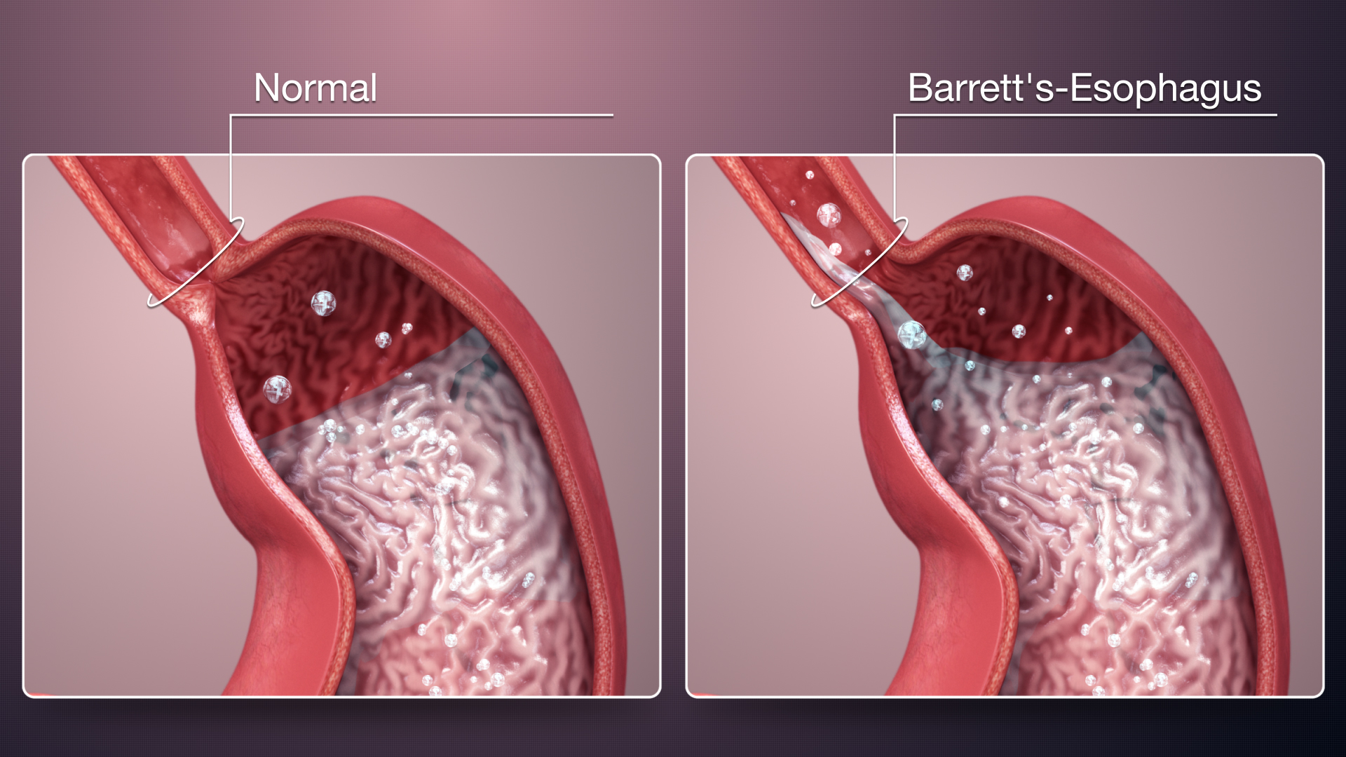
The primary cause of Barrett’s esophagus is chronic gastroesophageal reflux disease (GERD). GERD occurs when the lower esophageal sphincter, a muscle that acts as a valve between the esophagus and stomach, becomes weak or relaxes inappropriately, allowing stomach acid to flow back into the esophagus. This acid reflux leads to irritation and inflammation of the esophageal lining over time, triggering changes in the tissue.

Additionally, obesity is strongly associated with the development of Barrett’s esophagus. Excess body weight increases intra-abdominal pressure, which can push stomach contents upward into the esophagus, exacerbating acid reflux.

Smoking is another risk factor for Barrett’s esophagus. Smoking weakens the lower esophageal sphincter and reduces saliva production, which normally helps neutralize stomach acid in the esophagus.

A hiatal hernia, where a portion of the stomach protrudes through the diaphragm into the chest cavity, can also contribute to Barrett’s esophagus. This anatomical abnormality can disrupt the normal functioning of the lower esophageal sphincter, allowing acid reflux to occur more frequently.

Dietary factors may play a role as well. Consuming spicy, acidic, or fatty foods can exacerbate GERD symptoms and increase the likelihood of Barrett’s esophagus development.

Furthermore, genetic predisposition may contribute to an individual’s susceptibility to Barrett’s esophagus. Certain genetic factors may affect the function of the esophageal lining or the body’s response to acid reflux, influencing the development of this condition.

Overall, Barrett’s esophagus is a multifactorial condition influenced by a combination of lifestyle factors, anatomical abnormalities, and genetic predisposition. Understanding these causes is crucial for implementing preventive measures and managing the condition effectively. (Nursing Paper Example on Barrett’s Esophagus: Understanding, Diagnosis, and Treatment)

Signs and Symptoms

Barrett’s esophagus often does not present with specific symptoms, and many individuals with the condition may remain asymptomatic. However, when symptoms do occur, they are typically associated with gastroesophageal reflux disease (GERD), which often precedes and accompanies Barrett’s esophagus.

Nursing Paper Example on Barrett's Esophagus: Understanding, Diagnosis, and Treatment

Heartburn is one of the most common symptoms experienced by individuals with Barrett’s esophagus. It is characterized by a burning sensation in the chest, often occurring after meals or when lying down.

Regurgitation, where stomach contents flow back into the mouth or throat, is another common symptom of GERD and Barrett’s esophagus. This can lead to a sour or bitter taste in the mouth and may be accompanied by a sensation of fluid moving up the chest.

Difficulty swallowing, known as dysphagia, may also occur in individuals with Barrett’s esophagus. This symptom can range from a sensation of food getting stuck in the throat to pain or discomfort while swallowing.

Other symptoms associated with Barrett’s esophagus include:

  1. Chronic cough: Persistent coughing, particularly at night or after eating, can be a symptom of GERD and Barrett’s esophagus.
  2. Hoarseness: Irritation of the throat due to acid reflux can lead to hoarseness or changes in voice quality.
  3. Chest pain: While less common, some individuals with Barrett’s esophagus may experience chest pain that can mimic heart-related conditions.
  4. Unintentional weight loss: Severe or chronic GERD symptoms, including those associated with Barrett’s esophagus, may lead to decreased appetite and unintentional weight loss over time.
  5. Nausea and vomiting: In some cases, individuals with Barrett’s esophagus may experience nausea or vomiting, particularly after meals or when lying down.

Recognizing these signs and symptoms is important for prompt diagnosis and management of Barrett’s esophagus and associated complications. (Nursing Paper Example on Barrett’s Esophagus: Understanding, Diagnosis, and Treatment)

Etiology

The precise etiology of Barrett’s esophagus remains incompletely understood; however, several factors contribute to its development. Chronic gastroesophageal reflux disease (GERD) is the primary underlying cause of Barrett’s esophagus. GERD occurs when the lower esophageal sphincter (LES), a muscular valve between the esophagus and stomach, weakens or relaxes inappropriately, allowing stomach acid to reflux into the esophagus. This chronic exposure to gastric acid leads to inflammation and injury to the esophageal mucosa.

Obesity is a significant risk factor for the development of Barrett’s esophagus. Excessive body weight increases intra-abdominal pressure, which promotes the reflux of stomach contents into the esophagus. Additionally, adipose tissue secretes inflammatory cytokines that contribute to esophageal inflammation and injury.

Smoking is another established risk factor for Barrett’s esophagus. Cigarette smoke contains numerous toxic compounds that impair LES function and increase acid production in the stomach, exacerbating GERD symptoms.

A hiatal hernia, characterized by the protrusion of a portion of the stomach into the thoracic cavity through the esophageal hiatus of the diaphragm, predisposes individuals to Barrett’s esophagus. Hiatal hernias disrupt the normal anatomy of the LES, facilitating acid reflux into the esophagus.

Dietary factors also play a role in the development of Barrett’s esophagus. Consumption of acidic, spicy, or fatty foods exacerbates GERD symptoms and contributes to esophageal mucosal injury.

Furthermore, genetic predisposition may influence an individual’s susceptibility to Barrett’s esophagus. Variations in genes involved in the regulation of esophageal mucosal integrity and inflammation may predispose individuals to develop Barrett’s esophagus in response to environmental triggers such as GERD.

Overall, Barrett’s esophagus is a multifactorial condition influenced by the interplay of genetic susceptibility, lifestyle factors, and environmental exposures, particularly chronic GERD. Understanding the etiological factors underlying Barrett’s esophagus is crucial for implementing preventive measures and optimizing therapeutic interventions. (Nursing Paper Example on Barrett’s Esophagus: Understanding, Diagnosis, and Treatment)

Pathophysiology

The pathophysiology of Barrett’s esophagus involves a series of cellular changes in response to chronic gastroesophageal reflux disease (GERD). The persistent exposure of the esophageal mucosa to acidic gastric contents leads to inflammation and injury, initiating a cascade of cellular events.

Initially, the normal stratified squamous epithelium lining the esophagus undergoes metaplastic changes in response to chronic acid exposure. This process, known as intestinal metaplasia, involves the replacement of squamous epithelial cells with specialized columnar epithelial cells resembling those found in the intestine. These columnar cells are more resistant to acid-induced injury but lack the protective mechanisms present in the stomach and intestine, rendering them vulnerable to further damage.

Over time, the metaplastic epithelium may progress to dysplasia, characterized by abnormal cellular growth and differentiation. Dysplastic changes can range from low-grade, characterized by mild cellular abnormalities, to high-grade, indicative of more severe cellular atypia. High-grade dysplasia is considered a precursor to esophageal adenocarcinoma and represents a significant risk factor for cancer development.

The pathophysiological mechanisms underlying the progression from Barrett’s esophagus to adenocarcinoma are complex and multifactorial. Chronic inflammation, oxidative stress, genetic alterations, and dysregulated signaling pathways contribute to the malignant transformation of Barrett’s epithelium.

Ultimately, Barrett’s esophagus serves as a precursor to esophageal adenocarcinoma, a lethal malignancy with a poor prognosis. Early detection and intervention are crucial for preventing cancer progression in individuals with Barrett’s esophagus.

Understanding the pathophysiology of Barrett’s esophagus provides insights into its clinical manifestations, complications, and therapeutic targets. Targeted interventions aimed at mitigating acid reflux, reducing inflammation, and inhibiting dysplastic progression may help prevent the development of esophageal adenocarcinoma in individuals with Barrett’s esophagus. (Nursing Paper Example on Barrett’s Esophagus: Understanding, Diagnosis, and Treatment)

DMS-5 Diagnosis

Barrett’s esophagus is not a psychiatric disorder and thus not classified or diagnosed using the Diagnostic and Statistical Manual of Mental Disorders, Fifth Edition (DSM-5). Instead, the diagnosis of Barrett’s esophagus is established through clinical evaluation and endoscopic examination with biopsy.

Clinical evaluation involves a comprehensive assessment of the patient’s medical history, including symptoms suggestive of gastroesophageal reflux disease (GERD) such as heartburn, regurgitation, and difficulty swallowing. Additionally, the presence of risk factors such as obesity, smoking, and a history of hiatal hernia may raise suspicion for Barrett’s esophagus.

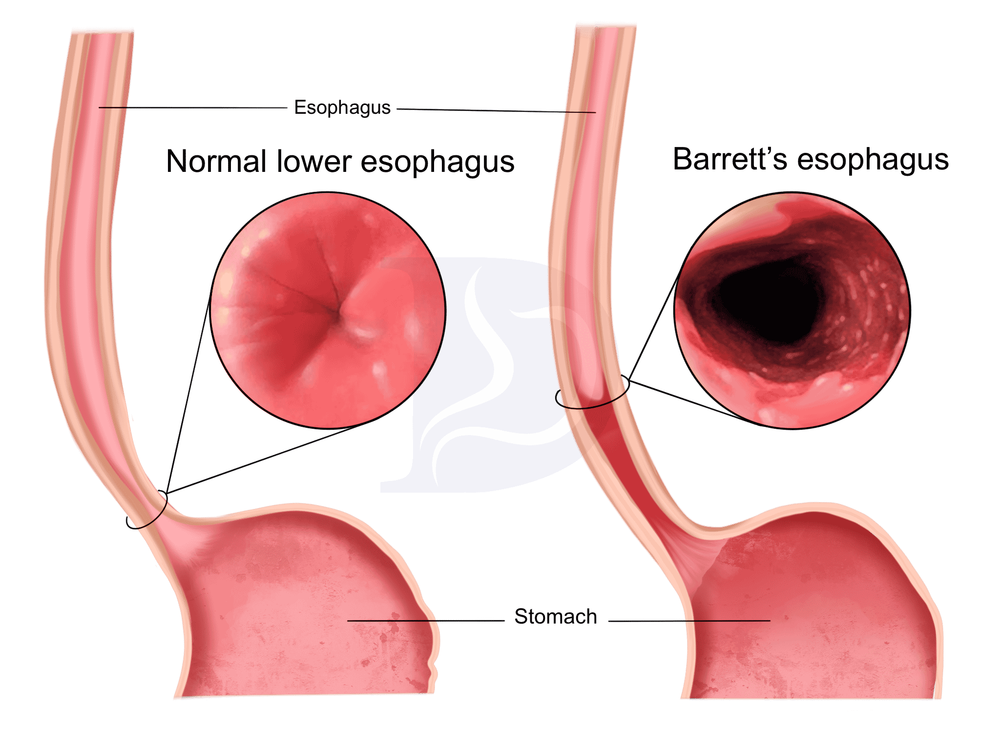
The definitive diagnosis of Barrett’s esophagus is made through upper endoscopy, also known as esophagogastroduodenoscopy (EGD). During this procedure, a flexible endoscope is passed through the mouth into the esophagus, allowing direct visualization of the esophageal mucosa. Characteristic findings suggestive of Barrett’s esophagus include the presence of salmon-pink or reddish mucosa extending into the esophagus, typically in the distal portion near the gastroesophageal junction.

Biopsies are obtained from suspicious areas of the esophageal mucosa during endoscopy to confirm the diagnosis of Barrett’s esophagus. Histopathological examination of these biopsies reveals the presence of specialized intestinal metaplasia, characterized by the replacement of squamous epithelium with columnar epithelium containing goblet cells. Goblet cells are a hallmark feature of intestinal metaplasia and are not typically present in normal esophageal epithelium.

The diagnosis of Barrett’s esophagus is established based on the presence of intestinal metaplasia in the esophageal mucosa on histopathological examination of biopsy specimens obtained during endoscopy. Once diagnosed, regular surveillance endoscopy with biopsy is recommended to monitor for dysplastic changes and early detection of esophageal adenocarcinoma in individuals with Barrett’s esophagus. (Nursing Paper Example on Barrett’s Esophagus: Understanding, Diagnosis, and Treatment)

Treatment Regimens and Patient Education

Treatment for Barrett’s esophagus focuses on managing gastroesophageal reflux disease (GERD) symptoms, reducing acid reflux, and preventing complications such as esophageal adenocarcinoma. Additionally, patient education plays a crucial role in empowering individuals to make lifestyle modifications and adhere to treatment regimens effectively.

Lifestyle Modifications:

Patients with Barrett’s esophagus are advised to make several lifestyle modifications to reduce acid reflux and alleviate symptoms:

  • Dietary Changes: Avoiding trigger foods such as spicy, acidic, or fatty foods can help reduce GERD symptoms. Eating smaller, more frequent meals and avoiding lying down immediately after eating can also minimize reflux.
  • Weight Management: Maintaining a healthy weight through diet and exercise is important, as obesity is a significant risk factor for Barrett’s esophagus and GERD.
  • Smoking Cessation: Quitting smoking is essential, as smoking exacerbates GERD symptoms and increases the risk of esophageal adenocarcinoma.
  • Elevating the Head of the Bed: Sleeping with the head of the bed elevated can help reduce nighttime reflux.

Medications:

  • Proton Pump Inhibitors (PPIs): PPIs such as omeprazole, lansoprazole, and esomeprazole are commonly prescribed to reduce stomach acid production and alleviate GERD symptoms. These medications help heal esophageal inflammation and may slow the progression of Barrett’s esophagus.
  • Histamine H2 Receptor Antagonists: H2 blockers such as ranitidine and famotidine reduce stomach acid production and may be used in combination with PPIs for symptom management.

Surgical Interventions:

In some cases, surgical interventions may be considered to address severe GERD symptoms or complications of Barrett’s esophagus, such as dysplasia or early-stage esophageal adenocarcinoma. Surgical procedures such as fundoplication aim to strengthen the lower esophageal sphincter and prevent acid reflux.

Patient Education:

Patient education is crucial for promoting understanding, adherence to treatment regimens, and lifestyle modifications. Key aspects of patient education include:

  • Understanding the Condition: Patients should be educated about the nature of Barrett’s esophagus, its association with GERD, and the risk of esophageal adenocarcinoma.
  • Medication Adherence: Patients should be instructed on the importance of adhering to medication regimens, including PPIs, and informed about potential side effects and interactions.
  • Lifestyle Modifications: Patients should receive guidance on dietary changes, weight management, smoking cessation, and other lifestyle modifications to minimize reflux and manage symptoms effectively.
  • Importance of Surveillance: Patients with Barrett’s esophagus should understand the importance of regular surveillance endoscopy with biopsy for monitoring disease progression and detecting dysplasia or early-stage cancer.

By empowering patients with knowledge about their condition and providing guidance on lifestyle modifications and treatment regimens, healthcare providers can optimize outcomes and improve the quality of life for individuals with Barrett’s esophagus. (Nursing Paper Example on Barrett’s Esophagus: Understanding, Diagnosis, and Treatment)

Conclusion

Barrett’s esophagus is a condition characterized by changes in the tissue lining of the esophagus due to chronic exposure to stomach acid, primarily resulting from gastroesophageal reflux disease (GERD). While often asymptomatic, Barrett’s esophagus is a significant precursor to esophageal adenocarcinoma, emphasizing the importance of early detection and intervention. Understanding the causes, signs and symptoms, etiology, pathophysiology, DSM-5 diagnosis, treatment regimens, and patient education surrounding Barrett’s esophagus is crucial for effective management. By implementing lifestyle modifications, adhering to medication regimens, and undergoing regular surveillance, individuals with Barrett’s esophagus can reduce their risk of disease progression and complications. Patient education plays a vital role in empowering individuals to make informed decisions about their health and actively participate in their care. Through a comprehensive approach that combines medical treatment, lifestyle modifications, and patient education, healthcare providers can improve outcomes and enhance the quality of life for patients with Barrett’s esophagus. (Nursing Paper Example on Barrett’s Esophagus: Understanding, Diagnosis, and Treatment)

References

https://www.ncbi.nlm.nih.gov/books/NBK430979/

 
Do you need a similar assignment done for you from scratch? Order now!
Use Discount Code "Newclient" for a 15% Discount!

Nursing Paper Example on Bacterial Meningitis: Understanding the Threat

Nursing Paper Example on Bacterial Meningitis: Understanding the Threat

Bacterial meningitis stands as a formidable adversary in the realm of infectious diseases, characterized by inflammation of the protective membranes enveloping the brain and spinal cord. This condition warrants our attention due to its swift onset and potential for grave complications. Understanding its nuances—from causative agents to clinical manifestations—is imperative in navigating its complexities. Through this paper, we embark on a journey to unravel the intricacies of bacterial meningitis, exploring its causes, signs, and symptoms, delving into its etiology and pathophysiology, dissecting its DSM-5 diagnosis, and elucidating treatment regimens and patient education strategies. By shedding light on this menacing ailment, we strive to equip ourselves with knowledge and awareness, empowering individuals and communities to confront bacterial meningitis head-on, thus fortifying our defenses against this insidious threat to public health. (Nursing Paper Example on Bacterial Meningitis: Understanding the Threat)

Nursing Paper Example on Bacterial Meningitis: Understanding the Threat

Causes

Bacterial meningitis emerges from a confluence of factors, primarily orchestrated by a group of nefarious bacteria adept at infiltrating the body’s defenses. Among the most notorious perpetrators are Neisseria meningitidis, Streptococcus pneumoniae, and Haemophilus influenzae type b. These microbial miscreants gain entry into the bloodstream through various portals, including respiratory droplets expelled during coughing or sneezing, direct contact with contaminated surfaces, or intimate contact with an infected individual.

Once in the bloodstream, these bacterial invaders embark on a treacherous journey, traversing the circulatory system with stealthy precision until they reach their ultimate destination: the brain and spinal cord. Here, they encounter the formidable blood-brain barrier, a fortress-like defense mechanism designed to safeguard the sanctity of the central nervous system. Despite its formidable nature, this barrier proves penetrable to certain bacterial species, allowing them to breach its defenses and initiate their malevolent assault on the meninges—the protective membranes enveloping the brain and spinal cord.

Several predisposing factors can facilitate the insidious entry of these bacteria into the bloodstream. Respiratory infections, such as sinusitis or otitis media, provide convenient portals of entry, allowing bacteria easy access to the bloodstream. Similarly, individuals with compromised immune systems due to underlying medical conditions or immunosuppressive medications are more susceptible to bacterial meningitis.

Moreover, trauma or injury to the skull, such as skull fractures or surgical procedures involving the head or neck, can compromise the integrity of the blood-brain barrier, creating an opportune pathway for bacterial invasion. Additionally, certain lifestyle factors, such as smoking or living in crowded environments, may increase the risk of exposure to bacterial pathogens responsible for meningitis.

In essence, bacterial meningitis is a multifaceted disease driven by the interplay of bacterial pathogens, host susceptibility factors, and environmental influences. Understanding the intricacies of its causative factors is paramount in devising effective preventive strategies and therapeutic interventions to combat this formidable foe. (Nursing Paper Example on Bacterial Meningitis: Understanding the Threat)

Signs and Symptoms

Bacterial meningitis is characterized by a diverse array of signs and symptoms that reflect the profound impact of inflammation on the central nervous system. The clinical presentation of bacterial meningitis can vary widely, depending on factors such as age, immune status, and the specific bacterial pathogen involved.

Nursing Paper Example on Bacterial Meningitis: Understanding the Threat

One of the hallmark features of bacterial meningitis is the sudden onset of symptoms, often accompanied by a rapid progression of illness. Fever is a common early sign, often accompanied by chills and rigors, reflecting the body’s immune response to the invading pathogens. Additionally, individuals may experience a severe headache, described as intense and unrelenting, which may be exacerbated by changes in body position.

A stiff neck, or nuchal rigidity, is another classic symptom of bacterial meningitis, resulting from inflammation of the meninges and irritation of the spinal nerves. This stiffness can make it difficult for individuals to flex their necks forward, a characteristic finding observed during physical examination.

Sensitivity to light, known as photophobia, is another prominent feature of bacterial meningitis, as the inflamed meninges become hypersensitive to external stimuli such as bright lights. This symptom often manifests as discomfort or pain in the eyes when exposed to light.

In addition to these cardinal symptoms, individuals with bacterial meningitis may experience a constellation of neurological manifestations, including confusion, altered mental status, and seizures. In infants and young children, the clinical presentation may be more subtle and nonspecific, with symptoms such as irritability, poor feeding, lethargy, and a bulging fontanelle.

Prompt recognition and evaluation of these signs and symptoms are crucial for early diagnosis and initiation of appropriate treatment. Timely intervention can help prevent serious complications and improve outcomes for individuals affected by bacterial meningitis. (Nursing Paper Example on Bacterial Meningitis: Understanding the Threat)

Etiology

Bacterial meningitis unfolds as a complex interplay between microbial invaders, host vulnerabilities, and environmental factors, culminating in a cascade of events that precipitate inflammation of the meninges—the protective membranes enveloping the brain and spinal cord.

Respiratory infections serve as common precursors to bacterial meningitis, providing a gateway for microbial entry into the bloodstream. Conditions such as sinusitis, otitis media, or pneumonia create conducive environments for bacterial proliferation, facilitating their dissemination to the central nervous system.

Individuals with compromised immune systems are particularly susceptible to bacterial meningitis, as their diminished defenses offer little resistance against invading pathogens. Underlying medical conditions, such as HIV/AIDS, diabetes mellitus, or malignancies, weaken the immune response, rendering individuals more vulnerable to infectious threats.

Trauma or injury to the head or neck region can compromise the integrity of the blood-brain barrier, paving the way for bacterial infiltration into the central nervous system. Skull fractures, penetrating head injuries, or neurosurgical procedures disrupt the protective barriers that normally shield the brain and spinal cord from microbial assault, increasing the risk of bacterial meningitis.

Certain lifestyle factors and environmental influences also contribute to the etiology of bacterial meningitis. Crowded living conditions, such as dormitories or military barracks, facilitate the transmission of respiratory pathogens among individuals in close proximity. Smoking, alcohol abuse, and illicit drug use can impair immune function and respiratory health, exacerbating the risk of bacterial infections.

Overall, bacterial meningitis arises from a complex interplay of microbial, host, and environmental factors. Understanding the multifaceted etiology of this condition is crucial in implementing preventive measures and targeted interventions to mitigate its impact on public health. (Nursing Paper Example on Bacterial Meningitis: Understanding the Threat)

Pathophysiology

The pathophysiology of bacterial meningitis is characterized by a series of intricate events that unfold within the central nervous system, orchestrated by the insidious invasion of pathogenic bacteria and the host’s robust inflammatory response.

Following the breach of the blood-brain barrier by bacterial pathogens, typically originating from a primary infection site such as the respiratory tract or bloodstream, these microbial invaders gain access to the cerebrospinal fluid (CSF) and the meninges—the delicate membranes enveloping the brain and spinal cord.

Once within the CSF and meninges, bacteria unleash a torrent of virulence factors and toxins, triggering a robust immune response characterized by the release of pro-inflammatory cytokines, chemokines, and other mediators of inflammation. These inflammatory mediators recruit immune cells, such as neutrophils and macrophages, to the site of infection, amplifying the inflammatory cascade and contributing to tissue damage.

The inflammatory response within the meninges leads to a constellation of clinical manifestations, including fever, headache, and nuchal rigidity. The release of pro-inflammatory cytokines also disrupts the blood-brain barrier, facilitating the infiltration of immune cells and circulating inflammatory mediators into the brain parenchyma, further exacerbating neuronal injury and tissue damage.

Moreover, the inflammatory cascade induces an increase in intracranial pressure, compromising cerebral blood flow and leading to cerebral edema—a hallmark feature of severe bacterial meningitis. Cerebral edema exacerbates neuronal injury, impairs neurological function, and contributes to the development of life-threatening complications such as seizures, cerebral herniation, and brainstem compression.

In summary, the pathophysiology of bacterial meningitis is characterized by a complex interplay of microbial virulence factors, host immune responses, and inflammatory mediators, culminating in neuronal injury, tissue damage, and the development of severe neurological complications. Understanding these intricate disease processes is crucial in guiding therapeutic interventions and improving outcomes for individuals affected by bacterial meningitis. (Nursing Paper Example on Bacterial Meningitis: Understanding the Threat)

DSM-5 Diagnosis

The diagnosis of bacterial meningitis relies on a combination of clinical assessment, laboratory investigations, and neuroimaging studies, guided by established criteria outlined in the Diagnostic and Statistical Manual of Mental Disorders, Fifth Edition (DSM-5).

Clinically, individuals with suspected bacterial meningitis typically present with a constellation of symptoms, including fever, headache, nuchal rigidity, and altered mental status. These clinical features raise suspicion for meningitis and warrant further evaluation.

Laboratory investigations play a pivotal role in confirming the diagnosis of bacterial meningitis. Cerebrospinal fluid (CSF) analysis, obtained via lumbar puncture, is a cornerstone diagnostic test. CSF analysis typically reveals an elevated white blood cell count, predominantly consisting of neutrophils, indicative of an inflammatory response. Additionally, CSF protein levels are often elevated, reflecting disruption of the blood-brain barrier, while glucose levels may be decreased due to increased utilization by inflammatory cells.

Furthermore, blood cultures are essential for identifying the causative organism responsible for bacterial meningitis. Positive blood cultures provide definitive evidence of bacteremia, supporting the diagnosis of bacterial meningitis.

Neuroimaging studies, such as computed tomography (CT) or magnetic resonance imaging (MRI) of the brain, may be performed to assess for complications of bacterial meningitis, such as cerebral edema, hydrocephalus, or intracranial hemorrhage. These imaging modalities help guide management decisions and prognostication.

Based on the aforementioned clinical and laboratory findings, a diagnosis of bacterial meningitis can be established according to DSM-5 criteria. The presence of compatible clinical symptoms, along with supportive laboratory and imaging findings, confirms the diagnosis and guides appropriate management strategies.

In summary, the DSM-5 provides a structured framework for the diagnosis of bacterial meningitis, integrating clinical assessment, laboratory investigations, and neuroimaging studies to facilitate accurate identification and management of this potentially life-threatening condition. (Nursing Paper Example on Bacterial Meningitis: Understanding the Threat)

Treatment Regimens and Patient Education: Combatting Bacterial Meningitis

Effective management of bacterial meningitis requires a multi-faceted approach encompassing prompt initiation of antibiotic therapy, supportive care, and patient education to optimize outcomes and minimize complications.

Treatment Regimens:

  1. Antibiotic Therapy: Prompt initiation of empiric antibiotic therapy is essential in suspected cases of bacterial meningitis to prevent complications and improve outcomes. Empiric antibiotic selection is guided by local epidemiology and the patient’s age, immune status, and risk factors. Commonly used antibiotics include third-generation cephalosporins (e.g., ceftriaxone or cefotaxime) in combination with vancomycin to provide broad-spectrum coverage against common bacterial pathogens.
  2. Supportive Care: Supportive measures aim to alleviate symptoms, maintain vital functions, and prevent complications. Intravenous fluids are administered to ensure hydration and maintain electrolyte balance. Antipyretics such as acetaminophen or ibuprofen are prescribed to reduce fever and alleviate discomfort. Corticosteroids, such as dexamethasone, may be used adjunctively to mitigate inflammation and reduce the risk of neurologic sequelae, particularly in cases of suspected or confirmed bacterial meningitis caused by Streptococcus pneumoniae.
  3. Monitoring and Surveillance: Close monitoring of clinical status, vital signs, and neurological status is imperative in managing bacterial meningitis. Serial neurologic examinations and frequent assessments of consciousness, pupil reactivity, and motor function are conducted to detect changes indicative of worsening intracranial pressure or neurological compromise. Serial lumbar punctures may be performed to monitor CSF parameters and assess treatment response.

Patient Education:

  1. Vaccination: Educating patients and caregivers about the importance of vaccination against bacterial meningitis is paramount in preventing future occurrences. Encouraging adherence to recommended vaccination schedules, particularly for meningococcal conjugate vaccines and pneumococcal vaccines, can significantly reduce the risk of bacterial meningitis.
  2. Hygiene Practices: Promoting good hygiene practices, such as frequent handwashing with soap and water, covering coughs and sneezes, and avoiding close contact with individuals who are sick, can help prevent the spread of respiratory pathogens implicated in bacterial meningitis.
  3. Seeking Medical Attention: Emphasizing the importance of seeking prompt medical attention in cases of suspected bacterial meningitis is crucial. Educating patients and caregivers about the warning signs and symptoms of bacterial meningitis, such as fever, severe headache, neck stiffness, altered mental status, and rash, empowers them to recognize early signs of infection and seek timely medical evaluation and treatment.

Comprehensive management of bacterial meningitis entails a combination of antibiotic therapy, supportive care, and patient education. By implementing timely treatment regimens and empowering individuals with knowledge about preventive measures, we can effectively combat bacterial meningitis and improve outcomes for affected individuals. (Nursing Paper Example on Bacterial Meningitis: Understanding the Threat)

Conclusion

Bacterial meningitis poses a significant threat to public health, characterized by its rapid onset, potential for severe complications, and profound impact on individuals and communities. Understanding its causes, recognizing its signs and symptoms, and implementing timely treatment regimens are crucial steps in mitigating its burden. Through vaccination, promotion of good hygiene practices, and empowering individuals with knowledge about preventive measures, we can collectively combat bacterial meningitis. Furthermore, ongoing research, education, and public health initiatives are essential to enhance awareness, improve diagnosis, and optimize treatment outcomes. By fostering collaboration among healthcare professionals, policymakers, and communities, we can strive to reduce the incidence and impact of bacterial meningitis, ensuring a healthier and safer future for all. (Nursing Paper Example on Bacterial Meningitis: Understanding the Threat)

References

https://www.ncbi.nlm.nih.gov/books/NBK470351/

 
Do you need a similar assignment done for you from scratch? Order now!
Use Discount Code "Newclient" for a 15% Discount!

Nursing Paper Example on Bell’s Palsy: Understanding its Causes, Symptoms, and Management

Nursing Paper Example on Bell’s Palsy: Understanding its Causes, Symptoms, and Management

Bell’s palsy, a form of facial paralysis, manifests as sudden weakness or drooping on one side of the face, affecting facial expressions and functions such as smiling and closing the eye. Named after Scottish anatomist Charles Bell, who first described it in the 19th century, Bell’s palsy is a relatively common condition, accounting for the majority of cases of facial paralysis worldwide. Despite its prevalence, the exact cause of Bell’s palsy remains elusive, with viral infections, particularly herpes simplex virus, being implicated in its pathogenesis. This condition can occur at any age but is most commonly observed in individuals between the ages of 15 and 60. Although Bell’s palsy typically resolves spontaneously within a few weeks to months, it can significantly impact the quality of life due to its effects on facial appearance and function. Understanding the causes, symptoms, and management of Bell’s palsy is crucial for timely diagnosis and effective treatment interventions. (Nursing Paper Example on Bell’s Palsy: Understanding its Causes, Symptoms, and Management)

Nursing Paper Example on Bell's Palsy: Understanding its Causes, Symptoms, and Management

Causes

The precise cause of Bell’s palsy remains uncertain, but it is widely believed to be related to inflammation and swelling of the facial nerve, known as the seventh cranial nerve. This inflammation disrupts the normal function of the nerve, leading to paralysis or weakness of the facial muscles on one side of the face. While the exact trigger for this inflammation is not fully understood, several factors have been proposed as potential causes or contributing factors to the development of Bell’s palsy.

Viral infections, particularly herpes simplex virus (HSV), are frequently associated with the onset of Bell’s palsy. It is theorized that viral replication within the facial nerve or surrounding tissue triggers an inflammatory response, leading to nerve damage and dysfunction. Other viruses, including herpes zoster virus (which causes shingles), Epstein-Barr virus (associated with infectious mononucleosis), and cytomegalovirus, have also been implicated in some cases.

Additionally, certain conditions and risk factors may predispose individuals to developing Bell’s palsy. These include diabetes mellitus, hypertension, pregnancy, upper respiratory tract infections, autoimmune diseases, and a family history of the condition. While the exact mechanism by which these factors contribute to Bell’s palsy is not fully understood, it is believed that they may exacerbate inflammation or compromise the immune response, increasing susceptibility to nerve damage.

Furthermore, anatomical factors such as the course of the facial nerve as it passes through the temporal bone and its proximity to other structures within the skull may also play a role in the development of Bell’s palsy. Compression or irritation of the facial nerve due to these anatomical features could potentially lead to nerve dysfunction and the characteristic symptoms of Bell’s palsy.

In summary, while viral infections, particularly HSV, are commonly associated with Bell’s palsy, a combination of genetic predisposition, environmental factors, and anatomical considerations may contribute to its onset. Further research is needed to elucidate the exact mechanisms underlying this condition and identify potential preventive strategies. (Nursing Paper Example on Bell’s Palsy: Understanding its Causes, Symptoms, and Management)

Signs and Symptoms

Bell’s palsy presents with distinctive signs and symptoms, typically characterized by sudden onset unilateral facial weakness or paralysis. The onset of symptoms is often rapid, occurring for hours to days, and may progress gradually or reach maximum severity within a short time frame.

Nursing Paper Example on Bell's Palsy: Understanding its Causes, Symptoms, and Management

Common manifestations of Bell’s palsy include asymmetry of facial expressions, with one side of the face appearing droopy or slack compared to the other side. This asymmetry can affect various facial movements, including smiling, raising the eyebrows, and closing the eye on the affected side. Patients may also experience difficulty in controlling facial muscles, leading to an inability to fully close one eye or difficulty with tasks such as blowing out candles or whistling.

In addition to facial weakness or paralysis, individuals with Bell’s palsy may report other associated symptoms, including facial pain or discomfort, particularly around the ear or jaw on the affected side. Some patients may also experience altered sensations, such as tingling or numbness, in the facial region.

One of the hallmark signs of Bell’s palsy is the presence of unilateral facial drooping or weakness that worsens with facial movements but improves or resolves during periods of rest. This fluctuating nature of symptoms distinguishes Bell’s palsy from other causes of facial paralysis, such as stroke or traumatic injury, which typically present with more persistent deficits.

Furthermore, Bell’s palsy may be accompanied by other sensory or motor disturbances, such as impaired taste perception on the anterior two-thirds of the tongue or increased sensitivity to sounds (hyperacusis) on the affected side. These additional symptoms can vary in severity and may not be present in all cases of Bell’s palsy. (Nursing Paper Example on Bell’s Palsy: Understanding its Causes, Symptoms, and Management)

Etiology

The etiology of Bell’s palsy is multifactorial and involves a complex interplay of genetic, environmental, and immunological factors. While the exact cause of the condition remains elusive, several hypotheses have been proposed to explain its pathogenesis.

One of the leading theories suggests that Bell’s palsy is triggered by viral infections, particularly herpes simplex virus (HSV). It is believed that viral replication within the facial nerve or surrounding tissue induces an inflammatory response, leading to edema and compression of the nerve. This inflammatory cascade ultimately results in nerve dysfunction and the characteristic facial paralysis observed in Bell’s palsy. Other viruses, including herpes zoster virus (which causes shingles), Epstein-Barr virus, and cytomegalovirus, have also been implicated in some cases.

In addition to viral infections, genetic predisposition may play a role in the development of Bell’s palsy. Studies have identified certain genetic polymorphisms associated with an increased risk of developing the condition, suggesting a hereditary component. However, the specific genes involved and their exact role in Bell’s palsy remain poorly understood.

Environmental factors, such as cold weather, upper respiratory tract infections, and trauma to the face or head, have also been implicated as potential triggers for Bell’s palsy. These factors may exacerbate inflammation or compromise the immune response, increasing susceptibility to nerve damage.

Furthermore, anatomical considerations may contribute to the development of Bell’s palsy. The facial nerve travels through a narrow bony canal within the temporal bone, leaving it susceptible to compression or irritation from surrounding structures. Additionally, the proximity of the facial nerve to other cranial nerves and blood vessels within the skull may increase the risk of nerve injury in certain individuals.

In summary, Bell’s palsy is likely the result of a complex interplay between viral infections, genetic predisposition, environmental factors, and anatomical considerations. Further research is needed to elucidate the specific mechanisms underlying the development of this condition and identify potential targets for therapeutic intervention. (Nursing Paper Example on Bell’s Palsy: Understanding its Causes, Symptoms, and Management)

Pathophysiology

Bell’s palsy is characterized by inflammation and dysfunction of the facial nerve, also known as the seventh cranial nerve, which controls the muscles responsible for facial expressions. The exact pathophysiological mechanisms underlying Bell’s palsy are not fully understood, but several key processes are thought to have contributed to its development.

The initial trigger for Bell’s palsy is believed to be viral infections, particularly herpes simplex virus (HSV), which leads to inflammation of the facial nerve and surrounding tissues. Viral replication within the nerve or adjacent structures triggers an immune response, resulting in the release of pro-inflammatory cytokines and the recruitment of immune cells to the site of infection.

This inflammatory cascade causes edema and swelling of the facial nerve, leading to compression within the narrow confines of the facial canal as it passes through the temporal bone. The compression of the nerve disrupts its normal function, impairing the transmission of signals from the brain to the facial muscles.

As a result, patients with Bell’s palsy experience weakness or paralysis of the facial muscles on one side of the face, leading to characteristic symptoms such as drooping of the mouth, difficulty closing one eye, and asymmetry of facial expressions.

In addition to inflammation and compression, ischemia (reduced blood flow) may also contribute to nerve damage in Bell’s palsy. The swelling and edema within the facial canal can compress blood vessels supplying the facial nerve, leading to impaired blood flow and ischemic injury to the nerve fibers.

Overall, the pathophysiology of Bell’s palsy involves a complex interplay of viral infection, inflammation, compression, and ischemia, ultimately resulting in dysfunction of the facial nerve and the characteristic clinical features of the condition. Further research is needed to fully elucidate the underlying mechanisms and identify potential targets for therapeutic intervention. (Nursing Paper Example on Bell’s Palsy: Understanding its Causes, Symptoms, and Management)

DSM-5 Diagnosis

Bell’s palsy is typically diagnosed clinically, relying on a thorough evaluation of symptoms and physical examination findings rather than specific criteria outlined in the Diagnostic and Statistical Manual of Mental Disorders, Fifth Edition (DSM-5). The DSM-5 primarily focuses on psychiatric and psychological disorders, whereas Bell’s palsy is a neurological condition characterized by facial paralysis.

Clinicians assess patients for the sudden onset of unilateral facial weakness or paralysis, which is a hallmark feature of Bell’s palsy. Other associated symptoms may include difficulty closing one eye, drooling, altered taste perception, and facial pain or discomfort. The absence of other neurological deficits or signs of stroke, coupled with the rapid progression of symptoms over hours to days, further supports the diagnosis of Bell’s palsy.

Diagnostic tests such as electromyography (EMG) and nerve conduction studies (NCS) may be used to confirm the diagnosis and assess the severity of nerve damage. Imaging studies such as magnetic resonance imaging (MRI) or computed tomography (CT) scans are typically reserved for cases where alternative diagnoses or underlying structural abnormalities are suspected.

While the DSM-5 does not provide specific diagnostic criteria for Bell’s palsy, clinicians need to differentiate this condition from other causes of facial paralysis, such as stroke, Ramsay Hunt syndrome, and tumors affecting the facial nerve. Additionally, a comprehensive medical history, including assessment of risk factors such as viral infections, diabetes, and pregnancy, may aid in the diagnostic process.

Overall, the diagnosis of Bell’s palsy relies on clinical judgment and a thorough evaluation of symptoms and physical examination findings. Prompt recognition and appropriate management are essential to optimize outcomes and prevent complications associated with facial paralysis. (Nursing Paper Example on Bell’s Palsy: Understanding its Causes, Symptoms, and Management)

Treatment Regimens

The management of Bell’s palsy aims to alleviate symptoms, promote nerve recovery, and prevent complications. While the condition often resolves spontaneously within a few weeks to months, various treatment options may be considered to expedite recovery and improve outcomes.

  1. Corticosteroids: Oral corticosteroids, such as prednisone, are commonly prescribed to reduce inflammation and edema of the facial nerve in Bell’s palsy. These medications are typically initiated within the first 72 hours of symptom onset and may be tapered for 10 to 14 days. Corticosteroids have been shown to accelerate recovery and improve facial function in some patients.
  2. Antiviral Medications: In cases where viral infection is suspected or confirmed, antiviral medications, such as acyclovir or valacyclovir, may be prescribed in combination with corticosteroids. While the evidence supporting the use of antiviral therapy in Bell’s palsy is limited, some studies suggest a potential benefit in certain subsets of patients, particularly those with severe or atypical presentations.
  3. Eye Care: Patients with Bell’s palsy are at risk of developing corneal exposure and dry eye due to incomplete closure of the affected eye. Lubricating eye drops or ointments may be recommended to prevent corneal abrasions and maintain ocular moisture. In severe cases, temporary eyelid taping or the use of protective eyewear, such as moisture chamber goggles, may be necessary to protect the eye during sleep or daytime activities.
  4. Physical Therapy: Physical therapy techniques, including facial exercises, massage, and electrical stimulation, may be employed to improve muscle strength, coordination, and mobility in individuals with Bell’s palsy. These interventions can help facilitate nerve regeneration and restore facial function, particularly in cases of incomplete recovery or persistent symptoms.
  5. Supportive Measures: Patients with Bell’s palsy may benefit from supportive measures to alleviate discomfort and facilitate daily activities. This may include using moist heat packs or gentle massage to relieve facial pain or stiffness, practicing relaxation techniques to reduce stress and anxiety, and modifying dietary habits to accommodate taste disturbances or difficulty chewing. (Nursing Paper Example on Bell’s Palsy: Understanding its Causes, Symptoms, and Management)

Patient Education

Educating patients about Bell’s palsy is essential to promote understanding, compliance with treatment regimens, and optimal self-care. Key aspects of patient education include:

  1. Nature of the Condition: Providing information about the self-limiting nature of Bell’s palsy and the expected course of recovery, which typically occurs within weeks to months, can help alleviate anxiety and uncertainty.
  2. Importance of Eye Care: Emphasizing the importance of eye protection and regular eye care, including the use of lubricating eye drops and protective eyewear, to prevent corneal injury and maintain visual health in individuals with incomplete eyelid closure.
  3. Treatment Goals and Expectations: Discussing treatment options, including the potential benefits and risks of corticosteroids and antiviral medications, and setting realistic expectations regarding the timeline and outcomes of therapy.
  4. Facial Exercises and Self-Care: Providing guidance on performing facial exercises, practicing good oral hygiene, and implementing supportive measures at home to promote facial muscle strength, mobility, and comfort.
  5. Follow-Up and Monitoring: Advising patients to follow up with their healthcare provider regularly to monitor progress, assess treatment response, and address any concerns or complications that may arise during treatment.

By empowering patients with knowledge and resources to actively participate in their care, healthcare providers can facilitate better outcomes and enhance the overall management of Bell’s palsy. (Nursing Paper Example on Bell’s Palsy: Understanding its Causes, Symptoms, and Management)

Conclusion

Bell’s palsy presents as sudden unilateral facial paralysis, often attributed to viral infections and inflammation of the facial nerve. While the exact cause remains unclear, timely diagnosis and appropriate management are crucial for optimizing outcomes. Treatment regimens may include corticosteroids, antiviral medications, eye care, physical therapy, and supportive measures to alleviate symptoms and promote nerve recovery. Patient education plays a vital role in empowering individuals with Bell’s palsy to understand the condition, adhere to treatment regimens, and implement self-care strategies. By providing comprehensive care and support, healthcare providers can effectively manage Bell’s palsy and improve the quality of life for affected individuals. Ongoing research efforts are needed to further elucidate the underlying mechanisms of Bell’s palsy and develop targeted therapies to enhance recovery and minimize long-term sequelae. (Nursing Paper Example on Bell’s Palsy: Understanding its Causes, Symptoms, and Management)

References

https://www.ncbi.nlm.nih.gov/books/NBK482290/

 
Do you need a similar assignment done for you from scratch? Order now!
Use Discount Code "Newclient" for a 15% Discount!

Nursing Paper Example on Bulimia Nervosa: Understanding and Treating the Cycle of Binge and Purge

Nursing Paper Example on Bulimia Nervosa: Understanding and Treating the Cycle of Binge and Purge

Bulimia nervosa is a debilitating eating disorder that affects millions of individuals worldwide, predominantly striking adolescents and young adults. This disorder is characterized by a destructive cycle of binge eating followed by purging behaviors, such as vomiting or excessive exercise, in an attempt to compensate for the consumed calories. While bulimia nervosa is often associated with distorted body image and weight concerns, its underlying causes are complex and multifaceted, involving a combination of genetic, psychological, environmental, and societal factors. Understanding the nature of bulimia nervosa is crucial for recognizing its signs and symptoms, implementing effective treatment strategies, and supporting individuals on their path to recovery. In this paper, we will delve into the causes, signs and symptoms, etiology, pathophysiology, DSM-5 diagnosis criteria, treatment regimens, patient education, and conclude with insights into addressing this pervasive eating disorder. By shedding light on bulimia nervosa, we aim to promote awareness and foster a compassionate understanding of this often misunderstood condition. (Nursing Paper Example on Bulimia Nervosa: Understanding and Treating the Cycle of Binge and Purge)

Nursing Paper Example on Bulimia Nervosa: Understanding and Treating the Cycle of Binge and Purge

Causes

The development of bulimia nervosa is influenced by a combination of genetic, psychological, environmental, and societal factors. Genetic predisposition plays a role, as individuals with a family history of eating disorders are more susceptible to developing bulimia. This suggests a hereditary component that may contribute to the disorder’s manifestation.

Psychological factors also contribute significantly to the development of bulimia nervosa. Low self-esteem, perfectionism, and body dissatisfaction are commonly observed traits in individuals with bulimia. These psychological factors often fuel a negative body image and a relentless pursuit of thinness, which can drive disordered eating behaviors.

Environmental factors, such as societal pressure to attain an idealized body shape, can also contribute to the development of bulimia nervosa. Media portrayal of thinness as the epitome of beauty and success can create unrealistic expectations and foster feelings of inadequacy in susceptible individuals.

Additionally, traumatic life events, such as childhood abuse, neglect, or dysfunctional family dynamics, may contribute to the onset of bulimia nervosa. These experiences can trigger maladaptive coping mechanisms, including disordered eating behaviors, as individuals attempt to regain a sense of control over their lives.

Furthermore, cultural factors can influence the development of bulimia nervosa. Societies that prioritize thinness and equate it with beauty and success may perpetuate disordered eating behaviors among susceptible individuals.

In summary, the causes of bulimia nervosa are multifaceted and involve a complex interplay of genetic, psychological, environmental, and societal factors. Understanding these underlying causes is crucial for developing effective prevention and intervention strategies to address this pervasive eating disorder. (Nursing Paper Example on Bulimia Nervosa: Understanding and Treating the Cycle of Binge and Purge)

Signs and Symptoms

Recognizing the signs and symptoms of bulimia nervosa is essential for early intervention and treatment. Individuals with bulimia often exhibit a range of behavioral, physical, and emotional indicators that signify their struggle with disordered eating.

Nursing Paper Example on Bulimia Nervosa: Understanding and Treating the Cycle of Binge and Purge

One of the hallmark signs of bulimia nervosa is recurrent episodes of binge eating, where individuals consume large amounts of food in a short period, often feeling a loss of control during these episodes. Binges are typically followed by compensatory behaviors to prevent weight gain, such as self-induced vomiting, misuse of laxatives or diuretics, fasting, or excessive exercise.

Frequent trips to the bathroom after meals may also indicate bulimia, as individuals may engage in purging behaviors to rid their bodies of consumed calories. Evidence of vomiting, including the smell of vomit or the presence of vomit residue, may be observed in bathrooms or other private areas.

Physical signs of bulimia nervosa may include swollen cheeks or jaw, caused by repeated episodes of self-induced vomiting. Additionally, calluses or scars on the knuckles or hands, known as Russell’s sign, may develop from the act of self-induced vomiting.

Dental problems such as enamel erosion, cavities, or tooth sensitivity may also be present due to frequent exposure to stomach acid from vomiting. Gastrointestinal issues, such as acid reflux or bloating, may occur as a result of purging behaviors.

Emotionally, individuals with bulimia nervosa may experience feelings of guilt, shame, or embarrassment related to their binge eating and purging behaviors. They may also exhibit mood swings, irritability, or depression as a result of their struggles with food and body image.

Overall, recognizing these signs and symptoms is crucial for identifying individuals who may be struggling with bulimia nervosa and facilitating timely intervention and support. (Nursing Paper Example on Bulimia Nervosa: Understanding and Treating the Cycle of Binge and Purge)

Etiology

The etiology of bulimia nervosa is multifaceted, involving a complex interplay of genetic, psychological, environmental, and sociocultural factors. Understanding these underlying influences is crucial for elucidating the development and maintenance of this eating disorder.

Genetic predisposition is believed to play a significant role in the etiology of bulimia nervosa. Research suggests that individuals with a family history of eating disorders, particularly bulimia nervosa, are at a higher risk of developing the disorder themselves. This familial clustering suggests a genetic susceptibility that may contribute to the disorder’s onset.

Psychological factors also contribute to the etiology of bulimia nervosa. Low self-esteem, perfectionism, and body dissatisfaction are commonly observed traits in individuals with bulimia. These psychological factors often interact with genetic predisposition, leading to a heightened vulnerability to the development of disordered eating behaviors.

Environmental factors, including family dynamics, peer influences, and societal norms, also play a role in the etiology of bulimia nervosa. Dysfunctional family dynamics, such as high levels of criticism or control, may contribute to the development of maladaptive coping mechanisms, including disordered eating behaviors. Peer influences, particularly during adolescence, can also shape attitudes and behaviors related to body image and weight control.

Sociocultural factors, such as societal pressure to attain an idealized body shape, also contribute to the etiology of bulimia nervosa. Media portrayal of thinness as the epitome of beauty and success can create unrealistic expectations and foster feelings of inadequacy in susceptible individuals. Additionally, cultural attitudes toward food, weight, and body image may influence the development and maintenance of disordered eating behaviors.

In summary, the etiology of bulimia nervosa is complex and multifaceted, involving a combination of genetic, psychological, environmental, and sociocultural factors. Understanding these underlying influences is crucial for developing effective prevention and intervention strategies to address this pervasive eating disorder. (Nursing Paper Example on Bulimia Nervosa: Understanding and Treating the Cycle of Binge and Purge)

Pathophysiology

The pathophysiology of bulimia nervosa involves dysregulation of appetite and satiety mechanisms, leading to recurrent episodes of binge eating followed by compensatory behaviors. This dysregulation is thought to arise from a complex interplay of biological, psychological, and environmental factors.

Neurotransmitter dysregulation, particularly involving serotonin, is believed to play a role in the pathophysiology of bulimia nervosa. Serotonin is a neurotransmitter involved in the regulation of mood, appetite, and impulse control. Dysregulation of serotonin pathways may contribute to disturbances in appetite regulation and mood, predisposing individuals to binge eating episodes.

Hypothalamic dysfunction may also play a role in the pathophysiology of bulimia nervosa. The hypothalamus is a brain region involved in the regulation of hunger, satiety, and energy balance. Dysfunction in hypothalamic circuits may lead to disruptions in appetite regulation, contributing to the development of disordered eating behaviors.

Psychological factors, such as low self-esteem, perfectionism, and body dissatisfaction, can further exacerbate the dysregulation of appetite and satiety mechanisms in individuals with bulimia nervosa. These psychological factors may influence eating behaviors and contribute to the cycle of binge eating and purging.

Environmental factors, including societal pressure to attain an idealized body shape and cultural attitudes toward food and weight, also play a role in the pathophysiology of bulimia nervosa. Societal and cultural influences can shape attitudes and behaviors related to food, body image, and weight control, contributing to the development and maintenance of disordered eating behaviors.

In summary, the pathophysiology of bulimia nervosa involves dysregulation of appetite and satiety mechanisms, influenced by a complex interplay of biological, psychological, and environmental factors. Understanding these underlying mechanisms is crucial for developing effective interventions to address this pervasive eating disorder. (Nursing Paper Example on Bulimia Nervosa: Understanding and Treating the Cycle of Binge and Purge)

DSM-5 Diagnosis

According to the Diagnostic and Statistical Manual of Mental Disorders, Fifth Edition (DSM-5), the diagnostic criteria for bulimia nervosa include specific criteria that must be met for a diagnosis to be made.

Firstly, recurrent episodes of binge eating must occur, characterized by eating a large amount of food in a discrete period while experiencing a sense of lack of control over eating during the episode. Binge-eating episodes are typically accompanied by feelings of guilt, shame, or distress.

Secondly, compensatory behaviors are employed to prevent weight gain following binge eating episodes. These compensatory behaviors may include self-induced vomiting, misuse of laxatives, diuretics, or other medications, fasting, or excessive exercise. These behaviors are intended to counteract the effects of binge eating and are engaged in on average at least once a week for three months.

Additionally, binge eating and compensatory behaviors are not exclusively associated with episodes of anorexia nervosa. Furthermore, the severity of bulimia nervosa is classified based on the frequency of compensatory behaviors:

  • Mild: An average of 1-3 episodes of inappropriate compensatory behaviors per week.
  • Moderate: An average of 4-7 episodes of inappropriate compensatory behaviors per week.
  • Severe: An average of 8-13 episodes of inappropriate compensatory behaviors per week.
  • Extreme: An average of 14 or more episodes of inappropriate compensatory behaviors per week.

The DSM-5 diagnosis of bulimia nervosa aims to provide a standardized framework for clinicians to assess and diagnose individuals experiencing symptoms of this eating disorder. By clearly defining the diagnostic criteria, the DSM-5 facilitates accurate diagnosis and appropriate treatment planning for individuals with bulimia nervosa. (Nursing Paper Example on Bulimia Nervosa: Understanding and Treating the Cycle of Binge and Purge)

Treatment Regimens and Patient Education for Bulimia Nervosa

Effective treatment for bulimia nervosa typically involves a multidisciplinary approach that addresses the complex biological, psychological, and social factors underlying the disorder. Treatment regimens may include psychotherapy, nutritional counseling, medication management, and medical monitoring.

Psychotherapy: Cognitive-behavioral therapy (CBT) is considered the first-line treatment for bulimia nervosa. CBT focuses on identifying and challenging maladaptive thoughts and behaviors related to food, body image, and weight. It helps individuals develop healthier coping strategies and address underlying psychological issues contributing to their eating disorder. Interpersonal psychotherapy (IPT) may also be beneficial, particularly for addressing interpersonal difficulties and relationship issues that may contribute to bulimia nervosa.

Nutritional Counseling: Nutritional counseling plays a crucial role in bulimia nervosa treatment by promoting a balanced and healthy approach to eating. Registered dietitians work with individuals to develop structured meal plans, normalize eating patterns, and address nutritional deficiencies that may arise from disordered eating behaviors. Education about portion control, mindful eating, and the importance of regular meals and snacks is emphasized.

Medication Management: Antidepressant medications, particularly selective serotonin reuptake inhibitors (SSRIs), may be prescribed to help alleviate symptoms of depression and anxiety commonly associated with bulimia nervosa. SSRIs have been shown to reduce the frequency of binge eating and purging episodes and improve overall mood and functioning in individuals with bulimia.

Medical Monitoring: Regular medical monitoring is essential for individuals with bulimia nervosa to assess and manage potential medical complications associated with the disorder. Medical professionals monitor vital signs, electrolyte levels, and cardiovascular health to identify and address any adverse effects of disordered eating behaviors. Nutritional supplementation and fluid replacement may be necessary in cases of severe malnutrition or dehydration.

Patient Education: Patient education is a critical component of bulimia nervosa treatment, as it empowers individuals to understand their condition and actively participate in their recovery. Patients are educated about the nature of bulimia nervosa, including its causes, signs, and symptoms. They learn about the potential consequences of untreated bulimia, such as electrolyte imbalances, dental problems, and gastrointestinal complications. Patients are also provided with practical strategies for managing urges to binge and purge, such as practicing mindfulness, developing healthy coping skills, and building a supportive network of friends and family.

In summary, effective treatment for bulimia nervosa involves a comprehensive approach that addresses the underlying biological, psychological, and social factors contributing to the disorder. Through psychotherapy, nutritional counseling, medication management, and patient education, individuals with bulimia nervosa can achieve recovery and improve their overall health and well-being. (Nursing Paper Example on Bulimia Nervosa: Understanding and Treating the Cycle of Binge and Purge)

Conclusion

Bulimia nervosa is a complex and debilitating eating disorder characterized by recurrent episodes of binge eating followed by compensatory behaviors. Its etiology involves a combination of genetic, psychological, environmental, and sociocultural factors. Recognizing the signs and symptoms, understanding the underlying causes, and implementing effective treatment regimens are crucial steps in addressing this pervasive disorder. By employing a multidisciplinary approach that includes psychotherapy, nutritional counseling, medication management, and medical monitoring, individuals with bulimia nervosa can achieve recovery and improve their overall health and well-being. Patient education plays a vital role in empowering individuals to understand their condition, develop healthier coping strategies, and actively participate in their recovery journey. Through increased awareness, early intervention, and ongoing support, we can break the cycle of binge eating and purging and help individuals overcome bulimia nervosa, leading to a healthier and more fulfilling life. (Nursing Paper Example on Bulimia Nervosa: Understanding and Treating the Cycle of Binge and Purge)

References

 
Do you need a similar assignment done for you from scratch? Order now!
Use Discount Code "Newclient" for a 15% Discount!

Comprehensive Nursing Paper Example on Bronchitis: Understanding the Common Respiratory Condition

Comprehensive Nursing Paper Example on Bronchitis: Understanding the Common Respiratory Condition

Bronchitis, a prevalent respiratory condition affecting millions worldwide annually, stands as a significant health concern due to its impact on individuals’ well-being. Characterized by inflammation of the bronchial tubes, bronchitis manifests in acute and chronic forms, each presenting unique challenges and implications for patient care. Despite its widespread occurrence, bronchitis often receives inadequate attention compared to other respiratory illnesses, yet its debilitating symptoms can significantly disrupt daily life and compromise respiratory function. Understanding the underlying causes, signs, symptoms, etiology, pathophysiology, DSM-5 diagnosis, treatment regimens, and patient education associated with bronchitis is crucial for healthcare professionals and individuals alike. By delving into the intricacies of bronchitis, we can develop a comprehensive understanding of this condition and implement effective strategies for prevention, management, and improvement of patients’ respiratory health. This paper aims to explore bronchitis comprehensively, shedding light on its multifaceted nature and emphasizing the importance of addressing this common ailment in clinical practice and public health initiatives. (Comprehensive Nursing Paper Example on Bronchitis: Understanding the Common Respiratory Condition)

Comprehensive Nursing Paper Example on Bronchitis: Understanding the Common Respiratory Condition

Causes

Bronchitis can be triggered by various factors, with the most common being viral and bacterial infections. Respiratory viruses such as influenza, rhinovirus, and respiratory syncytial virus (RSV) are frequent culprits, especially during colder months when respiratory illnesses are more prevalent. These viruses invade the respiratory tract, causing inflammation of the bronchial tubes and subsequent bronchitis symptoms.

In addition to infections, exposure to irritants plays a significant role in bronchitis development. Tobacco smoke, whether firsthand or secondhand, is a potent irritant known to inflame the bronchial tubes, leading to acute and chronic bronchitis in smokers and nonsmokers alike. Environmental pollutants, such as air pollution, industrial chemicals, and dust, can also irritate the airways and contribute to bronchitis.

Individual susceptibility to bronchitis varies, with certain risk factors predisposing individuals to the condition. Chronic exposure to irritants like tobacco smoke, air pollution, and occupational hazards increases the likelihood of developing chronic bronchitis. Furthermore, individuals with compromised immune systems, such as the elderly, young children, and those with underlying health conditions, are more susceptible to bronchitis infections.

Moreover, lifestyle factors play a significant role in bronchitis development. Poor hygiene practices, inadequate handwashing, and close contact with infected individuals can facilitate the spread of respiratory viruses, leading to bronchitis outbreaks in communities. Additionally, individuals with weakened immune systems due to factors such as malnutrition, stress, or chronic illnesses are at higher risk of contracting bronchitis.

Overall, bronchitis is a multifactorial condition influenced by various infectious, environmental, and lifestyle factors. Understanding these causes is essential for implementing preventive measures and managing bronchitis effectively, thereby reducing its impact on individuals’ respiratory health and overall well-being. (Comprehensive Nursing Paper Example on Bronchitis: Understanding the Common Respiratory Condition)

Signs and Symptoms

Bronchitis manifests with a range of signs and symptoms that can vary in severity and duration. The most common symptom is a persistent cough, which may be dry initially but often progresses to produce mucus as the condition worsens. This cough is typically accompanied by chest discomfort or tightness, which can range from mild to severe.

Excessive mucus production is a hallmark feature of bronchitis, leading to phlegm or sputum being expelled during coughing episodes. The color and consistency of the mucus can vary, ranging from clear or white to yellow or green, depending on the underlying cause of bronchitis.

In addition to coughing and mucus production, individuals with bronchitis may experience respiratory symptoms such as shortness of breath or wheezing. These symptoms result from inflammation and narrowing of the bronchial tubes, which restrict airflow and impair breathing.

Fatigue and malaise are common systemic symptoms associated with bronchitis, as the body expends energy to fight off the infection or inflammation. Individuals may feel tired or lethargic, with reduced energy levels and overall stamina.

Fever is another possible symptom of bronchitis, particularly in cases caused by bacterial infections. Elevated body temperature indicates an immune response to the underlying infection, although not all individuals with bronchitis will experience fever.

In acute cases of bronchitis, symptoms typically develop suddenly and may resolve within a few weeks with appropriate treatment and supportive care. Chronic bronchitis, on the other hand, is characterized by persistent or recurrent symptoms lasting for months or years, often accompanied by exacerbations or flare-ups.

Overall, recognizing the signs and symptoms of bronchitis is crucial for timely diagnosis and management, as early intervention can help alleviate symptoms, prevent complications, and promote recovery. (Comprehensive Nursing Paper Example on Bronchitis: Understanding the Common Respiratory Condition)

Etiology

Bronchitis can be classified into acute and chronic forms, each with distinct etiological factors contributing to its development.

Acute Bronchitis: Acute bronchitis is commonly caused by viral infections, with respiratory viruses such as influenza, rhinovirus, adenovirus, and respiratory syncytial virus (RSV) being the primary culprits. These viruses invade the respiratory tract, infecting the epithelial cells lining the bronchial tubes and triggering an inflammatory response. The resulting inflammation leads to swelling of the bronchial walls and increased mucus production, contributing to the characteristic symptoms of acute bronchitis.

In addition to viral infections, acute bronchitis can also be caused by bacterial infections, although less frequently. Bacterial pathogens such as Mycoplasma pneumoniae, Bordetella pertussis (pertussis), and Streptococcus pneumoniae can infect the bronchial tubes, leading to acute bronchitis with similar symptoms as viral infections.

Comprehensive Nursing Paper Example on Bronchitis: Understanding the Common Respiratory Condition

Chronic Bronchitis: Chronic bronchitis is primarily associated with long-term exposure to irritants, particularly tobacco smoke. Smoking is the leading cause of chronic bronchitis, with the toxic chemicals in cigarette smoke irritating the bronchial tubes and causing chronic inflammation. Prolonged exposure to tobacco smoke damages the cilia lining the bronchial tubes, impairing their ability to clear mucus and debris, and leading to mucus buildup and airway obstruction.

In addition to smoking, chronic bronchitis can also be caused by environmental pollutants such as air pollution, occupational exposure to dust and chemicals, and indoor air pollutants. These irritants can exacerbate bronchial inflammation and contribute to the development and progression of chronic bronchitis, particularly in individuals with pre-existing respiratory conditions or genetic predispositions.

Comprehensive Nursing Paper Example on Bronchitis: Understanding the Common Respiratory Condition

Overall, understanding the etiological factors underlying acute and chronic bronchitis is essential for implementing preventive measures and targeted interventions to reduce the burden of this respiratory condition. Efforts to mitigate exposure to respiratory viruses, tobacco smoke, and environmental pollutants are crucial in preventing bronchitis and promoting respiratory health. (Comprehensive Nursing Paper Example on Bronchitis: Understanding the Common Respiratory Condition)

Pathophysiology

Bronchitis is characterized by inflammation of the bronchial tubes, which play a crucial role in the pathophysiology of the condition. The inflammatory process involves a complex interplay of immune cells, cytokines, and inflammatory mediators, leading to structural changes in the airways and disruption of normal respiratory function.

In response to viral or bacterial infections, the immune system mounts an inflammatory response in the bronchial tubes, characterized by the infiltration of inflammatory cells such as neutrophils, lymphocytes, and macrophages. These cells release pro-inflammatory cytokines and mediators, including interleukins, tumor necrosis factor-alpha (TNF-alpha), and leukotrienes, which contribute to bronchial inflammation and tissue damage.

The inflammatory process results in swelling and edema of the bronchial walls, narrowing the airway lumen and impairing airflow. Additionally, increased mucus production by goblet cells in the bronchial epithelium leads to the accumulation of mucus in the airways, further obstructing airflow and contributing to cough and sputum production.

Chronic exposure to irritants such as tobacco smoke exacerbates the inflammatory response in the bronchial tubes, leading to chronic bronchitis. Prolonged inflammation and tissue damage result in structural changes in the airways, including hypertrophy and hyperplasia of the mucous glands, goblet cell metaplasia, and fibrosis of the bronchial walls. These changes contribute to airway remodeling and irreversible airflow obstruction characteristic of chronic bronchitis.

Overall, the pathophysiology of bronchitis involves a complex interplay of inflammatory processes, immune responses, and structural changes in the bronchial tubes, leading to airflow obstruction, mucus production, and respiratory symptoms. Understanding the underlying mechanisms of bronchitis is crucial for developing targeted therapeutic strategies aimed at alleviating inflammation, improving airway clearance, and promoting respiratory health. (Comprehensive Nursing Paper Example on Bronchitis: Understanding the Common Respiratory Condition)

DSM-5 Diagnosis

Bronchitis, while primarily a medical diagnosis based on clinical signs and symptoms, is not formally classified in the Diagnostic and Statistical Manual of Mental Disorders, Fifth Edition (DSM-5). However, it is essential to recognize the potential psychological impact of bronchitis on individuals’ mental health and well-being.

Individuals with bronchitis may experience psychological distress due to the physical discomfort and limitations imposed by the condition. Persistent coughing, chest discomfort, shortness of breath, and fatigue can significantly impact daily activities, leading to frustration, anxiety, and depression in some cases.

Moreover, chronic bronchitis, characterized by recurrent or persistent respiratory symptoms lasting for months or years, can have a profound effect on individuals’ quality of life. The chronic nature of the condition, coupled with the potential for exacerbations and flare-ups, may contribute to feelings of hopelessness, helplessness, and social isolation.

Furthermore, bronchitis can exacerbate existing mental health conditions or contribute to the development of new psychiatric symptoms. Individuals with pre-existing anxiety disorders or depression may experience worsening symptoms during bronchitis episodes, while others may develop psychological symptoms in response to the physical and emotional burden of the condition.

Although bronchitis itself is not classified in the DSM-5, healthcare professionals should be aware of the potential psychological impact of the condition and consider screening patients for mental health concerns. Integrating psychological support and counseling into the management of bronchitis can help address emotional distress, improve coping strategies, and enhance overall patient well-being.

While bronchitis is primarily a medical diagnosis, its potential psychological implications highlight the importance of a holistic approach to patient care that addresses both physical and emotional well-being. (Comprehensive Nursing Paper Example on Bronchitis: Understanding the Common Respiratory Condition)

Treatment Regimens and Patient Education

Effective management of bronchitis involves a combination of pharmacological and non-pharmacological interventions aimed at alleviating symptoms, promoting recovery, and preventing complications. Additionally, patient education plays a crucial role in empowering individuals to manage their condition effectively and adopt healthy lifestyle behaviors.

Pharmacological Interventions:

  1. Analgesics and Antipyretics: Over-the-counter pain relievers such as acetaminophen or ibuprofen may be recommended to alleviate chest discomfort and reduce fever associated with bronchitis.
  2. Cough Suppressants: Cough suppressants, such as dextromethorphan, can help reduce the frequency and intensity of coughing episodes, providing symptomatic relief.
  3. Expectorants: Expectorants, such as guaifenesin, help thin and loosen mucus, making it easier to expel from the airways during coughing.
  4. Bronchodilators: In cases of bronchitis with significant airway obstruction or wheezing, bronchodilators such as albuterol may be prescribed to relax the bronchial muscles and improve airflow.
  5. Antibiotics: Antibiotics are not routinely recommended for acute bronchitis caused by viral infections. However, in cases of bacterial bronchitis or secondary bacterial infections, antibiotics may be prescribed to target the underlying bacterial pathogens.

Non-Pharmacological Interventions:

  1. Rest and Hydration: Adequate rest and hydration are essential for supporting the body’s immune response and promoting recovery from bronchitis. Patients should be advised to get plenty of rest and drink fluids to stay hydrated.
  2. Humidifiers: Using a humidifier or steam inhalation can help soothe irritated airways and alleviate coughing and congestion by adding moisture to the air.
  3. Smoking Cessation: For individuals with chronic bronchitis or a history of smoking, smoking cessation is crucial to prevent further damage to the respiratory system and improve long-term outcomes.
  4. Avoidance of Irritants: Patients should be educated about avoiding exposure to tobacco smoke, air pollution, and other respiratory irritants that can exacerbate bronchitis symptoms and worsen respiratory function.
  5. Hand Hygiene: Practicing good hand hygiene, including frequent handwashing with soap and water, can help prevent the spread of respiratory viruses and reduce the risk of bronchitis infections.

Patient Education:

  1. Understanding Bronchitis: Patients should be educated about the causes, signs, and symptoms of bronchitis to help them recognize and manage the condition effectively.
  2. Medication Adherence: It is essential to educate patients about the proper use of prescribed medications, including dosage, frequency, and potential side effects.
  3. Lifestyle Modifications: Encouraging patients to adopt a healthy lifestyle, including regular exercise, balanced nutrition, and smoking cessation, can help improve respiratory health and reduce the risk of recurrent bronchitis episodes.
  4. Recognizing Complications: Patients should be informed about the potential complications of bronchitis, such as pneumonia or exacerbations of underlying respiratory conditions, and when to seek medical attention.
  5. Follow-Up Care: Patients should be advised to follow up with their healthcare provider for monitoring and management of bronchitis, especially in cases of chronic or recurrent symptoms.

The treatment of bronchitis involves a combination of pharmacological and non-pharmacological interventions tailored to individual patient needs. Patient education is crucial for empowering individuals to manage their condition effectively, prevent complications, and promote respiratory health and overall well-being. (Comprehensive Nursing Paper Example on Bronchitis: Understanding the Common Respiratory Condition)

Conclusion

Bronchitis is a common respiratory condition characterized by inflammation of the bronchial tubes, impacting individuals of all ages worldwide. Through an understanding of its causes, signs, symptoms, etiology, pathophysiology, DSM-5 diagnosis, treatment regimens, and patient education, healthcare professionals can effectively manage and alleviate the burden of this condition. By implementing preventive measures, promoting smoking cessation, and providing targeted interventions, individuals can reduce their risk of bronchitis and improve respiratory health. Additionally, patient education plays a crucial role in empowering individuals to recognize symptoms, adhere to treatment regimens, and adopt healthy lifestyle behaviors to manage bronchitis effectively. Overall, a comprehensive approach that addresses both the medical and psychological aspects of bronchitis is essential for promoting respiratory health and enhancing overall well-being in affected individuals. (Comprehensive Nursing Paper Example on Bronchitis: Understanding the Common Respiratory Condition)

References

https://www.ncbi.nlm.nih.gov/books/NBK448067/

 
Do you need a similar assignment done for you from scratch? Order now!
Use Discount Code "Newclient" for a 15% Discount!

Nursing Paper Example on Breast Cancer: Understanding, Detecting, and Treating

Nursing Paper Example on Breast Cancer: Understanding, Detecting, and Treating

Breast cancer, one of the most prevalent malignancies affecting women globally, is characterized by the abnormal growth of cells within the breast tissue. This disease poses a significant public health challenge, with millions of new cases diagnosed each year. Despite its widespread occurrence, advancements in research, early detection methods, and treatment modalities have led to improved outcomes for many patients. However, breast cancer remains a leading cause of cancer-related mortality among women worldwide. Thus, understanding its causes, identifying early signs and symptoms, implementing effective treatment regimens, and educating patients about the importance of proactive health management are crucial in combating this disease effectively. This paper aims to explore the various aspects of breast cancer, including its etiology, pathophysiology, diagnosis, treatment options, and the importance of patient education in improving outcomes and reducing the global burden of this malignancy. (Nursing Paper Example on Breast Cancer: Understanding, Detecting, and Treating)

Nursing Paper Example on Breast Cancer: Understanding, Detecting, and Treating

Causes

The development of breast cancer is influenced by a combination of genetic, hormonal, lifestyle, and environmental factors. Genetic mutations, particularly in genes such as BRCA1 and BRCA2, play a significant role in increasing susceptibility to breast cancer. These mutations can be inherited from one’s parents or occur spontaneously.

Hormonal influences also contribute to the development of breast cancer. Prolonged exposure to estrogen, either through early menstruation, late menopause, or hormone replacement therapy, can increase the risk of developing breast cancer. Conversely, factors that decrease estrogen exposure, such as early childbirth or breastfeeding, can lower the risk.

Lifestyle choices, including diet, exercise, alcohol consumption, and smoking, can impact breast cancer risk. A diet high in saturated fats and low in fruits and vegetables may increase the risk, while regular physical activity and a healthy diet rich in antioxidants may reduce the risk.

Environmental factors, such as exposure to radiation, certain chemicals (e.g., polycyclic aromatic hydrocarbons), and endocrine-disrupting compounds (e.g., bisphenol A), can also influence breast cancer risk. Additionally, factors like socioeconomic status and access to healthcare can affect screening rates and timely diagnosis, impacting treatment outcomes.

Moreover, age and gender are significant risk factors, as breast cancer predominantly affects women and the risk increases with age. However, men can also develop breast cancer, albeit at a much lower rate.

In summary, breast cancer is a multifactorial disease with various contributing factors. Genetic mutations, hormonal influences, lifestyle choices, environmental exposures, age, gender, and socioeconomic factors all play a role in shaping an individual’s risk of developing breast cancer. Understanding these factors is essential in implementing effective prevention strategies and personalized treatment approaches for individuals at risk or affected by this disease. (Nursing Paper Example on Breast Cancer: Understanding, Detecting, and Treating)

Signs and Symptoms

Recognizing the signs and symptoms of breast cancer is crucial for early detection and prompt treatment. While some symptoms may be subtle or go unnoticed, others can be more pronounced and indicative of underlying malignancy.

Nursing Paper Example on Breast Cancer: Understanding, Detecting, and Treating

The most common sign of breast cancer is the presence of a lump or mass in the breast tissue. These lumps may feel firm, irregular in shape, and may or may not be painful. However, not all breast lumps are cancerous, and benign conditions such as fibroadenomas or cysts can also cause lumps.

Changes in breast size or shape can also be indicative of breast cancer. This may manifest as asymmetry between the breasts or noticeable changes in contour.

Nipple abnormalities are another potential sign of breast cancer. These may include nipple discharge, especially if it is bloody or occurs spontaneously, as well as changes in nipple appearance such as inversion or retraction.

Skin changes on the breast can also signal the presence of breast cancer. These changes may include redness, swelling, dimpling, or thickening of the skin, often described as resembling the texture of an orange peel.

Breast pain is a less common symptom of breast cancer but can still occur. It is important to note that breast pain is more commonly associated with benign conditions such as mastitis or fibrocystic changes, but persistent or unexplained pain should be evaluated by a healthcare professional.

In some cases, breast cancer may spread to nearby lymph nodes, causing swelling or enlargement in the underarm or collarbone area.

Overall, being vigilant and aware of these signs and symptoms is crucial for early detection and timely intervention, which can significantly improve treatment outcomes and survival rates for individuals diagnosed with breast cancer. Regular self-exams, clinical breast exams, and mammography screenings are important tools in detecting breast cancer at an early, more treatable stage. (Nursing Paper Example on Breast Cancer: Understanding, Detecting, and Treating)

Etiology

Breast cancer’s etiology is multifactorial, involving a complex interplay of genetic, hormonal, environmental, and lifestyle factors. Understanding these underlying causes is essential for elucidating the mechanisms driving breast cancer development and identifying potential targets for prevention and treatment.

Genetic factors play a significant role in breast cancer etiology, with inherited mutations in genes such as BRCA1 and BRCA2 accounting for a small but notable proportion of cases. These mutations impair the body’s ability to repair DNA damage, leading to an increased risk of developing breast and ovarian cancer.

Hormonal influences also contribute to breast cancer development, particularly the role of estrogen. Prolonged exposure to estrogen, either through early menstruation, late menopause, or hormone replacement therapy, can stimulate the growth of breast tissue and increase the risk of cellular mutations.

Environmental factors, including exposure to ionizing radiation, certain chemicals, and endocrine-disrupting compounds, can also contribute to breast cancer etiology. Ionizing radiation, such as that from medical imaging or radiation therapy, can damage DNA and increase the risk of cancer development. Similarly, exposure to chemicals like polycyclic aromatic hydrocarbons (PAHs) and bisphenol A (BPA) has been associated with an increased risk of breast cancer.

Lifestyle factors, such as diet, physical activity, alcohol consumption, and smoking, also play a role in breast cancer etiology. A diet high in saturated fats and low in fruits and vegetables, coupled with sedentary behavior and excessive alcohol consumption, can contribute to weight gain and hormonal imbalances, increasing breast cancer risk.

Overall, breast cancer etiology is complex and multifaceted, involving a combination of genetic predisposition, hormonal influences, environmental exposures, and lifestyle choices. Identifying individuals at increased risk and implementing targeted prevention strategies is essential in reducing the incidence and mortality associated with this disease. (Nursing Paper Example on Breast Cancer: Understanding, Detecting, and Treating)

Pathophysiology

Breast cancer pathophysiology involves a series of complex molecular and cellular changes that drive the progression from normal breast tissue to malignant tumors. Understanding these underlying mechanisms is crucial for developing targeted therapies and improving treatment outcomes for patients with breast cancer.

The development of breast cancer often begins with genetic mutations that disrupt the normal regulatory mechanisms controlling cell growth and proliferation. Mutations in genes such as BRCA1, BRCA2, TP53, and PTEN can impair DNA repair processes, leading to the accumulation of genetic alterations and the initiation of oncogenic pathways.

These genetic alterations can lead to the dysregulation of key signaling pathways involved in cell growth and survival, such as the PI3K/AKT/mTOR pathway and the RAS/RAF/MEK/ERK pathway. Dysregulated signaling pathways promote uncontrolled cell proliferation, inhibit apoptosis, and enhance cellular invasion and metastasis, contributing to tumor growth and progression.

In addition to genetic mutations, epigenetic alterations, such as DNA methylation and histone modifications, can also play a role in breast cancer pathophysiology. These epigenetic changes can alter gene expression patterns and contribute to the development and progression of breast cancer.

The tumor microenvironment also plays a critical role in breast cancer pathophysiology. Stromal cells, immune cells, and extracellular matrix components interact with cancer cells to create a supportive niche that promotes tumor growth, invasion, and metastasis. Inflammatory cytokines, growth factors, and angiogenic factors secreted by tumor and stromal cells further contribute to tumor progression and treatment resistance.

Overall, breast cancer pathophysiology is characterized by a complex interplay of genetic, epigenetic, and microenvironmental factors that drive tumor growth, invasion, and metastasis. Understanding these underlying mechanisms is essential for developing effective targeted therapies and improving treatment outcomes for patients with breast cancer. (Nursing Paper Example on Breast Cancer: Understanding, Detecting, and Treating)

DMS-5 Diagnosis

It’s important to note that breast cancer is not classified or diagnosed using the Diagnostic and Statistical Manual of Mental Disorders, Fifth Edition (DSM-5), as it is not a mental disorder. Instead, breast cancer diagnosis relies on a combination of clinical evaluation, imaging studies, and pathological analysis.

The diagnostic process typically begins with a comprehensive physical examination, including a thorough breast examination by a healthcare provider. During the examination, the healthcare provider assesses for any signs or symptoms of breast cancer, such as lumps, changes in breast size or shape, nipple abnormalities, or skin changes.

Following the physical examination, imaging studies may be performed to further evaluate suspicious findings. Mammography is the primary imaging modality used for breast cancer screening and diagnosis, as it can detect abnormalities such as masses, calcifications, or architectural distortions within the breast tissue.

If imaging studies reveal suspicious findings, a biopsy may be recommended to obtain tissue samples for pathological analysis. Various biopsy techniques, including core needle biopsy and surgical biopsy, may be used to collect tissue samples from the breast lesion.

Pathological analysis of the biopsy samples, conducted by a pathologist, provides definitive confirmation of breast cancer diagnosis. The samples are examined under a microscope to assess the presence of cancer cells, their characteristics, and other relevant pathological features.

Once breast cancer is diagnosed, further testing may be performed to determine the extent of the disease, known as staging. Staging helps guide treatment decisions and prognosis by assessing the size of the tumor, lymph node involvement, and the presence of distant metastases.

In summary, breast cancer diagnosis relies on a comprehensive evaluation, including physical examination, imaging studies, and pathological analysis of tissue samples. While the DSM-5 does not play a role in breast cancer diagnosis, a multidisciplinary approach involving healthcare providers, radiologists, and pathologists is essential for accurate diagnosis and appropriate management of this disease. (Nursing Paper Example on Breast Cancer: Understanding, Detecting, and Treating)

Treatment Regimens and Patient Education

Effective management of breast cancer involves a multidisciplinary approach tailored to each patient’s specific characteristics, including tumor type, stage, hormone receptor status, and overall health. Treatment regimens typically consist of a combination of surgery, chemotherapy, radiation therapy, hormone therapy, and targeted therapy.

Surgery: Surgery is often the primary treatment for localized breast cancer and may involve either breast-conserving surgery (lumpectomy) or mastectomy. The choice of surgical procedure depends on factors such as tumor size, location, and patient preference.

Chemotherapy: Chemotherapy may be recommended to shrink tumors before surgery (neoadjuvant therapy), eliminate residual cancer cells after surgery (adjuvant therapy), or treat metastatic disease. Chemotherapy drugs are administered either intravenously or orally and work by targeting rapidly dividing cancer cells throughout the body.

Radiation Therapy: Radiation therapy uses high-energy beams to destroy cancer cells and shrink tumors. It is commonly used after breast-conserving surgery to reduce the risk of local recurrence and may also be used in certain cases of mastectomy.

Hormone Therapy: Hormone therapy, also known as endocrine therapy, targets hormone receptor-positive breast cancer by blocking the effects of estrogen or reducing its production in the body. This treatment is typically prescribed for patients whose tumors express hormone receptors (estrogen receptor-positive or progesterone receptor-positive).

Targeted Therapy: Targeted therapy drugs specifically target molecular pathways involved in cancer growth and progression. For example, drugs like trastuzumab (Herceptin) target the HER2 protein in HER2-positive breast cancer, while other targeted therapies may inhibit specific signaling pathways implicated in tumor growth.

In addition to these treatment modalities, patient education plays a crucial role in optimizing treatment outcomes and promoting overall well-being. Patient education encompasses various aspects, including understanding the disease, treatment options, potential side effects, and self-care strategies.

Patients should be informed about the importance of adhering to their treatment plan and attending regular follow-up appointments to monitor treatment response and address any concerns. Additionally, patients should be educated about potential side effects of treatment, such as fatigue, nausea, hair loss, and changes in mood, and strategies to manage these symptoms effectively.

Furthermore, lifestyle modifications, such as maintaining a healthy diet, engaging in regular physical activity, and managing stress, can support overall health and well-being during and after treatment.

Empowering patients with knowledge and resources enables them to actively participate in their care and make informed decisions about their health. Supportive care services, including counseling, support groups, and resources for financial assistance, can also provide valuable support to patients and their families throughout their breast cancer journey. By addressing the physical, emotional, and informational needs of patients, comprehensive patient education contributes to improved treatment outcomes and quality of life. (Nursing Paper Example on Breast Cancer: Understanding, Detecting, and Treating)

Conclusion

Breast cancer remains a formidable health challenge, but advancements in research and treatment have significantly improved outcomes for many patients. Understanding the complex interplay of genetic, hormonal, environmental, and lifestyle factors contributing to breast cancer is essential for developing targeted prevention strategies and personalized treatment approaches. Early detection through regular screenings, prompt diagnosis, and comprehensive treatment regimens tailored to individual patient characteristics are critical in improving survival rates and quality of life. Moreover, patient education plays a pivotal role in empowering individuals to actively participate in their care, make informed decisions, and adopt healthy lifestyle practices. By continuing to invest in research, education, and support services, we can enhance our ability to prevent, diagnose, and treat breast cancer effectively, ultimately reducing its impact on individuals, families, and communities worldwide. (Nursing Paper Example on Breast Cancer: Understanding, Detecting, and Treating)

References

https://www.ncbi.nlm.nih.gov/books/NBK482286/

 
Do you need a similar assignment done for you from scratch? Order now!
Use Discount Code "Newclient" for a 15% Discount!

Nursing Paper Example on Huntington’s Disease: Understanding a Devastating Neurological Disorder

Nursing Paper Example on Huntington’s Disease: Understanding a Devastating Neurological Disorder

Huntington’s disease (HD) stands as a formidable challenge within the realm of neurological disorders, characterized by its relentless progression and multifaceted impact on individuals and their families. This hereditary condition, marked by a mutation in the HTT gene, presents a myriad of symptoms affecting movement, cognition, and behavior. As a progressive neurodegenerative disorder, HD exacts a heavy toll on both the physical and emotional well-being of those affected, necessitating a deeper understanding of its causes, symptoms, and management strategies. Through exploration of its underlying etiology, pathophysiology, diagnostic criteria, treatment options, and patient education, this paper aims to shed light on the complexities of HD. By delving into these aspects, we can gain valuable insights into the challenges faced by individuals living with HD and the importance of comprehensive care and support in mitigating its impact. (Nursing Paper Example on Huntington’s Disease: Understanding a Devastating Neurological Disorder)

Nursing Paper Example on Huntington's Disease: Understanding a Devastating Neurological Disorder

Causes

Huntington’s disease (HD) is primarily caused by a mutation in the HTT gene located on chromosome 4. This mutation involves an abnormal repetition of the CAG trinucleotide sequence within the gene, leading to the production of an altered form of the huntingtin protein. Normally, this protein plays a crucial role in neuronal function and survival, but the mutated version results in its abnormal accumulation within neurons.

The inheritance pattern of HD follows an autosomal dominant pattern, meaning that a person needs only one copy of the mutated gene from either parent to develop the disease. As a result, each child of an affected parent has a 50% chance of inheriting the mutated gene and eventually developing HD.

The number of CAG repeats in the HTT gene correlates with the age of onset and severity of HD symptoms. Individuals with fewer repeats tend to develop symptoms later in life and have a milder form of the disease, while those with a higher number of repeats typically experience an earlier onset and more severe symptoms.

Although the exact mechanism by which the mutated huntingtin protein leads to neuronal dysfunction and death is not fully understood, research suggests that it disrupts various cellular processes within neurons. This disruption includes impaired protein degradation, mitochondrial dysfunction, excitotoxicity, and altered neurotransmitter signaling.

Furthermore, studies indicate that the mutated huntingtin protein may interfere with the function of other proteins and cellular pathways involved in neuronal survival, ultimately contributing to the progressive neurodegeneration observed in HD.

Overall, the underlying cause of Huntington’s disease lies in the abnormal expansion of the CAG repeats within the HTT gene, resulting in the production of a mutated huntingtin protein and subsequent neuronal dysfunction and death. (Nursing Paper Example on Huntington’s Disease: Understanding a Devastating Neurological Disorder)

Signs and Symptoms

Huntington’s disease (HD) manifests with a wide array of symptoms that affect various aspects of an individual’s physical and cognitive functioning. These symptoms typically begin to appear between the ages of 30 and 50, although onset can occur at any age, including childhood or late adulthood.

Nursing Paper Example on Huntington's Disease: Understanding a Devastating Neurological Disorder

One of the hallmark features of HD is involuntary movements, known as chorea. These movements are characterized by jerky, random, and uncontrollable motions that affect the limbs, face, and trunk. Chorea tends to worsen as the disease progresses, leading to difficulties in coordination and balance.

Cognitive impairment is another prominent feature of HD, affecting memory, judgment, and executive function. Individuals may experience difficulties with planning, organizing, and completing tasks, as well as a decline in overall cognitive abilities. Additionally, changes in personality and behavior are common, with individuals exhibiting impulsivity, irritability, and mood swings. Psychiatric symptoms such as depression and anxiety are also prevalent in individuals with HD.

Motor symptoms beyond chorea include rigidity, dystonia, and bradykinesia, which can further impair mobility and coordination. As the disease advances, individuals may experience difficulties with speech and swallowing, leading to aspiration pneumonia and other complications.

Furthermore, HD often leads to significant functional decline, impacting activities of daily living and reducing independence. Individuals may require assistance with basic tasks such as dressing, eating, and personal hygiene.

Overall, the signs and symptoms of Huntington’s disease encompass a broad spectrum of motor, cognitive, and psychiatric impairments that progressively worsen over time. Early recognition and management of these symptoms are crucial in providing optimal care and support for individuals living with HD. (Nursing Paper Example on Huntington’s Disease: Understanding a Devastating Neurological Disorder)

Etiology

The etiology of Huntington’s disease (HD) is rooted in a genetic mutation that affects the HTT gene, located on chromosome 4. This mutation involves an abnormal expansion of the CAG trinucleotide repeat sequence within the gene, resulting in the production of an altered form of the huntingtin protein.

The normal function of the huntingtin protein is essential for neuronal health and survival, but the mutated version leads to detrimental consequences within neurons. Specifically, the abnormal huntingtin protein undergoes misfolding and aggregation, forming insoluble clumps within neurons.

Nursing Paper Example on Huntington's Disease: Understanding a Devastating Neurological Disorder

These protein aggregates disrupt various cellular processes, including protein degradation pathways, mitochondrial function, and intracellular signaling pathways. This disruption ultimately leads to impaired neuronal function and eventual cell death, particularly in regions of the brain involved in movement control, cognition, and behavior.

Furthermore, research suggests that the mutated huntingtin protein may exert toxic effects on neighboring neurons through mechanisms such as excitotoxicity, oxidative stress, and inflammation. These secondary processes contribute to the progressive neurodegeneration observed in HD.

The exact mechanisms by which the mutated huntingtin protein leads to neuronal dysfunction and death are complex and multifaceted. Studies continue to explore the intricate molecular pathways involved in HD pathogenesis, with the goal of identifying potential targets for therapeutic intervention.

Additionally, while the presence of the mutated HTT gene is necessary for the development of HD, other genetic and environmental factors may modulate disease onset and progression. Factors such as genetic modifiers, epigenetic changes, and lifestyle factors may influence the age of symptom onset and the severity of symptoms in individuals with HD.

Overall, the etiology of Huntington’s disease encompasses a complex interplay of genetic, molecular, and environmental factors that contribute to the pathogenesis of this devastating neurodegenerative disorder. (Nursing Paper Example on Huntington’s Disease: Understanding a Devastating Neurological Disorder)

Pathophysiology

The pathophysiology of Huntington’s disease (HD) revolves around the progressive dysfunction and degeneration of neurons within specific regions of the brain, particularly the basal ganglia and cerebral cortex. This neurodegenerative process is driven by the accumulation of mutant huntingtin protein, resulting from the abnormal expansion of the CAG trinucleotide repeat sequence within the HTT gene.

The mutated huntingtin protein undergoes misfolding and aggregation, forming insoluble clumps within neurons. These protein aggregates disrupt various cellular processes, including protein degradation pathways, mitochondrial function, and intracellular signaling pathways.

One key aspect of HD pathophysiology is the disruption of neurotransmitter signaling within neuronal circuits. The basal ganglia, which are critical for motor control, exhibit altered dopamine neurotransmission due to dysfunction in the cortico-striatal pathway. This imbalance in neurotransmitter signaling contributes to the characteristic motor symptoms of HD, including chorea and dystonia.

Moreover, the cerebral cortex, responsible for higher cognitive functions, is also affected by HD pathology. Neuronal loss and atrophy in cortical regions lead to cognitive decline and psychiatric symptoms, including impairments in memory, executive function, and emotional regulation.

In addition to neuronal dysfunction, HD pathophysiology involves widespread cellular damage and neuroinflammation. Reactive gliosis, microglial activation, and cytokine release contribute to a neurotoxic environment, further exacerbating neuronal injury and degeneration.

Furthermore, emerging evidence suggests that non-cell autonomous mechanisms, involving interactions between neurons, glia, and other cell types, play a crucial role in HD pathophysiology. These interactions contribute to the spreading of pathology throughout the brain and the amplification of neurodegenerative processes.

Overall, the pathophysiology of Huntington’s disease encompasses a complex interplay of molecular, cellular, and circuit-level dysfunctions, ultimately leading to progressive neurodegeneration and the clinical manifestations of the disease. Understanding these underlying mechanisms is essential for the development of targeted therapeutic strategies aimed at slowing or halting disease progression. (Nursing Paper Example on Huntington’s Disease: Understanding a Devastating Neurological Disorder)

DSM-5 Diagnosis

The diagnosis of Huntington’s disease (HD) follows specific criteria outlined in the Diagnostic and Statistical Manual of Mental Disorders, Fifth Edition (DSM-5). These criteria help clinicians identify individuals with HD based on their clinical presentation and genetic testing results.

  1. Motor Abnormalities: The DSM-5 criteria for HD require the presence of motor abnormalities consistent with the disease. These may include chorea, dystonia, or other involuntary movements that interfere with daily functioning.
  2. Cognitive Decline: HD often involves cognitive impairment, which may manifest as difficulties with memory, executive function, and other cognitive domains. Clinically significant cognitive decline must be present to meet the diagnostic criteria.
  3. Psychiatric Symptoms: Individuals with HD commonly experience psychiatric symptoms such as depression, anxiety, irritability, and impulsivity. These symptoms contribute to the overall clinical picture and are considered in the diagnostic evaluation.
  4. Family History: A family history of HD is a crucial component of the diagnostic process. Since HD is an autosomal dominant disorder, a positive family history increases the likelihood of the diagnosis.
  5. Genetic Testing: Confirmatory genetic testing is typically performed to establish the presence of the mutated HTT gene. A positive genetic test result in combination with the clinical symptoms is necessary to confirm the diagnosis of HD.

It is important to note that the diagnosis of HD requires careful clinical assessment by a qualified healthcare professional, including a detailed medical history, neurological examination, and consideration of relevant laboratory and imaging studies. Additionally, genetic counseling and testing play a significant role in confirming the diagnosis and providing information about the inheritance pattern and implications for family members. (Nursing Paper Example on Huntington’s Disease: Understanding a Devastating Neurological Disorder)

Treatment Regimens and Patient Education:

While there is currently no cure for Huntington’s disease (HD), treatment focuses on managing symptoms, improving quality of life, and providing comprehensive care and support for patients and their families. A multidisciplinary approach involving healthcare professionals from various specialties is essential in addressing the complex needs of individuals with HD.

1. Medication Management:

  • Medications such as tetrabenazine and deutetrabenazine are commonly prescribed to help control chorea and other involuntary movements associated with HD.
  • Antipsychotic medications may be used to manage psychiatric symptoms such as depression, anxiety, and psychosis.
  • Antidepressants and mood stabilizers may also be prescribed to address mood disturbances and behavioral symptoms.

2. Physical and Occupational Therapy:

  • Physical therapy can help maintain mobility, improve balance, and manage motor symptoms.
  • Occupational therapy focuses on adapting the environment and developing strategies to maximize independence in activities of daily living.

3. Speech and Swallowing Therapy:

  • Speech therapy may be beneficial for individuals experiencing difficulties with speech and swallowing.
  • Speech therapists can provide exercises and techniques to improve communication and swallowing function.

4. Nutritional Support:

  • As swallowing difficulties can lead to malnutrition and dehydration, nutritional counseling and support are essential in managing these issues.
  • Speech therapists and dietitians can work together to develop strategies to ensure adequate nutrition and hydration.

5. Psychological Support and Counseling:

  • Psychologists or counselors can provide individual and family counseling to address emotional and psychological challenges associated with HD.
  • Support groups and peer support networks can offer valuable emotional support and practical advice for individuals and families coping with HD.

Patient Education:

  • Educating patients and their families about the progressive nature of HD, available treatments, and the importance of regular medical follow-up is crucial.
  • Providing information about community resources, support services, and advocacy organizations can help patients and families access additional support and assistance.
  • Genetic counseling and testing should be offered to individuals at risk of HD to provide information about the inheritance pattern, genetic testing options, and family planning considerations.
  • Encouraging open communication and collaboration between healthcare providers, patients, and families can facilitate shared decision-making and ensure that individual preferences and goals are considered in treatment planning.

A comprehensive approach to the management of Huntington’s disease involves a combination of pharmacological interventions, rehabilitative therapies, nutritional support, psychological counseling, and patient education. By addressing the diverse needs of patients and their families, healthcare professionals can optimize care and support individuals living with HD in maintaining their quality of life and well-being. (Nursing Paper Example on Huntington’s Disease: Understanding a Devastating Neurological Disorder)

Conclusion

Huntington’s disease (HD) presents a significant challenge in the field of neurology, affecting individuals and their families on physical, cognitive, and emotional levels. This essay has explored the causes, signs, etiology, pathophysiology, DSM-5 diagnosis, treatment regimens, and patient education related to HD. Understanding the genetic underpinnings and complex molecular pathways involved in HD pathogenesis is essential for developing targeted therapies aimed at slowing disease progression and improving symptom management. Moreover, a multidisciplinary approach to treatment, encompassing medication management, rehabilitative therapies, nutritional support, psychological counseling, and patient education, is crucial in addressing the diverse needs of individuals with HD. By prioritizing comprehensive care and support, healthcare professionals can empower patients and their families to navigate the challenges of living with HD while maintaining optimal quality of life. Continued research and advancements in the field offer hope for improved outcomes and a better understanding of this debilitating neurodegenerative disorder.

References

https://www.ncbi.nlm.nih.gov/books/NBK559166/

 
Do you need a similar assignment done for you from scratch? Order now!
Use Discount Code "Newclient" for a 15% Discount!

Nursing Paper Example on Narcolepsy: Understanding a Chronic Sleep Disorder

Nursing Paper Example on Narcolepsy: Understanding a Chronic Sleep Disorder

Narcolepsy is a chronic neurological disorder that profoundly impacts the sleep-wake cycle, leading to excessive daytime sleepiness and uncontrollable bouts of sleep. It affects approximately 1 in 2,000 people worldwide, making it a relatively rare but significant condition. Individuals with narcolepsy often experience sudden and overwhelming urges to sleep during the day, which can disrupt their daily activities and overall quality of life. Despite its prevalence, narcolepsy is frequently misunderstood, leading to misconceptions and stigma surrounding the condition. Therefore, it is essential to delve into the causes, symptoms, diagnosis, and management of narcolepsy to better comprehend its complexities and support those affected by it. By raising awareness and understanding this disorder, we can work towards improving the lives of individuals living with narcolepsy and promoting better sleep health for all. (Nursing Paper Example on Narcolepsy: Understanding a Chronic Sleep Disorder)

Nursing Paper Example on Narcolepsy: Understanding a Chronic Sleep Disorder

Causes

The exact cause of narcolepsy remains a topic of ongoing research, yet evidence suggests a multifactorial etiology involving genetic predispositions and environmental triggers. Genetic factors play a significant role, as narcolepsy tends to cluster within families, indicating a hereditary component. Specific gene variants, particularly those related to the human leukocyte antigen (HLA) complex, have been implicated in increasing susceptibility to narcolepsy.

Moreover, environmental factors such as viral infections or exposure to toxins may trigger or exacerbate narcolepsy in genetically predisposed individuals. For instance, certain infections, such as the H1N1 influenza virus, have been linked to an increased risk of developing narcolepsy. Additionally, exposure to toxins or environmental pollutants may disrupt the delicate balance of neurotransmitters involved in regulating sleep-wake cycles, potentially contributing to the onset of narcolepsy symptoms.

Another leading hypothesis implicates autoimmune mechanisms in the pathogenesis of narcolepsy. Research suggests that narcolepsy may result from an autoimmune response targeting specific neurons in the brain responsible for producing hypocretin (also known as orexin), a neurotransmitter crucial for regulating wakefulness and REM sleep. Autoimmune destruction of these hypocretin-producing neurons in the hypothalamus leads to a deficiency of hypocretin, disrupting the normal sleep-wake cycle and manifesting as narcolepsy symptoms.

Overall, while the precise interplay between genetic predispositions and environmental triggers in narcolepsy development requires further elucidation, it is evident that a complex interplay of factors contributes to the onset and progression of this debilitating sleep disorder. Understanding these underlying causes is essential for improving diagnostic accuracy, developing targeted treatments, and ultimately enhancing the quality of life for individuals living with narcolepsy. (Nursing Paper Example on Narcolepsy: Understanding a Chronic Sleep Disorder)

Signs and Symptoms

Narcolepsy is characterized by a constellation of symptoms that profoundly disrupt the sleep-wake cycle and daily functioning. The hallmark symptom of narcolepsy is excessive daytime sleepiness (EDS), which manifests as an overwhelming and persistent urge to sleep during the day, regardless of the individual’s prior sleep duration or quality. This pervasive drowsiness can significantly impair cognitive function, concentration, and overall productivity, leading to difficulties in performing daily activities such as work, school, or driving.

In addition to EDS, individuals with narcolepsy may experience cataplexy, a sudden loss of muscle tone triggered by strong emotions such as laughter, surprise, or anger. Cataplexy episodes range in severity from mild muscle weakness to complete collapse, often resembling symptoms of sudden weakness or paralysis. These episodes can be debilitating and may lead to falls or injuries, further impacting the individual’s quality of life.

Another characteristic symptom of narcolepsy is sleep paralysis, which involves temporary inability to move or speak upon waking up or falling asleep. Sleep paralysis episodes are typically brief but can be accompanied by vivid hallucinations or a sense of impending danger, causing significant distress to affected individuals.

Additionally, individuals with narcolepsy may experience hypnagogic hallucinations, vivid and often frightening hallucinations that occur upon falling asleep, or hypnopompic hallucinations, which occur upon waking up. These hallucinations can be visual, auditory, or tactile in nature and may contribute to feelings of fear or confusion upon awakening.

Overall, the combination of excessive daytime sleepiness, cataplexy, sleep paralysis, and hallucinations constitutes the hallmark symptoms of narcolepsy. Recognizing and understanding these symptoms is crucial for accurate diagnosis and effective management of this chronic sleep disorder. (Nursing Paper Example on Narcolepsy: Understanding a Chronic Sleep Disorder)

Nursing Paper Example on Narcolepsy: Understanding a Chronic Sleep Disorder

Etiology

Narcolepsy is a complex neurological disorder with a multifaceted etiology that involves genetic, immunological, and environmental factors. While the exact cause remains elusive, research has shed light on several key mechanisms underlying the development of narcolepsy.

Genetic predisposition plays a significant role in narcolepsy susceptibility, as evidenced by familial clustering and genome-wide association studies. Specific genetic variations, particularly within the human leukocyte antigen (HLA) complex, have been implicated in increasing the risk of developing narcolepsy. Notably, the HLA-DQB1*06:02 allele is strongly associated with narcolepsy type 1 (with cataplexy), highlighting the genetic basis of this disorder.

Immunological factors also contribute to narcolepsy pathogenesis, particularly through autoimmune mechanisms targeting neurons in the hypothalamus. Research suggests that narcolepsy may result from an autoimmune response directed against hypocretin (orexin)-producing neurons, leading to a deficiency of hypocretin in the brain. This autoimmune destruction of hypocretinergic neurons disrupts the normal regulation of sleep-wake cycles, contributing to the characteristic symptoms of narcolepsy.

Furthermore, environmental triggers such as viral infections or exposure to toxins may interact with genetic and immunological factors to precipitate narcolepsy onset. For instance, certain infections, including the H1N1 influenza virus, have been associated with an increased risk of narcolepsy development, possibly due to molecular mimicry or dysregulated immune responses triggered by the infection.

Overall, narcolepsy etiology is complex and multifactorial, involving a combination of genetic predisposition, immunological dysregulation, and environmental triggers. Further research is needed to elucidate the intricate interplay between these factors and develop targeted interventions for the prevention and treatment of this debilitating sleep disorder. Understanding the etiological factors underlying narcolepsy is essential for advancing our knowledge of the disorder and improving clinical management strategies. (Nursing Paper Example on Narcolepsy: Understanding a Chronic Sleep Disorder)

Pathophysiology

Narcolepsy’s pathophysiology revolves around the dysregulation of neurotransmitters and disrupted sleep-wake cycles, primarily linked to a deficiency of hypocretin (orexin). Hypocretin, a neuropeptide produced in the hypothalamus, plays a crucial role in promoting wakefulness and regulating REM sleep.

In narcolepsy, there’s a notable decrease in hypocretin levels due to the destruction of hypocretin-producing neurons. This destruction is believed to result from an autoimmune process where the body’s immune system mistakenly attacks these neurons. Consequently, the reduced hypocretin levels disrupt the balance between wakefulness and sleep, contributing to excessive daytime sleepiness and abnormal REM sleep patterns.

One of the key abnormalities in narcolepsy pathophysiology is the rapid and frequent occurrence of REM sleep. Individuals with narcolepsy experience rapid transitions into REM sleep upon sleep onset and fragmented REM sleep throughout the night. This REM sleep dysregulation is associated with vivid dreams, sleep paralysis, and hallucinations, which are characteristic symptoms of narcolepsy.

Furthermore, disturbances in other neurotransmitter systems, such as serotonin, dopamine, and norepinephrine, are also observed in narcolepsy. These neurotransmitter imbalances contribute to various symptoms, including excessive daytime sleepiness, cataplexy, and hallucinations, further complicating the disorder’s pathophysiology.

Overall, narcolepsy’s pathophysiology is complex and involves the interplay of multiple mechanisms, including autoimmune-mediated destruction of hypocretin-producing neurons and dysregulation of neurotransmitter systems. Understanding these underlying pathophysiological changes is crucial for developing effective treatment strategies aimed at alleviating symptoms and improving the quality of life for individuals living with narcolepsy. (Nursing Paper Example on Narcolepsy: Understanding a Chronic Sleep Disorder)

DSM-5 Diagnosis

The Diagnostic and Statistical Manual of Mental Disorders, Fifth Edition (DSM-5), provides criteria for diagnosing narcolepsy based on specific clinical features and symptoms. Diagnosis involves a comprehensive assessment of the individual’s medical history, sleep patterns, and physical examination, along with specialized sleep studies to confirm the presence of characteristic sleep abnormalities.

According to the DSM-5 criteria, narcolepsy is diagnosed based on the following key features:

  1. Excessive Daytime Sleepiness (EDS): Individuals must experience recurrent episodes of an irresistible need to sleep or daytime lapses into sleep occurring almost daily for at least three months. This excessive daytime sleepiness significantly impairs daily functioning and may be accompanied by sudden onset sleep episodes, often at inappropriate times.
  2. Cataplexy: Narcolepsy type 1 (with cataplexy) is diagnosed when cataplexy is present, characterized by sudden episodes of muscle weakness or paralysis triggered by strong emotions such as laughter, excitement, or anger. Cataplexy may range from mild muscle weakness to complete loss of muscle tone and can significantly impact daily activities.
  3. Other Narcolepsy Symptoms: In addition to excessive daytime sleepiness and cataplexy, individuals with narcolepsy may experience other symptoms such as sleep paralysis, hallucinations upon falling asleep or waking up (hypnagogic or hypnopompic hallucinations), and disrupted nighttime sleep with frequent awakenings.

Confirmation of narcolepsy diagnosis often involves polysomnography (PSG) and multiple sleep latency testing (MSLT). PSG helps identify abnormalities in sleep architecture, while MSLT evaluates daytime sleepiness and assesses the time taken to fall asleep during scheduled naps.

Overall, accurate diagnosis of narcolepsy according to DSM-5 criteria is essential for initiating appropriate treatment interventions and optimizing management strategies tailored to the individual’s specific symptoms and needs. (Nursing Paper Example on Narcolepsy: Understanding a Chronic Sleep Disorder)

Treatment Regimens and Patient Education for Narcolepsy

Effective management of narcolepsy involves a combination of pharmacological interventions, lifestyle modifications, and patient education to alleviate symptoms and improve overall quality of life.

Pharmacological Interventions: Medications are commonly prescribed to address the symptoms of narcolepsy, including excessive daytime sleepiness, cataplexy, and disrupted nighttime sleep. Stimulants such as modafinil and armodafinil are often the first-line treatment for excessive daytime sleepiness, helping individuals stay awake and alert during the day. Additionally, medications such as methylphenidate and dextroamphetamine may be used to manage daytime sleepiness and improve cognitive function.

For individuals with cataplexy, sodium oxybate (also known as gamma-hydroxybutyrate or GHB) is considered the most effective treatment. Sodium oxybate helps reduce the frequency and severity of cataplexy episodes and improves nighttime sleep quality. Antidepressant medications, particularly selective serotonin reuptake inhibitors (SSRIs) or selective norepinephrine reuptake inhibitors (SNRIs), may also be prescribed to manage cataplexy and other symptoms of narcolepsy.

Lifestyle Modifications: In addition to pharmacological interventions, lifestyle modifications can play a crucial role in managing narcolepsy symptoms. Establishing a regular sleep schedule with consistent bedtimes and wake-up times can help regulate the sleep-wake cycle and improve sleep quality. Avoiding caffeine, alcohol, and heavy meals close to bedtime can also promote better sleep hygiene.

Individuals with narcolepsy may benefit from scheduled short naps throughout the day to manage excessive daytime sleepiness and improve alertness. Strategic napping can help prevent sudden onset sleep episodes and minimize the impact of sleep disturbances on daily activities.

Patient Education: Patient education is an essential component of narcolepsy management, empowering individuals to understand their condition, adhere to treatment regimens, and make informed lifestyle choices. Patients should be educated about the chronic nature of narcolepsy and the importance of long-term treatment adherence to control symptoms effectively.

Furthermore, patients should be informed about potential side effects and risks associated with medications used to treat narcolepsy, as well as strategies to minimize these risks. Regular monitoring and communication with healthcare providers are essential to assess treatment efficacy and adjust interventions as needed.

Overall, comprehensive treatment regimens for narcolepsy encompass pharmacological interventions, lifestyle modifications, and patient education to optimize symptom management and enhance the overall well-being of affected individuals. By integrating these approaches, healthcare providers can support patients in effectively managing their condition and improving their quality of life. (Nursing Paper Example on Narcolepsy: Understanding a Chronic Sleep Disorder)

Conclusion

Narcolepsy is a complex neurological disorder characterized by excessive daytime sleepiness, cataplexy, and other disruptive symptoms that profoundly impact daily functioning and overall well-being. By delving into its causes, signs and symptoms, etiology, pathophysiology, DSM-5 diagnosis, treatment regimens, and patient education, we gain a comprehensive understanding of this chronic sleep disorder. Through recent advancements in research, we have uncovered key insights into the genetic, immunological, and environmental factors underlying narcolepsy’s development. Additionally, a multidisciplinary approach to treatment, including pharmacological interventions, lifestyle modifications, and patient education, offers effective strategies for managing symptoms and improving quality of life for individuals living with narcolepsy. Moving forward, continued research efforts and public awareness initiatives are essential to furthering our understanding of narcolepsy and enhancing support for affected individuals. By addressing the complexities of narcolepsy comprehensively, we can strive towards better management strategies and improved outcomes for those living with this challenging condition. (Nursing Paper Example on Narcolepsy: Understanding a Chronic Sleep Disorder)

References

https://www.ncbi.nlm.nih.gov/books/NBK459236/

 
Do you need a similar assignment done for you from scratch? Order now!
Use Discount Code "Newclient" for a 15% Discount!

Nursing Paper Example on Endometriosis: Understanding a Common yet Misunderstood Condition

Nursing Paper Example on Endometriosis: Understanding a Common yet Misunderstood Condition

Endometriosis is a chronic gynecological condition affecting millions of individuals worldwide, characterized by the abnormal growth of endometrial-like tissue outside the uterus. Despite its prevalence, endometriosis often goes undiagnosed or misdiagnosed due to a lack of awareness and understanding among healthcare professionals and the general public. This condition can cause debilitating symptoms such as pelvic pain, menstrual irregularities, and infertility, significantly impacting a person’s physical and emotional well-being. Moreover, the societal stigma surrounding menstruation and reproductive health further complicates discussions about endometriosis, leading to delays in seeking medical care and inadequate management of symptoms. Therefore, it is imperative to raise awareness about endometriosis, educate individuals about its signs and symptoms, and advocate for improved access to comprehensive healthcare services. By doing so, we can empower individuals affected by endometriosis to seek timely diagnosis and appropriate treatment, ultimately enhancing their quality of life. (Nursing Paper Example on Endometriosis: Understanding a Common yet Misunderstood Condition)

Nursing Paper Example on Endometriosis: Understanding a Common yet Misunderstood Condition

Causes

The exact cause of endometriosis remains elusive, but several factors are believed to contribute to its development. One widely accepted theory is retrograde menstruation, where menstrual blood containing endometrial cells flows backward through the fallopian tubes into the pelvic cavity instead of exiting the body. These displaced cells then adhere to pelvic organs, leading to the formation of endometrial-like tissue implants.

Furthermore, hormonal imbalances play a significant role in the pathogenesis of endometriosis. Estrogen, in particular, promotes the growth of endometrial tissue. Therefore, conditions associated with excess estrogen, such as early menarche, late menopause, or hormonal disorders, may increase the risk of developing endometriosis.

Genetic factors also contribute to endometriosis susceptibility. Studies have shown a higher prevalence of the condition among individuals with a family history of endometriosis, suggesting a genetic predisposition. Specific gene variants involved in hormone metabolism, inflammation, and immune response have been implicated in endometriosis pathogenesis.

Moreover, immune system dysfunction may allow endometrial cells to evade immune surveillance and implant in ectopic locations. Normally, the immune system recognizes and eliminates abnormal cells, but in individuals with endometriosis, this process may be impaired, facilitating the survival and proliferation of ectopic endometrial tissue.

Other potential contributors to endometriosis include environmental factors, such as exposure to endocrine-disrupting chemicals, which may mimic or interfere with hormonal function. Additionally, anatomical abnormalities, such as a retroverted uterus or obstructed menstrual flow, could predispose individuals to retrograde menstruation and subsequent endometriosis.

While these factors are believed to play a role in endometriosis development, the interplay between genetics, hormones, immune function, and environmental influences likely varies among individuals. Further research is needed to elucidate the complex etiology of endometriosis and identify additional contributing factors. (Nursing Paper Example on Endometriosis: Understanding a Common yet Misunderstood Condition)

Signs and Symptoms

Endometriosis presents with a wide range of signs and symptoms, which can vary in severity and often overlap with other gynecological conditions. The most common symptom is pelvic pain, which may be chronic and debilitating, affecting daily activities and quality of life. This pain is often cyclical, worsening during menstruation, but it can also occur at other times throughout the menstrual cycle.

Menstrual irregularities are also prevalent among individuals with endometriosis. This may include heavy menstrual bleeding, prolonged periods, or irregular menstrual cycles. Some individuals may experience bleeding between periods or spotting, further adding to the complexity of diagnosis.

Pain during intercourse, known as dyspareunia, is another hallmark symptom of endometriosis. This pain can be deep and may persist even after sexual activity has ended, causing emotional distress and impacting intimate relationships.

In addition to pelvic pain and menstrual irregularities, endometriosis can manifest as gastrointestinal symptoms. These may include bloating, constipation, diarrhea, nausea, or rectal pain, particularly during menstruation. These symptoms are often attributed to the presence of endometrial implants on the bowel or rectum, leading to inflammation and irritation.

Infertility is a significant concern for individuals with endometriosis, affecting approximately one-third to one-half of all cases. The presence of endometriosis can interfere with ovulation, fertilization, implantation, and embryo development, making it challenging to achieve pregnancy.

Furthermore, endometriosis can have a profound impact on mental health and overall well-being. Chronic pain, fertility concerns, and the disruptive nature of the condition may lead to feelings of frustration, anxiety, depression, and reduced quality of life.

Recognizing these signs and symptoms is crucial for early diagnosis and intervention, as timely management can alleviate pain, improve fertility outcomes, and enhance overall quality of life for individuals with endometriosis. (Nursing Paper Example on Endometriosis: Understanding a Common yet Misunderstood Condition)

Etiology

Endometriosis is a complex condition with a multifactorial etiology involving genetic, hormonal, immunological, and environmental factors. While the exact cause remains unclear, several theories have been proposed to explain the development of endometriosis.

One prominent theory is retrograde menstruation, where menstrual blood containing endometrial cells flows backward through the fallopian tubes into the pelvic cavity. These displaced endometrial cells then adhere to pelvic organs and tissues, leading to the formation of endometriotic lesions.

Hormonal imbalances, particularly estrogen dominance, play a crucial role in the pathogenesis of endometriosis. Estrogen promotes the growth and proliferation of endometrial tissue, contributing to the development and progression of endometriotic lesions. Conversely, progesterone, which typically inhibits endometrial growth, may be insufficient to counteract the effects of estrogen in individuals with endometriosis.

Genetic predisposition also influences the development of endometriosis, with a higher prevalence observed among individuals with a family history of the condition. Specific gene variants related to hormone metabolism, inflammation, and immune response have been implicated in endometriosis susceptibility.

Furthermore, immunological dysfunction may contribute to the establishment and maintenance of endometriotic lesions. In individuals with endometriosis, alterations in immune cell function and cytokine production may impair the immune system’s ability to recognize and eliminate ectopic endometrial tissue, allowing its growth and survival.

Environmental factors, such as exposure to endocrine-disrupting chemicals, may also influence endometriosis risk. These chemicals can interfere with hormonal signaling pathways, potentially exacerbating hormonal imbalances and promoting the development of endometriotic lesions.

Overall, the etiology of endometriosis is likely multifaceted, involving a complex interplay of genetic, hormonal, immunological, and environmental factors. Further research is needed to better understand the underlying mechanisms driving the development and progression of this debilitating condition. (Nursing Paper Example on Endometriosis: Understanding a Common yet Misunderstood Condition)

Nursing Paper Example on Endometriosis: Understanding a Common yet Misunderstood ConditionPathophysiology

The pathophysiology of endometriosis involves the aberrant growth and function of endometrial-like tissue outside the uterus, leading to inflammation, pain, and tissue damage. This condition is characterized by the presence of endometriotic lesions, which can vary in size, location, and appearance.

Endometriotic lesions typically consist of glands and stroma resembling endometrial tissue, which respond to hormonal fluctuations throughout the menstrual cycle. However, unlike normal endometrial tissue, these lesions are located outside the uterus, commonly found on pelvic organs such as the ovaries, fallopian tubes, pelvic peritoneum, and rectovaginal septum.

During the menstrual cycle, hormonal changes trigger the proliferation and differentiation of endometrial cells within these lesions, leading to the formation of cysts, adhesions, and scar tissue. This cyclical growth and shedding of ectopic endometrial tissue contribute to the inflammatory response and tissue damage associated with endometriosis.

The presence of endometriotic lesions can also disrupt normal anatomical structures and physiological processes within the pelvis. For example, adhesions and scar tissue may distort pelvic anatomy, leading to organ dysfunction, bowel and bladder symptoms, and pelvic pain. Additionally, the inflammatory environment created by endometriotic lesions can promote nerve hypersensitivity, further exacerbating pain perception.

Moreover, endometriosis is associated with alterations in immune function and inflammatory pathways. Dysregulation of immune cell activity, cytokine production, and inflammatory mediators may contribute to the persistence and progression of endometriotic lesions by impairing the immune system’s ability to recognize and eliminate ectopic endometrial tissue.

Overall, the pathophysiology of endometriosis is complex and multifaceted, involving hormonal, inflammatory, and immunological mechanisms that contribute to the development and maintenance of endometriotic lesions and associated symptoms. Understanding these underlying processes is essential for developing targeted treatment strategies and improving outcomes for individuals with endometriosis. (Nursing Paper Example on Endometriosis: Understanding a Common yet Misunderstood Condition)

DMS-5 Diagnosis

The Diagnostic and Statistical Manual of Mental Disorders, Fifth Edition (DSM-5), does not specifically provide diagnostic criteria for endometriosis as it primarily focuses on mental health disorders. However, the diagnosis of endometriosis is typically made based on clinical presentation, medical history, physical examination findings, and imaging studies, with laparoscopic surgery often required for definitive confirmation.

The American College of Obstetricians and Gynecologists (ACOG) and the European Society of Human Reproduction and Embryology (ESHRE) have established guidelines for diagnosing endometriosis. According to these guidelines, a thorough medical history should be obtained, including detailed information about menstrual patterns, pelvic pain symptoms, and any associated comorbidities. Additionally, a pelvic examination may reveal tender nodules or masses suggestive of endometriotic lesions.

Imaging studies, such as transvaginal ultrasound or magnetic resonance imaging (MRI), may be used to evaluate pelvic anatomy and detect the presence of endometriotic lesions, although these modalities have limited sensitivity and specificity for diagnosing endometriosis.

In cases where clinical suspicion remains high despite negative imaging findings, laparoscopic surgery may be performed to visualize the pelvic organs and obtain tissue samples for histological examination. The presence of endometriotic lesions, characterized by the presence of endometrial-like tissue outside the uterus, confirms the diagnosis of endometriosis.

Overall, while the DSM-5 does not provide specific diagnostic criteria for endometriosis, healthcare providers use a combination of clinical evaluation, imaging studies, and surgical findings to diagnose this condition accurately. Early recognition and diagnosis are crucial for initiating appropriate management strategies and improving outcomes for individuals with endometriosis. (Nursing Paper Example on Endometriosis: Understanding a Common yet Misunderstood Condition)

Treatment Regimens and Patient Education

Management of endometriosis aims to alleviate symptoms, improve quality of life, and preserve fertility. Treatment regimens often involve a multidisciplinary approach tailored to the individual’s symptoms, severity of disease, and reproductive goals.

  1. Pain Management: Nonsteroidal anti-inflammatory drugs (NSAIDs) are commonly used to alleviate pelvic pain and discomfort associated with endometriosis. These medications help reduce inflammation and provide symptomatic relief during menstruation and throughout the menstrual cycle.
  2. Hormonal Therapies: Hormonal therapies are frequently prescribed to suppress menstruation and hormonal fluctuations, thereby inhibiting the growth of endometrial tissue. Options include combined hormonal contraceptives (birth control pills, patches, or vaginal rings), progestins, gonadotropin-releasing hormone (GnRH) agonists, and danazol. These medications can help alleviate pelvic pain, reduce menstrual flow, and improve quality of life.
  3. Surgical Interventions: Laparoscopic surgery is often recommended for individuals with severe symptoms or fertility concerns. Surgical options include laparoscopic excision or ablation of endometriotic lesions, ovarian cystectomy for endometriomas, and lysis of adhesions to restore pelvic anatomy. In cases where fertility preservation is a priority, conservative surgical approaches may be employed to remove endometriotic lesions while preserving reproductive organs.
  4. Assisted Reproductive Technologies (ART): For individuals experiencing infertility due to endometriosis, ART procedures such as in vitro fertilization (IVF) may be considered. IVF involves retrieving eggs from the ovaries, fertilizing them with sperm in a laboratory, and transferring the resulting embryos into the uterus for implantation.
  5. Patient Education: Educating patients about endometriosis is essential for empowering them to participate in their care and make informed decisions about treatment options. Patients should be provided with information about the condition’s etiology, signs and symptoms, diagnostic methods, and available treatment modalities. Additionally, patients should be educated about lifestyle modifications, such as regular exercise, stress management, and dietary changes, which may help alleviate symptoms and improve overall well-being.

Furthermore, patients should be encouraged to maintain open communication with their healthcare providers, report any changes in symptoms promptly, and actively participate in shared decision-making regarding their treatment plan. By fostering patient education and engagement, healthcare providers can support individuals with endometriosis in managing their condition effectively and optimizing their quality of life. (Nursing Paper Example on Endometriosis: Understanding a Common yet Misunderstood Condition)

Conclusion

Endometriosis is a complex gynecological condition characterized by the growth of endometrial-like tissue outside the uterus, leading to chronic pelvic pain, menstrual irregularities, and infertility. While its exact cause remains unclear, factors such as retrograde menstruation, hormonal imbalances, genetic predisposition, and immunological dysfunction contribute to its development. Early recognition and accurate diagnosis of endometriosis are crucial for implementing effective management strategies and improving patient outcomes. Treatment regimens may include pain management with NSAIDs, hormonal therapies to suppress menstruation, surgical interventions to remove endometriotic lesions, and assisted reproductive technologies for infertility. Furthermore, patient education plays a pivotal role in empowering individuals with endometriosis to actively participate in their care and make informed decisions about treatment options. By fostering a multidisciplinary approach and promoting patient-centered care, healthcare providers can better support individuals with endometriosis in managing their condition and enhancing their quality of life. (Nursing Paper Example on Endometriosis: Understanding a Common yet Misunderstood Condition)

References

https://www.ncbi.nlm.nih.gov/books/NBK567777/

 
Do you need a similar assignment done for you from scratch? Order now!
Use Discount Code "Newclient" for a 15% Discount!

Nursing Paper Example on Ankylosing Spondylitis: A Comprehensive Overview

Nursing Paper Example on Ankylosing Spondylitis: A Comprehensive Overview

Ankylosing spondylitis (AS) is a chronic inflammatory condition that primarily affects the spine and sacroiliac joints, belonging to the broader category of spondyloarthritis. It is characterized by persistent low back pain and stiffness, typically beginning in early adulthood and disproportionately affecting men. While the exact cause of AS remains elusive, a combination of genetic predisposition, particularly the presence of the human leukocyte antigen (HLA)-B27 gene, and environmental factors is believed to contribute to its development. The inflammation associated with AS can lead to structural damage and fusion of the spine, resulting in significant disability and reduced quality of life for affected individuals. Early recognition and management of AS are crucial to alleviate symptoms, preserve joint function, and improve long-term outcomes. This paper aims to explore the causes, signs and symptoms, etiology, pathophysiology, diagnostic criteria, treatment regimens, patient education, and conclusion regarding ankylosing spondylitis. (Nursing Paper Example on Ankylosing Spondylitis: A Comprehensive Overview)

Nursing Paper Example on Ankylosing Spondylitis: A Comprehensive Overview

Causes

The precise cause of ankylosing spondylitis (AS) remains elusive, yet a combination of genetic and environmental factors is believed to contribute to its onset. Among the genetic factors, the human leukocyte antigen (HLA)-B27 gene has a profound association with AS, as it is present in over 90% of individuals with the condition. The HLA-B27 gene plays a crucial role in the immune system’s function, and its presence in AS patients suggests an autoimmune component to the disease.

Environmental factors also play a significant role in triggering AS in genetically susceptible individuals. Bacterial infections, particularly those affecting the gastrointestinal tract, have been implicated as potential environmental triggers. It is hypothesized that bacterial antigens may cross-react with HLA-B27, leading to an aberrant immune response and chronic inflammation in susceptible individuals.

Furthermore, lifestyle factors such as smoking have been identified as potential risk factors for AS. Smoking not only increases the risk of developing AS but also exacerbates disease severity and progression. The exact mechanism by which smoking contributes to AS pathogenesis is not fully understood, but it is thought to involve its pro-inflammatory effects on the immune system.

Additionally, hormonal factors may play a role in AS susceptibility, as the condition disproportionately affects men compared to women. Some studies suggest that sex hormones, particularly testosterone, may influence the development and progression of AS. However, further research is needed to elucidate the exact mechanisms underlying hormonal influences on AS.

In summary, ankylosing spondylitis is a complex disease with a multifactorial etiology involving genetic predisposition, environmental triggers, lifestyle factors such as smoking, and hormonal influences. Understanding the interplay between these factors is crucial for elucidating the underlying mechanisms of AS and developing targeted interventions for its prevention and treatment. (Nursing Paper Example on Ankylosing Spondylitis: A Comprehensive Overview)

Signs and Symptoms

Ankylosing spondylitis (AS) manifests with a range of signs and symptoms that primarily affect the axial skeleton, including the spine and sacroiliac joints, as well as peripheral joints. The hallmark symptom of AS is chronic low back pain and stiffness, which typically begins insidiously and worsens over time. The pain is often worse in the morning or after periods of inactivity and improves with movement and exercise. Additionally, individuals with AS may experience pain and stiffness in other joints, such as the hips, shoulders, and knees, which can significantly impact daily activities and mobility.

Nursing Paper Example on Ankylosing Spondylitis: A Comprehensive Overview

Another common symptom of AS is fatigue, which can be debilitating and affect various aspects of daily life, including work, social activities, and leisure pursuits. Fatigue in AS is often multifactorial, resulting from a combination of chronic pain, inflammation, poor sleep quality, and psychological factors such as stress and depression.

In severe cases, ankylosing spondylitis can lead to spinal deformities and impaired mobility. As the disease progresses, chronic inflammation of the spinal vertebrae can cause them to fuse together, resulting in a rigid, stooped posture known as kyphosis. This can lead to limitations in spinal mobility, making it difficult for individuals with AS to perform activities of daily living and participate in physical activities.

Other less common symptoms of AS may include inflammation of the eyes (uveitis), inflammatory bowel disease (such as Crohn’s disease or ulcerative colitis), and cardiovascular complications (such as aortitis or conduction abnormalities). These extra-articular manifestations can vary in severity and may require specialized treatment and management in addition to addressing the underlying AS symptoms.

Overall, recognizing the signs and symptoms of ankylosing spondylitis is crucial for early diagnosis and initiation of appropriate treatment to help alleviate pain, improve mobility, and enhance quality of life for affected individuals. (Nursing Paper Example on Ankylosing Spondylitis: A Comprehensive Overview)

Etiology

Ankylosing spondylitis (AS) is a complex disease with a multifaceted etiology involving genetic, environmental, and immunological factors. The strongest genetic association with AS is the presence of the human leukocyte antigen (HLA)-B27 gene, which is found in over 90% of individuals with the condition. HLA-B27 is a major histocompatibility complex (MHC) class I molecule that plays a crucial role in the immune system’s ability to recognize and respond to foreign antigens. Although the exact mechanism by which HLA-B27 contributes to AS pathogenesis remains unclear, it is thought to involve aberrant immune responses to self-antigens, leading to chronic inflammation and tissue damage in susceptible individuals.

Environmental factors also play a significant role in AS development, particularly in triggering disease onset in genetically predisposed individuals. Bacterial infections, particularly those affecting the gastrointestinal tract, have been implicated as potential environmental triggers for AS. It is hypothesized that bacterial antigens may cross-react with HLA-B27, triggering an exaggerated immune response and chronic inflammation in susceptible individuals.

Furthermore, lifestyle factors such as smoking have been identified as potential risk factors for AS. Smoking not only increases the risk of developing AS but also exacerbates disease severity and progression. The exact mechanism by which smoking contributes to AS pathogenesis is not fully understood, but it is thought to involve its pro-inflammatory effects on the immune system.

Overall, the etiology of ankylosing spondylitis is complex and multifactorial, involving a combination of genetic predisposition, environmental triggers, and immunological dysregulation. Understanding the interplay between these factors is crucial for elucidating the underlying mechanisms of AS and developing targeted interventions for its prevention and treatment. (Nursing Paper Example on Ankylosing Spondylitis: A Comprehensive Overview)

Pathophysiology

The pathophysiology of ankylosing spondylitis (AS) revolves around chronic inflammation of the axial skeleton, primarily affecting the sacroiliac joints and the spine. The initial trigger for the inflammatory process in AS is not fully understood, but it is believed to involve a combination of genetic predisposition, environmental factors, and immune dysregulation.

In genetically susceptible individuals, such as those carrying the human leukocyte antigen (HLA)-B27 gene, environmental triggers such as bacterial infections may activate the immune system. This results in an exaggerated inflammatory response characterized by the infiltration of immune cells, particularly T lymphocytes, into the affected joints.

The inflammatory process in AS primarily targets the entheses, which are the sites where ligaments and tendons attach to bone. Chronic inflammation at the entheses leads to fibrocartilage proliferation, bone erosion, and eventual new bone formation, a process known as syndesmophyte formation. This process contributes to the characteristic radiographic findings of AS, including sacroiliitis, vertebral squaring, and syndesmophytes.

Furthermore, the inflammatory cytokines released during the inflammatory process, such as tumor necrosis factor-alpha (TNF-α) and interleukin-17 (IL-17), play a crucial role in perpetuating the inflammatory cascade and tissue damage in AS. These cytokines promote the recruitment and activation of immune cells, as well as the production of matrix metalloproteinases (MMPs), which contribute to joint destruction and remodeling.

Over time, chronic inflammation and tissue damage in AS can lead to spinal fusion and structural deformities, resulting in reduced spinal mobility and impaired quality of life for affected individuals. Understanding the underlying pathophysiology of AS is essential for developing targeted therapies aimed at mitigating inflammation, preserving joint function, and improving long-term outcomes for patients with this chronic inflammatory condition. (Nursing Paper Example on Ankylosing Spondylitis: A Comprehensive Overview)

DMS-5 Diagnosis

Diagnosing ankylosing spondylitis (AS) typically involves a comprehensive evaluation based on clinical presentation, imaging studies, and laboratory tests. According to the Diagnostic and Statistical Manual of Mental Disorders, Fifth Edition (DSM-5), the criteria for diagnosing AS include:

  1. Evidence of Sacroiliitis: Imaging studies such as X-rays or magnetic resonance imaging (MRI) showing evidence of sacroiliitis, which is inflammation of the sacroiliac joints. Sacroiliitis is a hallmark feature of AS and is characterized by joint erosions, sclerosis, and joint space narrowing.
  2. Chronic Low Back Pain: Persistent low back pain and stiffness lasting for at least three months, which typically improves with exercise and worsens with rest. The pain is often insidious in onset and may be accompanied by morning stiffness, which improves with physical activity.
  3. Limitation of Spinal Mobility: Decreased spinal mobility in both the sagittal and frontal planes, as assessed by clinical examination. Patients with AS may have difficulty bending forward, backward, or to the sides due to inflammation and structural changes in the spine.
  4. Extra-articular Manifestations: Presence of extra-articular manifestations commonly associated with AS, such as uveitis (inflammation of the eye), inflammatory bowel disease (such as Crohn’s disease or ulcerative colitis), and cardiovascular complications (such as aortitis or conduction abnormalities).
  5. Laboratory Findings: Although not specific for AS, laboratory tests such as erythrocyte sedimentation rate (ESR) and C-reactive protein (CRP) may be elevated, indicating the presence of systemic inflammation.

By meeting these criteria, along with a thorough clinical assessment and exclusion of other potential causes of back pain, healthcare providers can confidently diagnose ankylosing spondylitis according to DSM-5 guidelines. (Nursing Paper Example on Ankylosing Spondylitis: A Comprehensive Overview)

Treatment Regimens and Patient Education

Treatment for ankylosing spondylitis (AS) aims to alleviate symptoms, reduce inflammation, preserve joint function, and improve overall quality of life. Management of AS typically involves a multimodal approach that includes pharmacological interventions, physical therapy, exercise, patient education, and lifestyle modifications.

Pharmacological Interventions: Nonsteroidal anti-inflammatory drugs (NSAIDs) are often the first-line treatment for managing pain and inflammation in AS. NSAIDs can help relieve symptoms and improve mobility by reducing inflammation in affected joints. In cases where NSAIDs are ineffective or poorly tolerated, disease-modifying antirheumatic drugs (DMARDs) such as sulfasalazine or methotrexate may be prescribed to suppress the underlying inflammatory process. Biologic agents, such as tumor necrosis factor-alpha (TNF-α) inhibitors or interleukin-17 (IL-17) inhibitors, are reserved for patients with moderate to severe AS who do not respond adequately to conventional treatments.

Physical Therapy and Exercise: Physical therapy plays a crucial role in AS management by improving joint flexibility, strength, and posture. A tailored exercise program, including stretching, strengthening, and aerobic exercises, can help reduce pain, improve mobility, and prevent spinal deformities. Patients should be encouraged to engage in regular physical activity to maintain joint function and overall physical health.

Patient Education: Patient education is essential in empowering individuals with AS to actively participate in their treatment and self-care. Patients should be educated about the chronic nature of AS and the importance of adhering to their treatment regimens. They should also be informed about strategies for managing pain and stiffness, such as proper posture, ergonomic modifications, and the use of heat or cold therapy. Additionally, patients should be educated about the potential complications of AS, including spinal deformities, cardiovascular complications, and extra-articular manifestations, and the importance of regular monitoring and follow-up with healthcare providers.

Lifestyle Modifications: Lifestyle modifications can also play a significant role in managing AS symptoms and improving overall quality of life. Patients should be encouraged to maintain a healthy weight, avoid smoking, and minimize alcohol consumption, as these factors can exacerbate inflammation and disease progression. Additionally, patients should be educated about ergonomic principles and proper body mechanics to prevent joint strain and injury.

A comprehensive treatment approach for ankylosing spondylitis involves a combination of pharmacological interventions, physical therapy, exercise, patient education, and lifestyle modifications. By addressing both the symptoms and underlying inflammatory process, healthcare providers can help improve outcomes and enhance quality of life for individuals with AS. (Nursing Paper Example on Ankylosing Spondylitis: A Comprehensive Overview)

Conclusion

Ankylosing spondylitis (AS) is a complex chronic inflammatory condition that primarily affects the spine and sacroiliac joints, leading to pain, stiffness, and impaired mobility. Through a deeper understanding of its causes, signs and symptoms, etiology, pathophysiology, DSM-5 diagnosis, treatment regimens, and patient education, healthcare providers can offer more effective management strategies for individuals with AS. By emphasizing a multimodal treatment approach that includes pharmacological interventions, physical therapy, exercise, patient education, and lifestyle modifications, healthcare providers can help alleviate symptoms, reduce inflammation, preserve joint function, and improve overall quality of life for patients with AS. Continued research into the underlying mechanisms of AS and the development of targeted therapies will further enhance our ability to manage this chronic inflammatory condition and improve outcomes for affected individuals. (Nursing Paper Example on Ankylosing Spondylitis: A Comprehensive Overview)

References

https://www.ncbi.nlm.nih.gov/books/NBK470173/

 
Do you need a similar assignment done for you from scratch? Order now!
Use Discount Code "Newclient" for a 15% Discount!