Posts

Nursing Paper Example on a Comprehensive Overview of Cerebral Palsy: Etiology, Diagnosis, Treatment, and Patient Empowerment

Nursing Paper Example on a Comprehensive Overview of Cerebral Palsy: Etiology, Diagnosis, Treatment, and Patient Empowerment

Cerebral palsy (CP) is a non-progressive neurological disorder affecting movement, posture, and muscle coordination. It is the most common childhood motor disability, with an estimated prevalence of 2 to 3 per 1000 live births globally. CP poses significant challenges to individuals, families, and communities due to its diverse manifestations and lifelong impact. From impaired movement and muscle stiffness to associated conditions like intellectual disabilities and seizures, CP manifests in various ways, making its management complex. Understanding its causes, signs, symptoms, etiology, pathophysiology, diagnostic criteria, treatment options, and patient education is crucial for effective management and support. This paper aims to provide a comprehensive overview of cerebral palsy, shedding light on its multifactorial nature and emphasizing the importance of a multidisciplinary approach in addressing its challenges and improving outcomes. (Nursing Paper Example on a Comprehensive Overview of Cerebral Palsy: Etiology, Diagnosis, Treatment, and Patient Empowerment)

Nursing Paper Example on a Comprehensive Overview of Cerebral Palsy: Etiology, Diagnosis, Treatment, and Patient Empowerment

Causes

Cerebral palsy (CP) stems from a multitude of factors, encompassing prenatal, perinatal, and postnatal origins. Prenatally, maternal infections, such as rubella or cytomegalovirus, can significantly increase the risk of CP. Genetic mutations and chromosomal abnormalities, though less frequent, are also implicated. Intrauterine growth restriction (IUGR) and maternal health issues like thyroid disorders or hypertension during pregnancy contribute to the prenatal landscape of CP.

Perinatal factors play a pivotal role in CP development. Birth asphyxia, resulting from oxygen deprivation during labor or delivery, remains a critical risk factor. Prematurity, defined as birth before 37 weeks of gestation, exposes infants to vulnerabilities in brain development, increasing CP risk. Low birth weight, often associated with prematurity, is another determinant, as it can lead to underdeveloped brain structures and heightened susceptibility to injury.

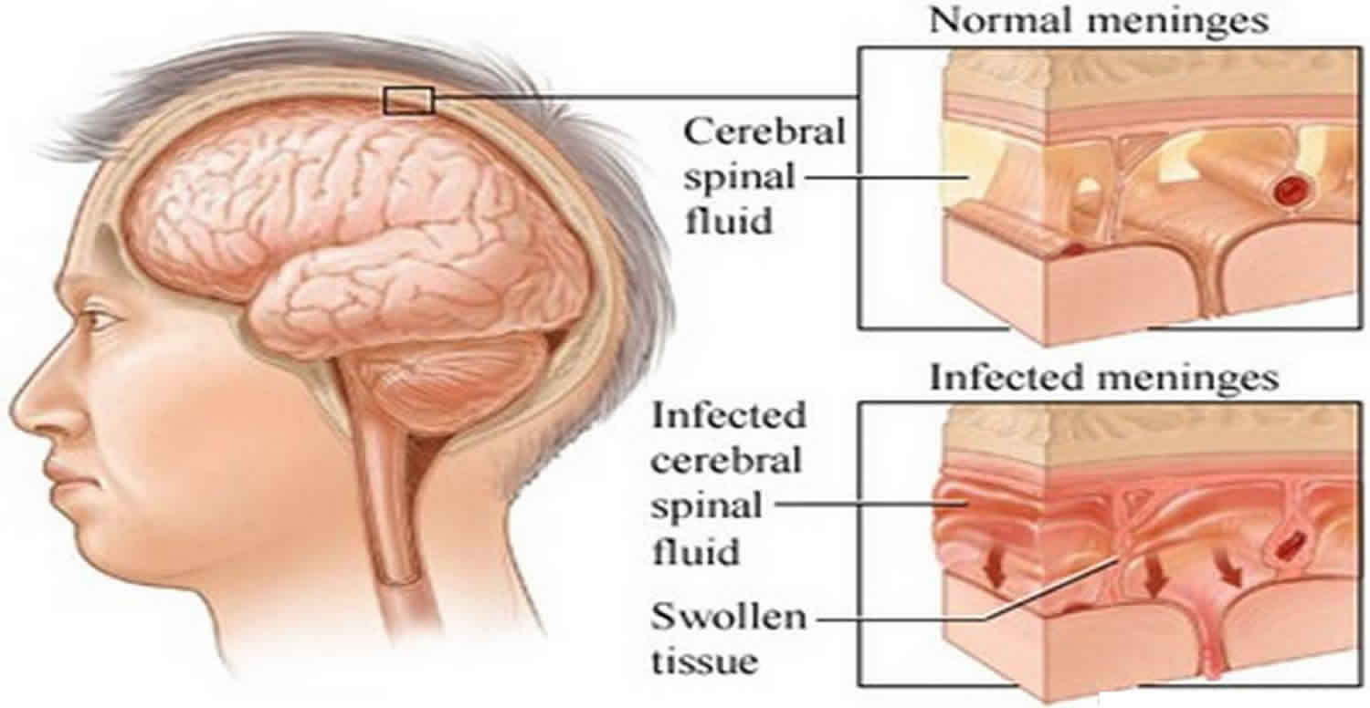
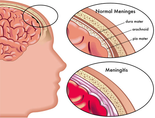
Postnatally, brain injuries during infancy or early childhood significantly contribute to CP. Infections like meningitis or encephalitis can cause inflammation and damage to brain tissue, impacting motor function. Traumatic brain injuries resulting from accidents or non-accidental trauma, such as shaken baby syndrome, are also potential triggers for CP. Additionally, strokes occurring in infancy or childhood can disrupt blood flow to the brain, leading to cerebral palsy.

While the exact mechanisms underlying CP causation remain intricate and multifaceted, understanding these diverse factors is crucial for prevention, early detection, and intervention strategies. Initiatives aimed at minimizing risk factors, promoting maternal health, and ensuring access to quality prenatal and perinatal care can play a pivotal role in reducing the incidence of cerebral palsy and improving outcomes for affected individuals. (Nursing Paper Example on a Comprehensive Overview of Cerebral Palsy: Etiology, Diagnosis, Treatment, and Patient Empowerment)

Signs and Symptoms

Cerebral palsy (CP) manifests in a spectrum of signs and symptoms, reflecting its heterogeneous nature and diverse impact on motor function and development. Impaired movement and coordination are hallmark features of CP, characterized by difficulties in controlling muscles and executing precise movements. Muscle stiffness or spasticity, often accompanied by involuntary muscle contractions, contributes to the overall motor impairment seen in CP.

Abnormal reflexes, such as exaggerated or absent reflex responses, are commonly observed in individuals with CP. These reflex abnormalities further hinder movement coordination and contribute to difficulties in achieving and maintaining posture and balance. As a result, individuals with CP may exhibit abnormal gait patterns, including toe-walking, scissoring of the legs, or asymmetrical movements.

In addition to motor impairments, CP frequently presents with associated conditions that impact overall functioning and quality of life. Intellectual disabilities, ranging from mild to severe, affect cognitive abilities and learning potential in varying degrees. Seizures, occurring in up to half of individuals with CP, pose additional challenges and require careful management to minimize their impact on daily activities.

Sensory impairments, including visual and auditory deficits, are also prevalent among individuals with CP, further complicating their ability to interact with their environment. Behavioral challenges, such as impulsivity, hyperactivity, or difficulties with social interactions, may arise due to the underlying neurological differences associated with CP.

The combination of motor impairments, associated conditions, and sensory and behavioral challenges underscores the complex nature of cerebral palsy and its wide-ranging impact on individuals’ lives. Early recognition of signs and symptoms, coupled with comprehensive multidisciplinary assessments, is crucial for timely intervention and support to optimize outcomes for individuals living with CP. (Nursing Paper Example on a Comprehensive Overview of Cerebral Palsy: Etiology, Diagnosis, Treatment, and Patient Empowerment)

Nursing Paper Example on a Comprehensive Overview of Cerebral Palsy: Etiology, Diagnosis, Treatment, and Patient Empowerment

Etiology

The etiology of cerebral palsy (CP) is multifactorial and often involves complex interactions between genetic, environmental, and developmental factors. Prenatal influences play a significant role in shaping CP risk, with maternal health and prenatal exposures impacting fetal brain development. Maternal infections, such as rubella or cytomegalovirus, can lead to neuroinflammation and disrupt normal brain development, increasing the likelihood of CP.

Genetic factors contribute to CP susceptibility, although their role is not always straightforward. Genetic mutations and chromosomal abnormalities may predispose individuals to CP, affecting neurodevelopmental processes critical for motor function and coordination. Additionally, variations in genes related to neuronal growth, synapse formation, and neurotransmitter regulation may influence CP risk.

Perinatal factors exert a substantial influence on CP etiology, with events occurring around the time of birth playing a pivotal role. Birth asphyxia, resulting from oxygen deprivation during labor or delivery, can lead to brain hypoxia and subsequent injury, contributing to CP development. Premature birth, defined as birth before 37 weeks of gestation, exposes infants to vulnerabilities in brain development, increasing CP risk.

Postnatal factors, including brain injuries and infections during infancy or early childhood, also contribute to CP etiology. Traumatic brain injuries, such as those resulting from accidents or non-accidental trauma, can cause structural damage to the brain, disrupting motor pathways and leading to CP. Infections like meningitis or encephalitis can result in neuroinflammation and neuronal damage, further exacerbating CP risk.

The interplay of these diverse etiological factors highlights the complexity of CP development and underscores the importance of a comprehensive approach to understanding its origins. Continued research into the genetic, environmental, and developmental determinants of CP is essential for elucidating its etiology and informing strategies for prevention, early intervention, and improved outcomes for affected individuals. (Nursing Paper Example on a Comprehensive Overview of Cerebral Palsy: Etiology, Diagnosis, Treatment, and Patient Empowerment)

Pathophysiology

The pathophysiology of cerebral palsy (CP) involves complex disruptions in brain development and function, affecting motor control, muscle tone, and coordination. While the precise mechanisms underlying CP pathogenesis vary among individuals, several key processes contribute to its manifestations.

Abnormalities in brain development, particularly during the prenatal and perinatal periods, play a central role in CP pathophysiology. Disturbances in neuronal migration, which occur during early brain development, can lead to structural abnormalities and impaired connectivity within the brain. Disruptions in white matter development, characterized by abnormalities in myelination and axonal connectivity, further impair the transmission of neural signals involved in motor control.

Neuroinflammation and oxidative stress are also implicated in CP pathophysiology, contributing to neuronal damage and functional impairments. Inflammatory responses triggered by prenatal infections, perinatal hypoxia-ischemia, or postnatal insults lead to the release of pro-inflammatory cytokines and reactive oxygen species, exacerbating neuronal injury and impairing neurodevelopmental processes.

Alterations in neurotransmitter systems, including imbalances in excitatory and inhibitory neurotransmission, contribute to motor dysfunction and spasticity in CP. Imbalances in neurotransmitters such as glutamate and gamma-aminobutyric acid (GABA) disrupt the delicate balance of neuronal signaling, leading to increased muscle tone and impaired motor control.

Structural abnormalities within specific brain regions, such as the basal ganglia and cerebellum, are commonly observed in individuals with CP and contribute to motor impairments. Dysfunction within these regions disrupts the coordination of voluntary movements and postural control, further exacerbating motor difficulties.

The cumulative effects of these pathophysiological processes result in the diverse manifestations of CP, including impaired movement, muscle stiffness, abnormal reflexes, and associated comorbidities. Understanding the underlying pathophysiology of CP is essential for developing targeted interventions aimed at mitigating its impact and improving outcomes for affected individuals. (Nursing Paper Example on a Comprehensive Overview of Cerebral Palsy: Etiology, Diagnosis, Treatment, and Patient Empowerment)

DSM-5 Diagnosis

The Diagnostic and Statistical Manual of Mental Disorders, Fifth Edition (DSM-5), provides criteria for diagnosing cerebral palsy (CP) based on motor impairments and evidence of neurological abnormalities. The DSM-5 criteria emphasize the importance of a comprehensive medical evaluation to accurately diagnose CP and differentiate it from other motor disorders.

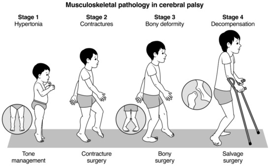
The primary criterion for CP diagnosis involves the presence of motor impairments that significantly interfere with movement and posture. These motor impairments typically manifest as abnormalities in muscle tone, coordination, and control, affecting both gross and fine motor skills. Examples include spasticity, muscle stiffness, involuntary movements, and difficulties with balance and coordination.

The onset of motor impairments is another key criterion for CP diagnosis according to DSM-5 guidelines. Motor symptoms must be evident before the age of 2 to meet the diagnostic criteria for CP. Early recognition of motor abnormalities and developmental delays is crucial for timely diagnosis and intervention.

In addition to motor impairments, the DSM-5 criteria for CP diagnosis require evidence of neurological abnormalities consistent with the disorder. Neurological assessments, including imaging studies such as magnetic resonance imaging (MRI) or computed tomography (CT) scans, may reveal structural brain abnormalities or signs of perinatal brain injury consistent with CP.

The DSM-5 also emphasizes the importance of ruling out other conditions that may mimic CP or coexist with it. Differential diagnosis may involve evaluating for other neurological disorders, musculoskeletal conditions, or developmental disabilities that share similar clinical features with CP.

A comprehensive evaluation incorporating clinical assessment, neurological examinations, developmental assessments, and imaging studies is essential for accurate CP diagnosis according to DSM-5 criteria. Timely and accurate diagnosis facilitates appropriate interventions, including early intervention services, rehabilitation therapies, and support services, to optimize outcomes for individuals living with CP. (Nursing Paper Example on a Comprehensive Overview of Cerebral Palsy: Etiology, Diagnosis, Treatment, and Patient Empowerment)

Treatment Regimens and Patient Education

The management of cerebral palsy (CP) involves a multidisciplinary approach aimed at addressing the diverse needs of individuals with CP and optimizing their overall function and quality of life. Treatment regimens for CP are tailored to the specific needs and challenges of each individual and may encompass various therapeutic interventions, support services, and patient education initiatives.

Therapeutic Interventions:

Physical therapy plays a central role in CP management, focusing on improving mobility, strength, flexibility, and motor function. Therapeutic exercises, stretching routines, and gait training are integral components of physical therapy programs tailored to the unique needs of individuals with CP.

Occupational therapy aims to enhance independence and participation in daily activities by addressing fine motor skills, sensory integration, and adaptive strategies. Occupational therapists work with individuals with CP to develop skills necessary for self-care, school, work, and recreational activities.

Speech therapy targets communication and swallowing difficulties commonly associated with CP. Speech-language pathologists assess and address speech articulation, language development, and oral motor function to improve communication and feeding skills.

Medications may be prescribed to manage spasticity, muscle stiffness, seizures, and associated comorbidities in individuals with CP. Common medications include muscle relaxants, antispasmodics, antiepileptic drugs, and medications to alleviate pain or discomfort.

Orthotic devices, such as braces, splints, or orthopedic footwear, may be recommended to improve posture, stability, and mobility in individuals with CP. Orthotics help support weakened muscles, correct alignment issues, and facilitate optimal movement patterns.

Patient Education:

Patient education is an essential component of CP management, empowering individuals with CP and their families to actively participate in treatment plans, access support services, and make informed decisions about their care. Education initiatives for individuals with CP may include:

  • Providing information about the nature of CP, including its causes, symptoms, and prognosis.
  • Educating individuals and families about available treatment options, including therapeutic interventions, medications, and surgical procedures.
  • Offering guidance on accessing support services, such as early intervention programs, special education services, and community resources.
  • Providing strategies for managing daily challenges associated with CP, including mobility limitations, communication difficulties, and behavioral issues.
  • Promoting self-advocacy skills and encouraging individuals with CP to express their needs, preferences, and goals in healthcare settings and beyond.

By incorporating therapeutic interventions and patient education initiatives into comprehensive treatment regimens, healthcare professionals can effectively support individuals with CP in achieving their maximum potential and enhancing their overall well-being.  (Nursing Paper Example on a Comprehensive Overview of Cerebral Palsy: Etiology, Diagnosis, Treatment, and Patient Empowerment)

Conclusion

Cerebral palsy (CP) presents a complex array of challenges requiring a comprehensive approach to management. From understanding its multifactorial etiology, diverse signs and symptoms, and intricate pathophysiology to utilizing the DSM-5 criteria for accurate diagnosis, the paper has shed light on various aspects of CP. Treatment regimens encompassing physical therapy, occupational therapy, speech therapy, medications, and orthotic devices are tailored to address the specific needs of individuals with CP. Moreover, patient education initiatives play a crucial role in empowering individuals and their families to actively participate in their care and make informed decisions. By incorporating these multidisciplinary interventions and educational efforts, healthcare professionals can enhance the quality of life for individuals living with CP, emphasizing the importance of early intervention, personalized care, and ongoing support to optimize outcomes and promote overall well-being. (Nursing Paper Example on a Comprehensive Overview of Cerebral Palsy: Etiology, Diagnosis, Treatment, and Patient Empowerment)

References

https://www.ncbi.nlm.nih.gov/books/NBK538147/

 
Do you need a similar assignment done for you from scratch? Order now!
Use Discount Code "Newclient" for a 15% Discount!

Nursing Paper Example on Chagas Disease: Causes, Symptoms, Diagnosis, Treatment, and Patient Education

Nursing Paper Example on Chagas Disease: Causes, Symptoms, Diagnosis, Treatment, and Patient Education

Chagas disease, or American trypanosomiasis, is a stealthy menace haunting tropical and subtropical regions. Discovered by Brazilian physician Carlos Chagas in 1909, this neglected tropical disease is caused by the protozoan parasite Trypanosoma cruzi. Despite its widespread prevalence, Chagas disease often eludes recognition, silently wreaking havoc on millions worldwide. Understanding its causes, signs and symptoms, etiology, pathophysiology, DSM-5 diagnosis, treatment regimens, and patient education is vital for combating its spread and alleviating its burden. From the humble beginnings of its discovery to the present-day challenges it poses to global health, unraveling the intricacies of Chagas disease is crucial in fostering awareness, enhancing surveillance, and ultimately, championing effective control measures against this formidable foe. (Nursing Paper Example on Chagas Disease: Causes, Symptoms, Diagnosis, Treatment, and Patient Education)

Nursing Paper Example on Chagas Disease: Causes, Symptoms, Diagnosis, Treatment, and Patient Education

Causes

Chagas disease, also known as American trypanosomiasis, is caused by the protozoan parasite Trypanosoma cruzi. This parasite is primarily transmitted to humans and other mammals through the feces of infected triatomine bugs, also known as “kissing bugs.” These bugs are prevalent in rural areas of Latin America, where poor housing conditions facilitate their proliferation. The transmission cycle begins when an infected triatomine bug feeds on the blood of a human or animal host. During feeding, the bug defecates near the site of the bite, releasing Trypanosoma cruzi parasites in its feces. The parasite then enters the host’s bloodstream through mucous membranes or breaks in the skin, initiating infection. Other modes of transmission include congenital transmission from mother to child, organ transplantation, and blood transfusion. Additionally, oral transmission can occur through the ingestion of contaminated food or beverages, particularly in endemic regions where triatomine bugs may contaminate fruits, juices, or other food items. Once inside the host, Trypanosoma cruzi undergoes complex developmental stages, including trypomastigotes, amastigotes, and epimastigotes, adapting to various environments within the host’s tissues. The parasite primarily targets cardiac muscle cells, smooth muscle cells in the gastrointestinal tract, and neurons in the nervous system, leading to the diverse clinical manifestations of Chagas disease. Understanding the intricate interplay between the parasite, the vector, and the host is crucial for devising effective control strategies aimed at interrupting the transmission cycle and preventing the spread of Chagas disease. Through targeted interventions focused on vector control, improved housing conditions, and public health education, we can work towards reducing the burden of this neglected tropical disease and protecting vulnerable populations from its devastating effects. (Nursing Paper Example on Chagas Disease: Causes, Symptoms, Diagnosis, Treatment, and Patient Education)

Signs and Symptoms

Chagas disease manifests through a spectrum of signs and symptoms, reflecting its dynamic progression from acute to chronic stages. In the acute phase, which typically lasts for weeks to months after initial infection, individuals may experience a myriad of nonspecific symptoms, including fever, fatigue, body aches, rash, diarrhea, and vomiting. However, it’s important to note that the majority of cases during this phase are asymptomatic, further complicating early detection and intervention efforts.

As the disease transitions into the chronic phase, which may unfold over years or even decades, the clinical picture evolves to encompass more severe and potentially life-threatening manifestations. Cardiac involvement, characterized by cardiomyopathy, arrhythmias, and heart failure, represents a hallmark of chronic Chagas disease, affecting up to 30% of infected individuals. These cardiac complications can manifest as palpitations, chest pain, shortness of breath, and peripheral edema, significantly impairing quality of life and increasing mortality risk.

Additionally, Chagas disease can precipitate gastrointestinal complications, primarily affecting the esophagus and colon. Megacolon, characterized by dilatation and hypomotility of the colon, can lead to symptoms such as constipation, abdominal distention, and fecal impaction. Similarly, megaesophagus, marked by esophageal dilatation and dysmotility, may present with dysphagia, regurgitation, and recurrent aspirations.

Beyond the cardiovascular and gastrointestinal domains, Chagas disease can also affect the nervous system, giving rise to neurologic manifestations such as peripheral neuropathy, meningoencephalitis, and stroke-like syndromes. These neurological complications, though less common than cardiac and gastrointestinal involvement, can contribute to significant morbidity and disability in affected individuals.

Overall, the signs and symptoms of Chagas disease encompass a diverse array of clinical presentations, spanning multiple organ systems and evolving. Early recognition and timely intervention are paramount in mitigating disease progression, preventing complications, and improving outcomes for those afflicted by this insidious parasitic infection. (Nursing Paper Example on Chagas Disease: Causes, Symptoms, Diagnosis, Treatment, and Patient Education)

Etiology

The etiology of Chagas disease is intricately intertwined with the life cycle of its causative agent, Trypanosoma cruzi, and the ecological dynamics of its vector, the triatomine bug. Trypanosoma cruzi, a protozoan parasite belonging to the Trypanosomatidae family, undergoes a complex developmental journey within the triatomine bug, also known as the “kissing bug” due to its biting behavior around the mouth and eyes. These bugs serve as vectors, transmitting the parasite to humans and other mammals through their feces during blood meals.

The transmission cycle begins when an infected triatomine bug feeds on the blood of a human or animal host. During feeding, the bug excretes feces containing Trypanosoma cruzi near the bite wound. The parasite then enters the host’s bloodstream through mucous membranes or breaks in the skin, initiating infection.

Once inside the host, Trypanosoma cruzi undergoes several developmental stages, including trypomastigotes, amastigotes, and epimastigotes, each adapted to specific environments within the host’s tissues. The parasite primarily targets cardiac muscle cells, smooth muscle cells in the gastrointestinal tract, and neurons in the nervous system, leading to the diverse clinical manifestations of Chagas disease.

The ecological and environmental factors play a crucial role in shaping the epidemiology of Chagas disease. Poor housing conditions, inadequate sanitation, and proximity to natural habitats favored by triatomine bugs contribute to the persistence of transmission cycles in endemic regions of Latin America. Additionally, human activities such as deforestation and urbanization can alter the distribution and abundance of triatomine bugs, further influencing the risk of Chagas disease transmission.

Understanding the complex interplay between the parasite, the vector, and the host is essential for devising effective control strategies aimed at interrupting the transmission cycle and preventing the spread of Chagas disease. By targeting key ecological and environmental determinants, alongside implementing vector control measures and promoting community-based interventions, we can strive towards reducing the burden of this neglected tropical disease and safeguarding the health of vulnerable populations. (Nursing Paper Example on Chagas Disease: Causes, Symptoms, Diagnosis, Treatment, and Patient Education)

Pathophysiology

The pathophysiology of Chagas disease is characterized by a cascade of events triggered by the invasion and persistence of Trypanosoma cruzi within the host’s tissues. Upon entry into the bloodstream, the parasite targets various organs and systems, including the heart, gastrointestinal tract, and nervous system, leading to progressive tissue damage and dysfunction.

In the acute phase of infection, Trypanosoma cruzi proliferates rapidly at the site of inoculation before disseminating throughout the host’s body. During this phase, the immune response plays a crucial role in controlling parasite replication and limiting tissue invasion. However, in some cases, the parasite manages to evade immune surveillance, establishing chronic infection.

As the disease progresses to the chronic phase, the ongoing presence of Trypanosoma cruzi triggers a persistent inflammatory response, leading to chronic myocarditis and cardiomyopathy. The parasite directly invades cardiac muscle cells, causing cellular damage and fibrosis, which impairs myocardial contractility and contributes to the development of cardiac complications such as arrhythmias, heart failure, and thromboembolic events.

In addition to cardiac involvement, Chagas disease can also affect the gastrointestinal tract, leading to disorders such as megaesophagus and megacolon. Trypanosoma cruzi invades the smooth muscle cells of the esophagus and colon, disrupting peristalsis and causing dilatation of these organs. This dysmotility results in symptoms such as dysphagia, regurgitation, constipation, and abdominal distention, significantly impacting the patient’s quality of life.

Furthermore, the parasite can infiltrate the nervous system, leading to neurologic manifestations such as peripheral neuropathy, meningoencephalitis, and stroke-like syndromes. Neuronal damage and inflammation contribute to the development of neurological deficits, including sensory and motor impairments, cognitive dysfunction, and autonomic disturbances.

Overall, the pathophysiology of Chagas disease is multifaceted, involving a complex interplay between the parasite, host immune response, and tissue-specific damage mechanisms. Understanding these underlying processes is essential for developing targeted therapeutic interventions and mitigating the long-term complications associated with this chronic parasitic infection. (Nursing Paper Example on Chagas Disease: Causes, Symptoms, Diagnosis, Treatment, and Patient Education)

DSM-5 Diagnosis

Diagnosing Chagas disease relies on a combination of clinical evaluation, laboratory testing, and imaging studies, guided by established diagnostic criteria outlined in the Diagnostic and Statistical Manual of Mental Disorders, Fifth Edition (DSM-5). The DSM-5 criteria provide a standardized framework for the identification and classification of mental disorders, including Chagas disease-related neurocognitive disorders.

The diagnosis of Chagas disease begins with a thorough medical history and physical examination to assess for risk factors, symptoms, and potential exposure to the parasite. Clinical suspicion may be heightened in individuals with a history of residence or travel to endemic regions, as well as those presenting with cardiac, gastrointestinal, or neurological symptoms consistent with Chagas disease.

Laboratory testing plays a pivotal role in confirming the diagnosis of Chagas disease, with serological assays serving as the mainstay of diagnostic evaluation. Enzyme-linked immunosorbent assay (ELISA) and indirect immunofluorescence assays (IFA) are commonly employed to detect specific antibodies against Trypanosoma cruzi in the patient’s blood serum. Positive serological results, indicative of past or current infection, are essential for establishing the diagnosis of Chagas disease.

In cases where the acute phase of infection is suspected, direct parasitological methods such as blood smear examination or polymerase chain reaction (PCR) testing may be utilized to detect the presence of Trypanosoma cruzi parasites in the patient’s blood. However, these methods are less sensitive and specific compared to serological assays, particularly during the chronic phase of infection.

Imaging studies, including electrocardiography (ECG) and echocardiography, may be employed to assess cardiac involvement and monitor disease progression in individuals with Chagas disease-related cardiomyopathy. These modalities enable the detection of cardiac abnormalities such as arrhythmias, conduction disturbances, ventricular dilatation, and systolic dysfunction, aiding in risk stratification and treatment planning.

In summary, the DSM-5 diagnosis of Chagas disease entails a comprehensive approach encompassing clinical evaluation, serological testing, and imaging studies to confirm the presence of Trypanosoma cruzi infection and assess its clinical manifestations. Early diagnosis and intervention are crucial for improving outcomes and reducing the burden of Chagas disease-related morbidity and mortality. (Nursing Paper Example on Chagas Disease: Causes, Symptoms, Diagnosis, Treatment, and Patient Education)

Treatment Regimens and Patient Education

Treatment of Chagas disease focuses on two main goals: eradicating the parasite and managing complications associated with chronic infection. Pharmacological therapy with antiparasitic medications, such as benznidazole or nifurtimox, represents the cornerstone of treatment, particularly in the acute and early chronic phases of the disease. These medications work by inhibiting the replication of Trypanosoma cruzi, thereby reducing parasite burden and preventing disease progression. However, it’s essential to note that treatment efficacy may vary depending on the stage of infection and individual factors such as age, comorbidities, and drug tolerability.

In addition to antiparasitic therapy, symptomatic management plays a crucial role in addressing complications related to Chagas disease, particularly cardiac and gastrointestinal manifestations. Patients with Chagas disease-related cardiomyopathy may require treatment with medications such as beta-blockers, angiotensin-converting enzyme (ACE) inhibitors, diuretics, and antiarrhythmic agents to manage heart failure, arrhythmias, and other cardiovascular symptoms. Similarly, individuals with gastrointestinal complications, such as megaesophagus or megacolon, may benefit from dietary modifications, prokinetic agents, laxatives, and surgical interventions to alleviate symptoms and improve gastrointestinal motility.

Patient education is paramount in the management of Chagas disease, empowering individuals with the knowledge and skills needed to prevent transmission, recognize early symptoms, and adhere to treatment regimens. Health education initiatives should emphasize the importance of vector control measures, such as improving housing conditions, using insecticide-treated bed nets, and avoiding outdoor sleeping areas to reduce exposure to triatomine bugs. Furthermore, promoting awareness about the potential risks of congenital transmission and blood transfusion-related transmission can help prevent secondary cases of Chagas disease in at-risk populations.

Moreover, educating patients about the importance of seeking timely medical evaluation and adhering to prescribed treatment regimens is essential for optimizing treatment outcomes and preventing disease progression. Patients should be counseled on the potential side effects and adverse reactions associated with antiparasitic medications, as well as strategies to manage these symptoms effectively. Additionally, healthcare providers should address the psychosocial aspects of Chagas disease, providing emotional support and connecting patients with relevant resources and support groups to cope with the challenges of living with a chronic, stigmatized condition.

A comprehensive approach to the management of Chagas disease encompasses pharmacological therapy, symptomatic management, and patient education. By integrating these components into clinical practice and public health initiatives, we can improve outcomes for individuals affected by Chagas disease and mitigate its impact on communities worldwide. (Nursing Paper Example on Chagas Disease: Causes, Symptoms, Diagnosis, Treatment, and Patient Education)

Conclusion

Chagas disease presents a multifaceted challenge requiring a comprehensive understanding of its causes, signs and symptoms, etiology, pathophysiology, DSM-5 diagnosis, treatment regimens, and patient education. Through the exploration of its intricate complexities, we have shed light on the stealthy nature of this neglected tropical disease and highlighted the importance of early recognition and intervention. By refining our understanding of its pathophysiological mechanisms, including the interplay between Trypanosoma cruzi and its host, and by emphasizing the DSM-5 diagnostic criteria, we can enhance our ability to identify and manage Chagas disease effectively. Furthermore, integrating pharmacological therapy with antiparasitic medications and symptomatic management, while prioritizing patient education and empowerment, is essential for improving treatment outcomes and reducing the burden of this chronic parasitic infection. Through collaborative efforts encompassing research, clinical practice, and public health initiatives, we can strive towards mitigating the impact of Chagas disease and improving the health and well-being of affected individuals and communities worldwide. (Nursing Paper Example on Chagas Disease: Causes, Symptoms, Diagnosis, Treatment, and Patient Education)

References

https://www.ncbi.nlm.nih.gov/books/NBK459272/

 
Do you need a similar assignment done for you from scratch? Order now!
Use Discount Code "Newclient" for a 15% Discount!

Nursing Paper Example on Chickenpox: Causes, Symptoms, Treatment, and Prevention

Nursing Paper Example on Chickenpox: Causes, Symptoms, Treatment, and Prevention

Chickenpox, also known as varicella, is a highly contagious viral infection characterized by a distinctive rash and fever. While commonly associated with childhood, this infectious disease can affect individuals of any age group. Despite the availability of a vaccine, chickenpox continues to pose a significant public health concern globally. Understanding the causes, symptoms, and treatment options is crucial for effective management and prevention of complications associated with this condition. With its characteristic rash and systemic symptoms, chickenpox presents unique challenges in terms of diagnosis and management. This paper aims to provide a comprehensive overview of chickenpox, including its causes, signs and symptoms, etiology, pathophysiology, DSM-5 diagnosis, treatment regimens, and patient education. By examining these aspects in detail, healthcare professionals and individuals alike can gain a deeper understanding of chickenpox and its impact on public health. (Nursing Paper Example on Chickenpox: Causes, Symptoms, Treatment, and Prevention)

Nursing Paper Example on Chickenpox: Causes, Symptoms, Treatment, and Prevention

Causes

Chickenpox is primarily caused by the varicella-zoster virus (VZV), a member of the herpesvirus family. This highly contagious virus is transmitted through respiratory droplets or direct contact with the fluid from the blisters of an infected individual. The virus can also spread through airborne transmission, making close contact with an infected person a significant risk factor for contracting the disease.

Individuals who have not been vaccinated or previously exposed to the virus are at risk of developing chickenpox upon exposure to an infected individual. The virus is most contagious during the early stages of the illness, typically from one to two days before the onset of the characteristic rash until all blisters have crusted over, which usually occurs about 5 to 7 days after the rash appears.

Children are particularly susceptible to chickenpox, as they often have close contact with other children in school or daycare settings where the virus can easily spread. However, adults who have not been previously infected or vaccinated are also at risk of developing chickenpox if exposed to the virus.

The varicella-zoster virus enters the body through the respiratory tract or mucous membranes, where it begins to replicate. After an incubation period of about 10 to 21 days following exposure, the virus causes symptomatic infection, leading to the characteristic clinical manifestations of chickenpox.

In recent years, widespread vaccination efforts have significantly reduced the incidence of chickenpox in many countries. Vaccination against chickenpox is highly effective in preventing infection and reducing the risk of complications associated with the disease. Routine immunization with the varicella vaccine is recommended for children, adolescents, and adults who have not been previously vaccinated or infected with the virus, highlighting the importance of vaccination in controlling the spread of chickenpox and protecting vulnerable populations from this infectious disease. (Nursing Paper Example on Chickenpox: Causes, Symptoms, Treatment, and Prevention)

Signs and Symptoms

Chickenpox typically presents with a characteristic rash and systemic symptoms, which can vary in severity among affected individuals. The hallmark sign of chickenpox is the development of a rash consisting of small, itchy blisters that appear in successive crops over several days. These blisters usually begin on the face and trunk before spreading to other parts of the body, including the scalp, arms, and legs.

Along with the rash, individuals with chickenpox may experience systemic symptoms such as fever, headache, fatigue, and loss of appetite. Fever is a common early symptom of chickenpox and may precede the appearance of the rash by a day or two. The rash typically progresses through different stages, starting as red bumps that evolve into fluid-filled blisters before crusting over and forming scabs. The entire process from the onset of the rash to the resolution of the lesions usually takes about 7 to 10 days.

The itching associated with the rash can vary in intensity and may be particularly distressing for some individuals. Scratching the blisters can lead to complications such as secondary bacterial infections or scarring, highlighting the importance of managing itching and promoting proper wound care.

In addition to the characteristic rash and systemic symptoms, individuals with chickenpox may experience other manifestations, including sore throat, cough, and swollen lymph nodes. In severe cases or individuals with weakened immune systems, complications such as pneumonia or encephalitis may occur, emphasizing the importance of prompt medical evaluation and management.

Overall, recognizing the signs and symptoms of chickenpox is essential for early diagnosis and appropriate management of the disease. Timely intervention can help alleviate symptoms, prevent complications, and reduce the risk of spreading the virus to others. (Nursing Paper Example on Chickenpox: Causes, Symptoms, Treatment, and Prevention)

Etiology

The etiology of chickenpox is attributed to the varicella-zoster virus (VZV), a highly contagious pathogen belonging to the herpesvirus family. Transmission of the virus occurs primarily through respiratory droplets expelled during coughing or sneezing by infected individuals. Direct contact with the fluid from the blisters of an infected person is another common mode of transmission, particularly during the active phase of the illness when the virus is shed from the skin lesions.

The varicella-zoster virus is capable of surviving outside the human body for a limited period, allowing it to spread via contaminated surfaces and objects. Additionally, airborne transmission of the virus can occur in settings where infected individuals share enclosed spaces, such as households, schools, and healthcare facilities.

Individuals who have not been previously infected with or vaccinated against the varicella-zoster virus are at risk of developing chickenpox upon exposure to the virus. Susceptibility to chickenpox is highest among young children who have not yet been vaccinated and adults who have not been previously exposed to the virus.

Following exposure, the varicella-zoster virus enters the body through the respiratory tract or mucous membranes, where it begins to replicate. The virus then disseminates to regional lymph nodes, leading to viremia and systemic spread throughout the body. This systemic dissemination results in the characteristic clinical manifestations of chickenpox, including the development of the characteristic rash and associated symptoms.

In recent years, widespread vaccination efforts have significantly reduced the incidence of chickenpox in many countries. Routine immunization with the varicella vaccine has proven highly effective in preventing infection and reducing the overall burden of chickenpox, highlighting the importance of vaccination in controlling the spread of the disease and protecting vulnerable populations from its complications. (Nursing Paper Example on Chickenpox: Causes, Symptoms, Treatment, and Prevention)

Pathophysiology

The pathophysiology of chickenpox involves a complex interplay between the varicella-zoster virus (VZV) and the host immune system. Following exposure to the virus, the initial site of infection is typically the respiratory epithelium or mucous membranes, where the virus gains entry into the body.

Once inside the host, the varicella-zoster virus undergoes a period of incubation, during which it replicates and begins to spread within the body. The virus then enters the bloodstream, leading to viremia and systemic dissemination to various organs and tissues.

The systemic spread of the virus results in the widespread distribution of viral particles throughout the body, contributing to the characteristic clinical manifestations of chickenpox. This includes the development of the characteristic rash, which is characterized by the formation of small, itchy blisters that appear in successive crops over several days.

The immune response plays a crucial role in the pathophysiology of chickenpox, as the host’s immune system mounts a coordinated defense against the invading virus. Activation of the innate and adaptive immune responses leads to the production of cytokines and antibodies, which help control viral replication and clear the infection.

In some cases, the immune response may also contribute to the development of complications associated with chickenpox. For example, severe cases of chickenpox may be characterized by excessive inflammation and tissue damage, leading to complications such as pneumonia or encephalitis.

Overall, the pathophysiology of chickenpox involves a complex interaction between the varicella-zoster virus and the host immune system. Understanding these mechanisms is crucial for developing effective treatment strategies and interventions aimed at controlling the spread of the virus and minimizing the risk of complications associated with chickenpox. (Nursing Paper Example on Chickenpox: Causes, Symptoms, Treatment, and Prevention)

DSM-5 Diagnosis

Chickenpox, also known as varicella, is a medical condition caused by the varicella-zoster virus (VZV) and is not diagnosed using the criteria outlined in the Diagnostic and Statistical Manual of Mental Disorders, Fifth Edition (DSM-5). Instead, the diagnosis of chickenpox is primarily based on clinical presentation, including the characteristic rash and associated symptoms.

Healthcare professionals typically rely on a combination of history, physical examination, and laboratory tests to diagnose chickenpox accurately. The hallmark sign of chickenpox is the development of a rash consisting of small, itchy blisters that appear in successive crops over several days. Along with the rash, individuals with chickenpox may experience systemic symptoms such as fever, headache, fatigue, and loss of appetite.

Laboratory tests, such as viral culture or polymerase chain reaction (PCR) testing, may be performed to confirm the presence of the varicella-zoster virus in suspected cases of chickenpox. However, these tests are not routinely recommended for diagnosis due to the characteristic clinical presentation of the disease.

It is essential to differentiate chickenpox from other viral exanthems, such as measles or rubella, which may present with similar symptoms. Healthcare professionals must consider the epidemiological context, vaccination status, and clinical features when making a diagnosis of chickenpox.

While chickenpox is primarily a clinical diagnosis, healthcare professionals may also consider the patient’s medical history, including vaccination status and previous exposure to the virus, when evaluating suspected cases. Additionally, healthcare providers should be aware of the potential for complications associated with chickenpox, such as pneumonia or encephalitis, and monitor patients accordingly.

In summary, chickenpox is diagnosed based on clinical presentation, including the characteristic rash and associated symptoms, rather than using the criteria outlined in the DSM-5. Healthcare professionals rely on a combination of history, physical examination, and laboratory tests to accurately diagnose and manage cases of chickenpox. (Nursing Paper Example on Chickenpox: Causes, Symptoms, Treatment, and Prevention)

Treatment Regimens and Patient Education

The management of chickenpox primarily involves supportive care to alleviate symptoms and prevent complications. While chickenpox is usually a self-limiting illness that resolves on its own within a few weeks, certain interventions can help alleviate discomfort and minimize the risk of complications, particularly in high-risk individuals such as pregnant women, newborns, and individuals with weakened immune systems.

Supportive care measures aim to alleviate symptoms such as fever and itching and promote proper wound care to prevent secondary bacterial infections and scarring. Over-the-counter antipyretic medications such as acetaminophen (Tylenol) may be recommended to reduce fever and discomfort. It is essential to follow dosing instructions carefully and avoid giving aspirin to children and teenagers with chickenpox due to the risk of Reye’s syndrome, a rare but serious condition.

To relieve itching associated with the rash, patients can apply calamine lotion or take oatmeal baths. Short nails and gentle scratching with clean hands or a soft cloth can help minimize skin damage and reduce the risk of infection. Avoiding exposure to extreme temperatures and wearing loose-fitting clothing made of breathable fabrics can also help prevent exacerbation of itching and discomfort.

In severe cases or individuals at high risk of complications, antiviral medications such as acyclovir may be prescribed to shorten the duration of illness and reduce the severity of symptoms. Antiviral therapy is most effective when initiated within 24 to 48 hours of the onset of rash but may still be beneficial if started later in the course of the illness, particularly in high-risk individuals.

Patient education plays a crucial role in the management of chickenpox, emphasizing the importance of good hygiene practices and proper wound care to prevent complications and reduce the risk of spreading the virus to others. Patients should be advised to wash their hands frequently with soap and water, especially after touching the rash or coming into contact with respiratory secretions.

Furthermore, patients should be educated about the importance of avoiding close contact with individuals who are susceptible to complications, such as pregnant women, newborns, and individuals with weakened immune systems. Vaccination against chickenpox is highly effective in preventing infection and reducing the risk of complications, underscoring the importance of routine immunization for individuals of all ages.

In summary, supportive care measures, including proper wound care and symptom management, are essential components of the treatment regimen for chickenpox. Patient education focusing on hygiene practices, avoidance of close contact with high-risk individuals, and the importance of vaccination is crucial for effective management and prevention of complications associated with this infectious disease. (Nursing Paper Example on Chickenpox: Causes, Symptoms, Treatment, and Prevention)

Conclusion

Chickenpox, caused by the varicella-zoster virus, presents with a characteristic rash and systemic symptoms, with a complex pathophysiology involving systemic spread and immune response. The diagnosis is primarily clinical, relying on characteristic symptoms and history, rather than DSM-5 criteria. Treatment involves supportive care, including antipyretics, antivirals in severe cases, and proper wound care, while patient education emphasizes hygiene, avoidance of high-risk individuals, and the importance of vaccination. With these interventions, complications can be minimized, and the spread of the virus can be controlled effectively. While chickenpox remains a concern, widespread vaccination efforts have significantly reduced its incidence, underscoring the importance of routine immunization in preventing this infectious disease and protecting vulnerable populations. Overall, understanding the causes, symptoms, treatment options, and preventive measures is essential for effective management and prevention of complications associated with chickenpox, highlighting the ongoing importance of public health efforts in combating this infectious disease. (Nursing Paper Example on Chickenpox: Causes, Symptoms, Treatment, and Prevention)

References

https://www.ncbi.nlm.nih.gov/books/NBK448191/

 
Do you need a similar assignment done for you from scratch? Order now!
Use Discount Code "Newclient" for a 15% Discount!

Nursing Paper Example on Chlamydia: Causes, Symptoms, Diagnosis, Treatment, and Public Health Implications

Nursing Paper Example on Chlamydia: Causes, Symptoms, Diagnosis, Treatment, and Public Health Implications

Chlamydia, caused by the bacterium Chlamydia trachomatis, is a prevalent global health concern, particularly affecting sexually active individuals. Despite often being asymptomatic, untreated chlamydia can lead to severe complications, making early detection and treatment crucial. This paper aims to provide a comprehensive overview of chlamydia, covering its causes, signs and symptoms, etiology, pathophysiology, DSM-5 diagnosis, treatment regimens, patient education, and implications for public health. Understanding the multifaceted aspects of chlamydia is vital for healthcare professionals and the general public alike, as it facilitates effective prevention strategies, timely diagnosis, and appropriate management. By delving into the intricacies of this sexually transmitted infection, we can work towards reducing its prevalence, mitigating its impact on individuals and communities, and promoting overall sexual health and well-being. (Nursing Paper Example on Chlamydia: Causes, Symptoms, Diagnosis, Treatment, and Public Health Implications)

Causes

Chlamydia is primarily transmitted through sexual contact with an infected individual, including vaginal, anal, or oral sex. The bacterium Chlamydia trachomatis infects the mucous membranes of the genital tract, urethra, rectum, throat, and eyes, leading to various complications if left untreated.

The transmission of chlamydia occurs when infected bodily fluids, such as vaginal secretions or semen, come into contact with mucous membranes or breaks in the skin of a susceptible individual. This can occur during penetrative sexual activities or through non-penetrative sexual practices that involve genital contact.

Additionally, chlamydia can be transmitted from an infected mother to her newborn during childbirth, leading to neonatal conjunctivitis and pneumonia. Inadequate prenatal care, lack of screening during pregnancy, and untreated maternal chlamydial infections contribute to the vertical transmission of the bacterium to the infant.

Several factors increase the risk of chlamydial transmission and acquisition. These include engaging in unprotected sexual activities with multiple partners, having a history of previous STIs, being sexually active at a young age, and engaging in high-risk sexual behaviors such as inconsistent condom use or substance abuse during sexual encounters.

Furthermore, individuals with chlamydia are often unaware of their infection status due to its asymptomatic nature, leading to unknowing transmission to sexual partners. Lack of awareness about chlamydia, stigma associated with STIs, and barriers to accessing sexual health services also contribute to the ongoing transmission of the infection within communities.

Efforts to prevent chlamydia transmission focus on promoting safer sexual practices, increasing awareness about STIs, encouraging regular STI screening, and ensuring access to comprehensive sexual health services. By addressing the underlying factors contributing to chlamydial transmission and implementing targeted prevention strategies, it is possible to reduce the burden of chlamydia and its associated complications on individuals and public health systems. (Nursing Paper Example on Chlamydia: Causes, Symptoms, Diagnosis, Treatment, and Public Health Implications)

Signs and Symptoms

Nursing Paper Example on Chlamydia: Causes, Symptoms, Diagnosis, Treatment, and Public Health Implications
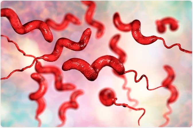
Chlamydia infection can manifest with a wide range of signs and symptoms, although many individuals infected with Chlamydia trachomatis remain asymptomatic, especially in the early stages of infection. When symptoms do occur, they typically appear within 1 to 3 weeks after exposure to the bacterium.

One of the most common symptoms of chlamydia is abnormal genital discharge, which may be accompanied by a foul odor or changes in color or consistency. In men, this discharge often presents as a clear or cloudy fluid from the urethra, while in women, it may be vaginal discharge that is different from their usual discharge.

Painful or burning sensation during urination, known as dysuria, is another hallmark symptom of chlamydia. This discomfort is caused by inflammation and irritation of the urethra or genital mucosa due to the infection.

Genital itching or irritation may also occur in individuals with chlamydia, particularly in women. This symptom is often attributed to inflammation of the genital tissues and can contribute to discomfort and distress.

Pelvic pain or lower abdominal pain is a common complaint among individuals with chlamydial infections, especially in cases where the infection has spread to the reproductive organs. This pain may be mild to severe and can interfere with daily activities and quality of life.

In some cases, chlamydia can infect the rectum or throat, leading to symptoms such as rectal pain, discharge, or discomfort during bowel movements, as well as sore throat or pharyngitis, respectively.

It is important to note that the absence of symptoms does not necessarily indicate the absence of infection, particularly in asymptomatic individuals. Therefore, regular screening for chlamydia is recommended for sexually active individuals, especially those at increased risk of infection or with a history of unprotected sexual activity. Early detection and treatment of chlamydia are essential for preventing the development of complications and reducing the risk of transmission to sexual partners. (Nursing Paper Example on Chlamydia: Causes, Symptoms, Diagnosis, Treatment, and Public Health Implications)

Etiology

Chlamydia trachomatis, the bacterium responsible for chlamydial infections, is a unique and highly adaptable pathogen that exhibits a complex life cycle and sophisticated mechanisms of host interaction. Belonging to the family Chlamydiaceae, Chlamydia trachomatis is an obligate intracellular bacterium, meaning it can only survive and replicate within the cytoplasm of host cells.

The biphasic developmental cycle of Chlamydia trachomatis is a key aspect of its etiology. This cycle involves two distinct forms of the bacterium: the infectious elementary body (EB) and the non-infectious reticulate body (RB). Upon entering a host cell, Chlamydia trachomatis initially exists in the form of EB, which is metabolically inactive and adapted for survival in the extracellular environment. The EB then undergoes a process of differentiation into the metabolically active RB, which replicates within the host cell.

Chlamydia trachomatis manipulates various cellular processes to facilitate its survival and replication within host cells. The bacterium employs strategies to evade the host immune response, including inhibition of apoptosis (programmed cell death) and modulation of host cell signaling pathways. Additionally, Chlamydia trachomatis alters intracellular trafficking pathways to create a niche for its replication, while also subverting host cell functions to acquire essential nutrients for growth.

The ability of Chlamydia trachomatis to establish a persistent infection within host cells contributes to its pathogenicity and chronicity. Persistent infection is characterized by the prolonged presence of viable Chlamydia trachomatis within host cells, despite the activation of host immune responses. This persistence can lead to the development of chronic inflammatory conditions, such as pelvic inflammatory disease (PID) in women and epididymitis in men, as well as long-term complications such as infertility.

Understanding the etiology of Chlamydia trachomatis infection is essential for developing effective prevention and treatment strategies. By elucidating the intricate mechanisms underlying chlamydial pathogenesis, researchers can identify potential targets for therapeutic intervention and enhance our ability to control the spread of this important human pathogen. (Nursing Paper Example on Chlamydia: Causes, Symptoms, Diagnosis, Treatment, and Public Health Implications)

Pathophysiology

Chlamydia trachomatis, the causative agent of chlamydia, exhibits a unique pathophysiology characterized by its obligate intracellular lifestyle and biphasic developmental cycle within host cells. Upon entry into the host cell, Chlamydia trachomatis manipulates various cellular processes to facilitate its survival and replication. The bacterium actively inhibits host cell apoptosis (programmed cell death) and modulates intracellular signaling pathways to create a favorable environment for its growth and proliferation.

Central to the pathophysiology of chlamydia is the bacterium’s biphasic developmental cycle, which involves two distinct forms: the infectious elementary body (EB) and the non-infectious reticulate body (RB). Following entry into the host cell, the EB differentiates into the metabolically active RB, which replicates within a membrane-bound inclusion within the host cell cytoplasm. The RBs undergo multiple rounds of replication, leading to the accumulation of bacterial progeny within the inclusion.

Chlamydia trachomatis exhibits a unique ability to modulate host cell functions to its advantage. The bacterium manipulates intracellular trafficking pathways to acquire essential nutrients and evade host immune surveillance. Additionally, Chlamydia trachomatis alters host cell gene expression and cytokine production, leading to the suppression of pro-inflammatory responses and the promotion of a chronic, low-grade inflammatory state within infected tissues.

The pathophysiological consequences of chlamydia infection extend beyond the local mucosal site to involve systemic immune dysregulation and tissue damage. Chronic chlamydial infections have been implicated in the development of inflammatory sequelae, including pelvic inflammatory disease (PID), infertility, and ectopic pregnancy in women, as well as epididymitis and prostatitis in men. Furthermore, chlamydia-induced inflammation can increase susceptibility to HIV acquisition and transmission.

Understanding the intricate pathophysiological mechanisms underlying chlamydia infection is essential for developing targeted therapeutic interventions and preventive strategies aimed at mitigating the long-term complications associated with this prevalent sexually transmitted infection. (Nursing Paper Example on Chlamydia: Causes, Symptoms, Diagnosis, Treatment, and Public Health Implications)

DSM-5 Diagnosis

The Diagnostic and Statistical Manual of Mental Disorders, Fifth Edition (DSM-5), does not include specific diagnostic criteria for chlamydia infection, as it primarily focuses on mental health disorders. However, healthcare providers rely on clinical assessment and laboratory testing to diagnose chlamydia and other sexually transmitted infections (STIs).

The diagnosis of chlamydia is primarily based on laboratory testing of genital, rectal, or pharyngeal specimens to detect the presence of Chlamydia trachomatis DNA or antigens. Nucleic acid amplification tests (NAATs) are the preferred diagnostic method due to their high sensitivity and specificity. NAATs can accurately detect Chlamydia trachomatis in various clinical specimens, including urine, vaginal swabs, cervical swabs, urethral swabs, rectal swabs, and pharyngeal swabs.

In clinical practice, healthcare providers typically screen asymptomatic individuals who are at increased risk of chlamydia infection, such as sexually active individuals under the age of 25, individuals with multiple sexual partners, and pregnant individuals. Additionally, symptomatic individuals presenting with signs and symptoms suggestive of chlamydia, such as genital discharge, dysuria, pelvic pain, or rectal symptoms, should undergo diagnostic testing for chlamydia and other STIs.

It is essential for healthcare providers to adhere to established clinical practice guidelines for chlamydia screening, testing, and management to ensure accurate diagnosis and appropriate treatment. Prompt diagnosis and treatment of chlamydia are critical for preventing the spread of infection, reducing the risk of complications, and promoting sexual health.

Although the DSM-5 does not include specific criteria for chlamydia diagnosis, healthcare providers need to consider the impact of STIs on mental health and well-being. Individuals diagnosed with chlamydia may experience psychological distress, anxiety, or depression related to their diagnosis and concerns about sexual health, underscoring the importance of comprehensive care and support for affected individuals. (Nursing Paper Example on Chlamydia: Causes, Symptoms, Diagnosis, Treatment, and Public Health Implications)

Treatment Regimens and Patient Education

The cornerstone of managing chlamydia infection involves prompt initiation of antibiotic therapy and comprehensive patient education to ensure treatment adherence, prevent reinfection, and promote sexual health.

Antibiotic Therapy:

The recommended first-line antibiotics for treating chlamydia are azithromycin and doxycycline, both of which are highly effective in eradicating Chlamydia trachomatis. Azithromycin is administered as a single oral dose, simplifying treatment adherence and enhancing patient convenience. On the other hand, doxycycline is typically prescribed as a twice-daily regimen for seven days. Both antibiotics work by inhibiting bacterial protein synthesis, thereby eliminating the bacterium from the body.

Healthcare providers must assess patient factors such as allergies, medication tolerability, and pregnancy status when selecting the appropriate antibiotic regimen. In cases where azithromycin or doxycycline is contraindicated or not tolerated, alternative antibiotics such as erythromycin or levofloxacin may be considered.

Patient Education:

Effective patient education plays a pivotal role in ensuring treatment success, preventing reinfection, and promoting overall sexual health. Healthcare providers should engage in open and nonjudgmental discussions with patients about chlamydia, emphasizing the following key points:

  1. Medication Adherence: Patients must be instructed to take the prescribed antibiotics exactly as directed, completing the full course of treatment even if symptoms improve before completion. Skipping doses or discontinuing treatment prematurely can lead to treatment failure and the development of antibiotic-resistant strains of Chlamydia trachomatis.
  2. Abstaining from Sexual Activity: During the course of antibiotic treatment, patients should abstain from sexual activity to prevent the transmission of chlamydia to their sexual partners. Partners should also be tested and treated simultaneously to prevent reinfection.
  3. Condom Use: Consistent and correct condom use is essential for reducing the risk of chlamydia transmission and other sexually transmitted infections. Patients should be educated on the proper use of condoms and encouraged to incorporate them into their sexual practices.
  4. Regular Screening: Patients who have been treated for chlamydia should undergo repeat testing approximately three months after completing treatment to confirm eradication of the infection. Additionally, individuals who engage in high-risk sexual behaviors or have multiple sexual partners should undergo regular screening for chlamydia and other STIs.
  5. Partner Notification: Patients diagnosed with chlamydia should inform their recent sexual partners about their infection to facilitate partner notification, testing, and treatment. This helps break the cycle of transmission and prevent the spread of chlamydia within sexual networks.

By providing comprehensive patient education, healthcare providers empower patients to take an active role in managing their chlamydia infection, reducing the risk of complications, and promoting sexual health and well-being. Open communication, nonjudgmental support, and personalized counseling are essential components of effective patient education initiatives aimed at addressing chlamydia and other sexually transmitted infections. (Nursing Paper Example on Chlamydia: Causes, Symptoms, Diagnosis, Treatment, and Public Health Implications)

Conclusion

Chlamydia, caused by the bacterium Chlamydia trachomatis, poses a significant public health challenge worldwide, particularly among sexually active individuals. This paper has provided a comprehensive overview of chlamydia, covering its causes, signs and symptoms, etiology, pathophysiology, DSM-5 diagnosis, treatment regimens, patient education, and implications for public health. By delving into the multifaceted aspects of this sexually transmitted infection, we have highlighted the importance of early detection, prompt treatment with appropriate antibiotics, and comprehensive patient education in preventing complications, reducing transmission rates, and promoting sexual health. The updated sections, including the expanded discussion on treatment regimens and patient education, underscore the critical role of healthcare providers in delivering patient-centered care and empowering individuals to make informed decisions regarding their sexual health. Through collaborative efforts among healthcare professionals, policymakers, and community stakeholders, we can work towards addressing the challenges posed by chlamydia and improving overall sexual health outcomes for individuals and populations globally. (Nursing Paper Example on Chlamydia: Causes, Symptoms, Diagnosis, Treatment, and Public Health Implications)

References

https://www.ncbi.nlm.nih.gov/books/NBK537286/

 
Do you need a similar assignment done for you from scratch? Order now!
Use Discount Code "Newclient" for a 15% Discount!

Nursing Paper Example on Cholera

Nursing Paper Example on Cholera

Cholera, often regarded as one of the most feared infectious diseases in history, continues to pose a significant public health threat in various parts of the world. Its sudden onset and rapid progression can lead to severe dehydration and death if not promptly treated. Understanding the intricacies of cholera, including its causes, symptoms, treatment, and preventive measures, is paramount in combating its spread and minimizing its impact on affected populations. Cholera, caused by the bacterium Vibrio cholerae, primarily affects regions with poor sanitation and hygiene practices, where contaminated water and food sources contribute to its transmission. The hallmark symptoms of cholera, including profuse watery diarrhea and vomiting, can quickly lead to dehydration and electrolyte imbalance if left untreated. This paper aims to provide comprehensive and accurate information on cholera, covering its causes, signs and symptoms, etiology, pathophysiology, diagnosis, treatment regimens, patient education, and preventive measures. By enhancing our understanding of cholera, we can better address the challenges it presents and work towards its prevention and control on a global scale. (Nursing Paper Example on Cholera)

Nursing Paper Example on Cholera

Causes

Cholera, a waterborne disease, is primarily caused by the bacterium Vibrio cholerae, with serogroups O1 and O139 being the most commonly associated with epidemic and pandemic outbreaks. The bacterium thrives in aquatic environments, particularly in areas with poor sanitation and inadequate sewage systems. Contaminated water sources, including rivers, lakes, and wells, serve as reservoirs for Vibrio cholerae, facilitating its transmission to humans through the consumption of contaminated water or food.

Human activities play a significant role in the spread of cholera. In regions lacking access to clean water and proper sanitation facilities, individuals are more likely to use contaminated water for drinking, cooking, and personal hygiene, increasing their risk of infection. Additionally, overcrowded living conditions and poor waste management contribute to the proliferation of Vibrio cholerae in communities, leading to outbreaks of cholera.

Natural disasters and humanitarian crises can exacerbate the spread of cholera by disrupting water and sanitation systems, displacing populations, and creating conditions conducive to the transmission of the bacterium. Floods, earthquakes, and conflicts can compromise infrastructure and access to essential services, amplifying the risk of cholera outbreaks in affected areas.

Climate variability and environmental factors also influence the incidence of cholera. Warmer temperatures and heavy rainfall promote the growth of Vibrio cholerae in aquatic environments, leading to increased contamination of water sources during certain seasons. Coastal regions and areas prone to flooding are particularly susceptible to cholera outbreaks due to their geographical characteristics and environmental conditions.

Addressing the root causes of cholera requires comprehensive approaches that focus on improving access to clean water and sanitation, promoting hygiene practices, and addressing socioeconomic factors that contribute to vulnerability. By addressing these underlying determinants, communities can reduce the risk of cholera transmission and mitigate the impact of outbreaks on public health. (Nursing Paper Example on Cholera)

Signs and Symptoms

The clinical presentation of cholera is characterized by sudden onset diarrhea, often described as “rice-water stool,” due to its appearance as a clear fluid containing particles resembling rice grains. This profuse watery diarrhea is a result of the cholera toxin produced by Vibrio cholerae, which leads to increased secretion of fluid and electrolytes into the intestinal lumen. The volume of fluid loss can be substantial, reaching up to 1 liter per hour in severe cases.

Alongside diarrhea, patients with cholera commonly experience vomiting, which may initially help to expel the ingested bacteria but can exacerbate dehydration if it persists. Dehydration is a hallmark feature of cholera and can rapidly progress to hypovolemic shock if not promptly treated. Signs of dehydration include sunken eyes, decreased skin turgor, dry mucous membranes, and reduced urine output.

Muscle cramps are another common symptom of cholera, resulting from electrolyte imbalances, particularly low levels of potassium (hypokalemia) and sodium (hyponatremia). These cramps can be severe and debilitating, further contributing to patient discomfort and weakness.

Other systemic manifestations of cholera may include rapid heart rate (tachycardia), low blood pressure (hypotension), and metabolic acidosis due to the loss of bicarbonate ions through diarrhea. These physiological changes reflect the body’s response to fluid and electrolyte disturbances caused by cholera toxin.

In severe cases, untreated cholera can lead to complications such as renal failure, circulatory collapse, and death within hours of symptom onset. Prompt recognition of the signs and symptoms of cholera is critical for initiating timely treatment and preventing life-threatening complications. Healthcare providers in endemic regions must maintain a high index of suspicion for cholera, particularly during outbreaks or in areas with poor sanitation and hygiene practices. Early diagnosis and intervention can significantly improve patient outcomes and reduce mortality associated with this devastating disease. (Nursing Paper Example on Cholera)

Etiology

The etiology of cholera encompasses a complex interplay of environmental, socioeconomic, and individual factors that contribute to the persistence and spread of the disease. Vibrio cholerae, the bacterium responsible for cholera, thrives in aquatic environments, particularly in regions with inadequate sanitation and hygiene infrastructure. Contaminated water sources, such as rivers, lakes, and wells, serve as reservoirs for Vibrio cholerae, facilitating its transmission to humans through the consumption of contaminated water or food.

Socioeconomic factors play a significant role in shaping the epidemiology of cholera. Poverty, inadequate access to clean water and sanitation facilities, overcrowded living conditions, and poor waste management practices increase the risk of cholera transmission in affected communities. Limited healthcare infrastructure and resources further exacerbate the burden of cholera in low-resource settings, where access to timely diagnosis and treatment may be limited.

Population density and mobility also influence the spread of cholera, particularly in urban areas and densely populated settlements where the risk of person-to-person transmission is heightened. Migration, displacement, and travel contribute to the dissemination of Vibrio cholerae across geographic regions, leading to outbreaks and the potential for pandemics.

Climate variability and environmental factors play a role in shaping the seasonal and geographic distribution of cholera. Warmer temperatures and heavy rainfall promote the growth of Vibrio cholerae in aquatic environments, leading to increased contamination of water sources during certain seasons. Coastal regions and areas prone to flooding are particularly susceptible to cholera outbreaks due to their geographical characteristics and environmental conditions.

Addressing the underlying determinants of cholera requires multifaceted interventions that target improvements in water and sanitation infrastructure, poverty alleviation, health education, and community engagement. By addressing the root causes of cholera transmission, communities can reduce the risk of outbreaks and mitigate the impact of the disease on public health. (Nursing Paper Example on Cholera)

Pathophysiology

The pathophysiology of cholera revolves around the actions of the cholera toxin, a key virulence factor produced by Vibrio cholerae. Upon ingestion of contaminated food or water, Vibrio cholerae colonizes the small intestine, where it adheres to the epithelial cells lining the intestinal mucosa. Once attached, Vibrio cholerae secretes cholera toxin, a protein complex consisting of two subunits: the A subunit, responsible for enzymatic activity, and the B subunit, which binds to specific receptors on the surface of intestinal cells.

The cholera toxin exerts its effects by activating adenylate cyclase within the intestinal epithelial cells, leading to increased intracellular levels of cyclic adenosine monophosphate (cAMP). Elevated cAMP levels stimulate the secretion of chloride ions (Cl-) into the intestinal lumen, followed by passive movement of sodium ions (Na+) and water. This process, known as the secretory diarrhea pathway, results in the rapid secretion of large volumes of fluid into the intestinal lumen, leading to the characteristic profuse watery diarrhea observed in cholera.

The loss of fluid and electrolytes through diarrhea leads to dehydration and electrolyte imbalances, particularly hypokalemia and hyponatremia. Dehydration can rapidly progress to hypovolemic shock if left untreated, leading to organ failure and death in severe cases.

In addition to its effects on intestinal epithelial cells, the cholera toxin can also exert systemic effects, including stimulation of the release of proinflammatory cytokines and activation of the complement system. These systemic effects contribute to the clinical manifestations of cholera, including systemic inflammation, hypotension, and metabolic acidosis.

Overall, the pathophysiology of cholera is characterized by the dysregulation of fluid and electrolyte balance within the intestinal tract, resulting from the actions of the cholera toxin produced by Vibrio cholerae. Understanding these mechanisms is essential for the development of effective treatment strategies aimed at correcting fluid and electrolyte imbalances and preventing life-threatening complications associated with severe cholera infection. (Nursing Paper Example on Cholera)

DMS-5 Diagnosis

Cholera is a severe gastrointestinal infection caused by the bacterium Vibrio cholerae and is primarily diagnosed based on clinical presentation and epidemiological factors. While the Diagnostic and Statistical Manual of Mental Disorders, Fifth Edition (DSM-5), is not typically used to diagnose infectious diseases like cholera, certain criteria can aid in identifying and categorizing the condition within a broader public health context.

The DSM-5 does not provide specific diagnostic criteria for infectious diseases; however, it does emphasize the importance of considering contextual factors such as environmental, social, and cultural influences in understanding the presentation and impact of diseases. In the case of cholera, these contextual factors play a crucial role in shaping the epidemiology and clinical manifestations of the disease.

Clinically, the diagnosis of cholera is primarily based on the presence of characteristic symptoms, including profuse watery diarrhea, vomiting, and dehydration, in individuals with a history of exposure to contaminated water or food sources. Laboratory confirmation through stool culture or rapid diagnostic tests may be performed to identify the presence of Vibrio cholerae in clinical specimens, particularly in outbreak settings or cases where confirmation is necessary for public health surveillance purposes.

Epidemiological factors, such as geographic location, recent travel history to endemic regions, and involvement in outbreaks, also contribute to the diagnosis of cholera. Public health authorities and healthcare providers rely on epidemiological surveillance data to monitor and control the spread of cholera, implement targeted interventions, and allocate resources effectively.

While the DSM-5 does not provide specific diagnostic criteria for cholera, its emphasis on considering environmental and contextual factors underscores the importance of a comprehensive approach to understanding and addressing infectious diseases within a broader public health framework. By incorporating clinical, laboratory, and epidemiological factors, healthcare providers can accurately diagnose and manage cholera cases and contribute to efforts aimed at preventing and controlling its spread. (Nursing Paper Example on Cholera)

Treatment Regimens and Patient Education

The treatment of cholera encompasses a multifaceted approach aimed at addressing dehydration, electrolyte imbalances, and bacterial eradication, coupled with patient education to enhance preventive measures and promote timely intervention.

Fluid and Electrolyte Replacement: The cornerstone of cholera treatment is rehydration therapy to replace fluid and electrolyte losses resulting from profuse diarrhea and vomiting. Oral rehydration solution (ORS), containing a precise balance of glucose and electrolytes, is the preferred method for mild to moderate cases. ORS is readily available, cost-effective, and easy to administer, making it suitable for use in resource-limited settings. For severe dehydration or cases with significant fluid loss, intravenous fluid therapy may be necessary to rapidly restore hydration and electrolyte balance.

Antibiotic Therapy: In addition to rehydration, antibiotic therapy may be indicated in severe cases or outbreaks to reduce the duration and severity of symptoms and limit bacterial shedding. Antibiotics such as azithromycin or doxycycline are commonly used due to their efficacy against Vibrio cholerae and ease of administration. However, antibiotic resistance patterns should be considered when selecting appropriate treatment regimens, and treatment decisions should be guided by local epidemiological data and antimicrobial susceptibility testing.

Patient Education: Patient education plays a crucial role in cholera management, focusing on preventive measures, recognition of dehydration symptoms, and early intervention. Communities at risk of cholera should be educated on the importance of access to clean water, proper sanitation and hygiene practices, and vaccination where available. Patients and caregivers should receive guidance on preparing and administering ORS, monitoring hydration status, and seeking medical care promptly if symptoms worsen or persist. Health education campaigns, community outreach programs, and collaboration with local stakeholders are essential for disseminating accurate information and empowering individuals to take proactive measures against cholera.

By integrating comprehensive treatment regimens with targeted patient education initiatives, healthcare providers can effectively manage cholera cases, prevent complications, and reduce the spread of the disease within affected communities. Empowering individuals with knowledge and resources not only improves health outcomes but also strengthens community resilience and response to cholera outbreaks, ultimately contributing to global efforts to control and eliminate this devastating infectious disease. (Nursing Paper Example on Cholera)

Conclusion

The treatment of cholera requires a multifaceted approach that encompasses fluid and electrolyte replacement, antibiotic therapy, and patient education. By addressing dehydration and electrolyte imbalances through oral rehydration solution (ORS) and intravenous fluid therapy, healthcare providers can effectively manage cholera cases and prevent life-threatening complications. The addition of antibiotic therapy, guided by local resistance patterns and epidemiological data, helps to shorten the duration and severity of symptoms and limit bacterial shedding, particularly in severe cases or outbreaks. Patient education plays a pivotal role in cholera management, empowering individuals with knowledge on preventive measures, recognition of dehydration symptoms, and the importance of seeking timely medical care. By integrating comprehensive treatment regimens with targeted patient education initiatives, healthcare providers can enhance cholera prevention and control efforts, improve health outcomes, and ultimately reduce the burden of this devastating infectious disease on affected communities. (Nursing Paper Example on Cholera)

References

https://www.ncbi.nlm.nih.gov/books/NBK470232/

 
Do you need a similar assignment done for you from scratch? Order now!
Use Discount Code "Newclient" for a 15% Discount!

Nursing Paper Example on Aseptic Meningitis: Understanding Causes, Symptoms, Diagnosis, and Treatment

Nursing Paper Example on Aseptic Meningitis: Understanding Causes, Symptoms, Diagnosis, and Treatment

Aseptic meningitis, characterized by inflammation of the meninges without bacterial involvement, poses significant challenges in diagnosis and management. Typically caused by viral infections, this condition manifests with symptoms like fever, headache, and neck stiffness. Understanding its causes, symptoms, and underlying mechanisms is crucial for effective patient care. This paper provides comprehensive insights into aseptic meningitis, aiming to enhance understanding among students and healthcare practitioners. By exploring the diverse etiologies, clinical presentations, pathophysiological mechanisms, and diagnostic criteria of aseptic meningitis, this paper seeks to equip readers with the knowledge necessary to recognize, diagnose, and manage this condition effectively. With a focus on simplicity and clarity, this paper elucidates the complexities of aseptic meningitis, offering a valuable resource for students seeking foundational knowledge in this area and healthcare practitioners striving to deliver high-quality care to patients affected by this condition. (Nursing Paper Example on Aseptic Meningitis: Understanding Causes, Symptoms, Diagnosis, and Treatment)

Nursing Paper Example on Aseptic Meningitis: Understanding Causes, Symptoms, Diagnosis, and Treatment

Causes

Aseptic meningitis, often triggered by viral infections, encompasses a spectrum of causative agents. Enteroviruses, particularly echoviruses and coxsackieviruses, stand out as predominant contributors to this condition. These viruses, ubiquitous in the environment, gain entry through the respiratory or gastrointestinal tract, subsequently disseminating to the central nervous system. Additionally, herpesviruses, including herpes simplex virus (HSV) and varicella-zoster virus (VZV), can precipitate aseptic meningitis, particularly during primary infection or reactivation.

Moreover, mumps virus, typically associated with parotitis, occasionally manifests with aseptic meningitis as a complication. Influenza viruses, notably influenza A and B, also pose a risk, especially during seasonal outbreaks. Beyond viral etiologies, certain medications such as nonsteroidal anti-inflammatory drugs (NSAIDs), antibiotics, and intravenous immunoglobulins (IVIG) have been implicated in drug-induced aseptic meningitis.

Furthermore, autoimmune disorders like systemic lupus erythematosus (SLE) and Behçet’s disease can trigger meningitis through immune-mediated mechanisms. Neoplastic disorders, particularly lymphomas and leukemias, may rarely infiltrate the meninges, causing aseptic meningitis as a paraneoplastic phenomenon.

Environmental factors, including overcrowding and poor sanitation, facilitate viral transmission, contributing to the incidence of aseptic meningitis. Individual susceptibility, influenced by genetic predispositions and immunocompromised states, also plays a pivotal role.

Understanding this diverse array of etiologies is paramount for accurate diagnosis and tailored management strategies. Clinicians must consider the specific infectious or non-infectious triggers, along with patient-specific factors, to formulate an effective approach to mitigating the impact of aseptic meningitis. (Nursing Paper Example on Aseptic Meningitis: Understanding Causes, Symptoms, Diagnosis, and Treatment)

Signs and Symptoms

Aseptic meningitis presents with a constellation of symptoms that overlap with those of bacterial meningitis, making accurate diagnosis challenging. The hallmark features of aseptic meningitis include fever, which is often abrupt in onset and may be accompanied by chills and rigors. Severe headache, typically diffuse and throbbing in nature, is another common symptom experienced by patients with aseptic meningitis.

Neck stiffness, or nuchal rigidity, is a characteristic finding resulting from meningeal irritation. This stiffness limits the patient’s ability to flex the neck forward, leading to discomfort and resistance upon attempted movement. Photophobia, an aversion to light, is frequently reported and contributes to the overall discomfort experienced by patients.

Nausea and vomiting are common gastrointestinal symptoms associated with aseptic meningitis, often attributed to meningeal inflammation and increased intracranial pressure. These symptoms can exacerbate the patient’s overall distress and contribute to dehydration if not adequately managed.

Patients with aseptic meningitis may also exhibit altered mental status, ranging from lethargy and confusion to frank delirium or coma in severe cases. Behavioral changes, including irritability and restlessness, may be observed, particularly in pediatric patients.

Additionally, neurological signs such as focal deficits, seizures, and cranial nerve abnormalities may manifest, depending on the extent of meningeal inflammation and involvement of adjacent structures.

Clinicians must meticulously evaluate these signs and symptoms in the context of the patient’s clinical presentation and medical history to differentiate aseptic meningitis from other conditions, including bacterial meningitis, encephalitis, and non-infectious causes of meningeal irritation. Early recognition and prompt initiation of appropriate treatment are essential for optimizing outcomes and preventing potential complications associated with aseptic meningitis. (Nursing Paper Example on Aseptic Meningitis: Understanding Causes, Symptoms, Diagnosis, and Treatment)

Etiology

The etiology of aseptic meningitis encompasses a diverse range of viral and non-viral factors. Viral infections, particularly those caused by enteroviruses, represent the most common etiological agents. Enteroviruses, including coxsackieviruses and echoviruses, are ubiquitous in the environment and commonly infect individuals through fecal-oral or respiratory routes. Other viral culprits associated with aseptic meningitis include herpesviruses, such as herpes simplex virus (HSV) and varicella-zoster virus (VZV), which can lead to meningitis during primary infection or reactivation.

Non-viral causes of aseptic meningitis include autoimmune disorders, such as systemic lupus erythematosus (SLE) and Behçet’s disease, which trigger meningitis through immune-mediated mechanisms. Certain medications, including nonsteroidal anti-inflammatory drugs (NSAIDs), antibiotics, and intravenous immunoglobulins (IVIG), have also been implicated in drug-induced aseptic meningitis.

Furthermore, neoplastic disorders, particularly lymphomas and leukemias, may infiltrate the meninges, leading to aseptic meningitis as a paraneoplastic phenomenon. Environmental factors, such as overcrowding and poor sanitation, facilitate viral transmission and contribute to the incidence of aseptic meningitis.

Individual susceptibility plays a crucial role in the development of aseptic meningitis, influenced by genetic predispositions and immunocompromised states. Immunocompromised individuals, including those with HIV/AIDS or undergoing immunosuppressive therapy, are at increased risk of developing aseptic meningitis due to impaired immune responses.

Understanding the diverse etiological factors contributing to aseptic meningitis is essential for accurate diagnosis and appropriate management strategies. Clinicians must consider the specific infectious or non-infectious triggers, along with patient-specific factors, to formulate an effective approach to mitigating the impact of aseptic meningitis. (Nursing Paper Example on Aseptic Meningitis: Understanding Causes, Symptoms, Diagnosis, and Treatment)

Pathophysiology

The pathophysiology of aseptic meningitis involves a cascade of inflammatory processes triggered by viral or non-viral etiologies. In viral aseptic meningitis, viral particles gain entry into the central nervous system (CNS) via various routes, including the bloodstream or direct neuronal spread. Once inside the CNS, viral replication occurs, leading to the release of viral antigens and activation of the immune response.

Nursing Paper Example on Aseptic Meningitis: Understanding Causes, Symptoms, Diagnosis, and Treatment

This immune response involves the activation of resident microglia and infiltration of peripheral immune cells, including lymphocytes and monocytes, into the meninges and cerebrospinal fluid (CSF). The release of pro-inflammatory cytokines, such as interleukin-1β (IL-1β) and tumor necrosis factor-alpha (TNF-α), further amplifies the inflammatory cascade, contributing to meningeal inflammation and increased permeability of the blood-brain barrier.

The influx of immune cells and cytokines into the CSF leads to characteristic changes, including elevated white blood cell count (pleocytosis) and increased protein levels, observed in the cerebrospinal fluid analysis of patients with aseptic meningitis. Additionally, disruption of the blood-brain barrier allows for the diffusion of inflammatory mediators and viral particles into the brain parenchyma, contributing to neuronal injury and dysfunction.

Non-viral causes of aseptic meningitis, such as autoimmune disorders and drug-induced reactions, involve immune-mediated mechanisms that result in meningeal inflammation and disruption of normal CNS homeostasis. Autoimmune reactions lead to the production of autoantibodies targeting components of the CNS, while drug-induced reactions may involve direct toxicity or immune-mediated hypersensitivity responses.

Overall, the pathophysiology of aseptic meningitis is characterized by a complex interplay of viral or non-viral triggers, immune-mediated inflammation, and disruption of CNS homeostasis, resulting in the characteristic clinical manifestations observed in affected individuals. Understanding these underlying mechanisms is crucial for guiding therapeutic interventions and preventing complications associated with aseptic meningitis. (Nursing Paper Example on Aseptic Meningitis: Understanding Causes, Symptoms, Diagnosis, and Treatment)

DMS-5 Diagnosis

The diagnosis of aseptic meningitis relies on a combination of clinical evaluation, laboratory tests, and imaging studies, with specific criteria outlined in the Diagnostic and Statistical Manual of Mental Disorders, Fifth Edition (DMS-5). According to DMS-5, the presence of characteristic symptoms such as fever, headache, and neck stiffness is essential for diagnosing aseptic meningitis. These symptoms typically manifest acutely and may be accompanied by additional signs such as photophobia, nausea, and altered mental status.

Laboratory tests play a crucial role in confirming the diagnosis of aseptic meningitis. Cerebrospinal fluid (CSF) analysis via lumbar puncture reveals findings consistent with meningeal inflammation, including elevated white blood cell count (pleocytosis), increased protein levels, and normal to mildly elevated glucose levels. Additionally, polymerase chain reaction (PCR) testing of CSF can detect viral nucleic acids, aiding in the identification of specific viral pathogens responsible for aseptic meningitis.

Imaging studies, such as magnetic resonance imaging (MRI) or computed tomography (CT) scans of the brain, may be performed to rule out other neurological conditions or complications associated with aseptic meningitis, such as cerebral edema or hydrocephalus.

The diagnosis of aseptic meningitis requires careful consideration of the patient’s clinical presentation, medical history, and laboratory findings, as well as the exclusion of bacterial meningitis through appropriate diagnostic tests. By adhering to the diagnostic criteria outlined in DMS-5 and utilizing a comprehensive approach to evaluation, healthcare practitioners can accurately diagnose aseptic meningitis and initiate timely management strategies to optimize patient outcomes. (Nursing Paper Example on Aseptic Meningitis: Understanding Causes, Symptoms, Diagnosis, and Treatment)

Treatment Regimens and Patient Education

The management of aseptic meningitis primarily focuses on supportive care, symptom relief, and addressing specific underlying causes if identified. Since aseptic meningitis is often caused by viral infections, treatment is aimed at alleviating symptoms and promoting recovery, as viral infections are typically self-limiting.

Patients diagnosed with aseptic meningitis are advised to get adequate rest and ensure proper hydration. Bed rest is often recommended during the acute phase of the illness to facilitate recovery and reduce the risk of complications. Hydration is crucial to prevent dehydration, especially in cases where patients experience nausea, vomiting, or fever. Encouraging fluid intake, including water, electrolyte solutions, and clear fluids, helps maintain hydration status and supports the body’s immune response.

Over-the-counter medications such as nonsteroidal anti-inflammatory drugs (NSAIDs) or acetaminophen may be used to alleviate fever, headache, and muscle pain. However, caution should be exercised when administering NSAIDs, particularly in patients with underlying renal impairment or gastrointestinal disorders.

In severe cases of aseptic meningitis or when complications arise, hospitalization may be necessary for close monitoring and intravenous administration of fluids and medications. Intravenous fluids help maintain hydration and electrolyte balance, while medications such as antiemetics may be prescribed to alleviate nausea and vomiting.

Patient education plays a crucial role in the management of aseptic meningitis, empowering individuals to actively participate in their care and recovery process. Patients should be informed about the nature of the illness, the expected course of symptoms, and potential complications. Clear instructions regarding medication use, including dosage, frequency, and possible side effects, should be provided to ensure compliance and safety.

Additionally, patients should be educated about the importance of follow-up appointments with healthcare providers to monitor their progress and address any concerns or new symptoms. Prevention strategies, including good hygiene practices such as handwashing and avoiding close contact with individuals who are sick, should also be emphasized to reduce the risk of viral transmission and recurrence of aseptic meningitis.

By providing comprehensive patient education and implementing appropriate treatment regimens, healthcare providers can effectively manage aseptic meningitis, promote symptom relief, and support patients toward optimal recovery. (Nursing Paper Example on Aseptic Meningitis: Understanding Causes, Symptoms, Diagnosis, and Treatment)

Conclusion

Aseptic meningitis presents a complex challenge in diagnosis and management, requiring a thorough understanding of its diverse etiologies, clinical manifestations, and treatment approaches. Through comprehensive insights into the causes, symptoms, pathophysiology, and diagnosis of aseptic meningitis, healthcare practitioners can better navigate the complexities of this condition and provide optimal care for affected individuals. The multifactorial nature of aseptic meningitis, encompassing viral and non-viral triggers, underscores the importance of tailored treatment regimens and patient education to address specific underlying causes and promote symptom relief. By emphasizing supportive care, symptom management, and preventive measures, healthcare providers can empower patients to actively participate in their recovery process and minimize the risk of complications. Through ongoing research and clinical advancements, further improvements in the diagnosis, treatment, and management of aseptic meningitis can be achieved, ultimately enhancing outcomes and reducing the burden of this condition on individuals and healthcare systems alike. (Nursing Paper Example on Aseptic Meningitis: Understanding Causes, Symptoms, Diagnosis, and Treatment)

References

https://www.ncbi.nlm.nih.gov/books/NBK557412/

 
Do you need a similar assignment done for you from scratch? Order now!
Use Discount Code "Newclient" for a 15% Discount!

Nursing Paper Example on Bunion: Understanding Causes, Symptoms, Diagnosis, Treatment, and Patient Education

Nursing Paper Example on Bunion: Understanding Causes, Symptoms, Diagnosis, Treatment, and Patient Education

Bunions, medically referred to as hallux valgus, represent a prevalent foot deformity affecting the joint at the base of the big toe. Characterized by the gradual deviation of the big toe towards the second toe, bunions can lead to considerable discomfort, pain, and difficulty in wearing shoes. Despite their common occurrence, bunions can significantly impact an individual’s quality of life, hindering mobility and causing discomfort during daily activities. As such, gaining a comprehensive understanding of bunions, including their causes, symptoms, diagnosis, treatment options, and patient education, is paramount for healthcare providers and individuals alike. This paper aims to delve into the various aspects of bunions, providing accurate and thorough information to enhance awareness, facilitate effective management strategies, and ultimately improve outcomes for individuals affected by this condition. (Nursing Paper Example on Bunion: Understanding Causes, Symptoms, Diagnosis, Treatment, and Patient Education)

Nursing Paper Example on Bunion: Understanding Causes, Symptoms, Diagnosis, Treatment, and Patient Education

Causes

Bunions develop due to a combination of genetic predisposition and external factors. One of the primary causes is an inherited foot type that makes individuals more susceptible to developing bunions. Certain foot shapes, such as having a low arch or flat feet, can contribute to abnormal foot mechanics, leading to the misalignment of the big toe joint over time.

External factors also play a significant role in bunion formation. Wearing tight, narrow shoes, particularly high heels, can exert excessive pressure on the big toe joint, forcing it out of alignment. Prolonged wearing of ill-fitting footwear can exacerbate this pressure, contributing to the development and progression of bunions.

Foot injuries and trauma can also increase the likelihood of developing bunions. Damage to the structures around the big toe joint, such as ligaments and tendons, can disrupt the normal alignment of the toe, leading to the formation of a bunion.

Additionally, certain medical conditions, such as inflammatory joint diseases like rheumatoid arthritis, can predispose individuals to developing bunions. These conditions can cause inflammation and damage to the joints, including the big toe joint, contributing to bunion formation.

Furthermore, repetitive stress on the feet, such as participating in activities that involve excessive pressure on the toes, can also increase the risk of developing bunions over time. Athletes and individuals who engage in activities that involve repetitive movements of the foot may be more susceptible to developing bunions due to the constant stress placed on the toe joint.

Overall, bunions develop as a result of a complex interplay between genetic factors, foot structure, footwear choices, foot injuries, and underlying medical conditions. Understanding these contributing factors is crucial for effective prevention and management of bunions. (Nursing Paper Example on Bunion: Understanding Causes, Symptoms, Diagnosis, Treatment, and Patient Education)

Signs and Symptoms

Bunions present with various signs and symptoms, ranging from mild discomfort to significant pain and functional impairment. The most prominent sign of a bunion is the visible bump or protrusion at the base of the big toe. This bump is caused by the misalignment of the big toe joint, which forces the bone to protrude outward, often creating a noticeable deformity.

Individuals with bunions commonly experience pain and tenderness around the affected joint. This discomfort may be exacerbated by pressure from footwear or movement of the toe. The pain can range from mild to severe and may interfere with daily activities such as walking or standing for extended periods.

Swelling and inflammation are also common symptoms of bunions. The soft tissues around the big toe joint may become swollen and tender due to irritation and friction caused by the misaligned toe rubbing against shoes. In some cases, the skin overlying the bunion may become red and irritated, further exacerbating discomfort.

As bunions progress, individuals may notice changes in the alignment of their toes. The big toe may begin to drift towards the second toe, causing overlapping or crowding of the toes. This can lead to the development of corns and calluses on the affected toes, as well as discomfort and difficulty finding shoes that fit properly.

Nursing Paper Example on Bunion: Understanding Causes, Symptoms, Diagnosis, Treatment, and Patient Education

In severe cases, bunions can cause a limited range of motion in the affected toe joint, making it difficult to bend or flex the toe. This restriction in movement can further impact mobility and may contribute to gait abnormalities.

Overall, the signs and symptoms of bunions can vary in severity and may worsen over time if left untreated. Early recognition and appropriate management are essential for alleviating discomfort and preventing further complications associated with this common foot deformity. (Nursing Paper Example on Bunion: Understanding Causes, Symptoms, Diagnosis, Treatment, and Patient Education)

Etiology

The development of bunions, known medically as hallux valgus, is multifactorial and influenced by a combination of genetic predisposition, structural abnormalities, and external factors.

Genetic factors play a significant role in bunion formation, with individuals having a family history of bunions being more likely to develop this condition. Inherited foot types, such as having a low arch or flat feet, can contribute to abnormal foot mechanics, leading to the misalignment of the big toe joint over time. Genetic factors may also influence the shape and structure of the bones in the foot, predisposing certain individuals to bunions.

Structural abnormalities in the foot can also contribute to the development of bunions. Conditions such as excessive pronation (rolling inward) of the foot, a long second toe, or a tight Achilles tendon can alter the biomechanics of the foot, placing increased stress on the big toe joint and leading to its misalignment.

External factors, particularly footwear choices, play a crucial role in bunion development. Wearing tight, narrow shoes, especially high heels, can exert excessive pressure on the big toe joint, forcing it out of alignment over time. Prolonged wearing of ill-fitting footwear can exacerbate this pressure, contributing to the development and progression of bunions.

Additionally, foot injuries and trauma can increase the risk of developing bunions. Damage to the structures around the big toe joint, such as ligaments and tendons, can disrupt the normal alignment of the toe, leading to the formation of a bunion.

Overall, bunions develop as a result of the interplay between genetic predisposition, structural abnormalities, and external factors such as footwear choices and foot injuries. Understanding these etiological factors is essential for implementing effective preventive measures and treatment strategies for bunions. (Nursing Paper Example on Bunion: Understanding Causes, Symptoms, Diagnosis, Treatment, and Patient Education)

Pathophysiology

The pathophysiology of bunions involves a complex interplay of biomechanical changes, structural abnormalities, and inflammatory processes that ultimately result in the characteristic deformity of the big toe joint.

Bunions typically develop gradually over time, beginning with alterations in foot biomechanics. Factors such as excessive pronation (rolling inward) of the foot, a long second toe, or a tight Achilles tendon can disrupt the normal alignment of the foot and place increased stress on the big toe joint. This increased stress leads to the gradual displacement of the big toe towards the second toe, resulting in the formation of a bunion.

Nursing Paper Example on Bunion: Understanding Causes, Symptoms, Diagnosis, Treatment, and Patient Education

As the big toe deviates laterally, the joint at its base becomes misaligned, causing the bones to protrude outward and form a bony prominence. This misalignment alters the distribution of weight across the foot, leading to further deformity and instability in the joint.

The structural changes associated with bunions also affect the surrounding soft tissues, including the ligaments, tendons, and joint capsule. These tissues become stretched and strained as they attempt to accommodate the misaligned joint, leading to inflammation, swelling, and pain.

Chronic inflammation of the soft tissues surrounding the bunion contributes to the development of secondary symptoms such as redness, tenderness, and swelling. Additionally, the constant friction and pressure from footwear can lead to the formation of corns and calluses over the bunion, further exacerbating discomfort.

Over time, untreated bunions can progress and become more severe, impacting the individual’s ability to walk, stand, and engage in daily activities. Severe bunions may also increase the risk of developing complications such as bursitis, arthritis, and hammertoe deformities.

Understanding the pathophysiology of bunions is crucial for developing effective treatment strategies aimed at addressing the underlying biomechanical abnormalities and inflammatory processes contributing to the condition. (Nursing Paper Example on Bunion: Understanding Causes, Symptoms, Diagnosis, Treatment, and Patient Education)

DSM-5 Diagnosis

The Diagnostic and Statistical Manual of Mental Disorders, Fifth Edition (DSM-5), does not provide specific diagnostic criteria for bunions, as they are a physical rather than psychiatric condition. However, healthcare providers may use clinical assessment and imaging studies to diagnose and evaluate the severity of bunions.

Clinically, bunions are diagnosed based on physical examination and medical history. Healthcare providers typically assess the appearance of the foot, looking for characteristic signs such as a visible bump at the base of the big toe and the deviation of the big toe towards the second toe. They may also palpate the affected area to assess for tenderness, swelling, and range of motion in the affected joint.

Imaging studies, such as X-rays, may be ordered to evaluate the severity of the bunion deformity and assess the alignment of the bones in the foot. X-rays can also help healthcare providers identify any underlying structural abnormalities or joint damage associated with the bunion.

The severity of bunions is often classified using the Hallux Valgus Angle (HVA) and the Intermetatarsal Angle (IMA), which are measured on X-rays. The HVA measures the angle between the first metatarsal bone and the proximal phalanx of the big toe, while the IMA measures the angle between the first and second metatarsal bones. These measurements help healthcare providers determine the degree of misalignment and deformity in the big toe joint.

Although bunions do not have specific DSM-5 diagnostic criteria, healthcare providers may use clinical assessment and imaging studies to diagnose and classify the severity of the condition. This information guides treatment decisions and helps healthcare providers develop personalized management plans for individuals with bunions. (Nursing Paper Example on Bunion: Understanding Causes, Symptoms, Diagnosis, Treatment, and Patient Education)

Treatment Regimens and Patient Education

Treatment for bunions aims to alleviate symptoms, slow the progression of the deformity, and improve foot function. The management of bunions typically involves a combination of conservative measures and, in some cases, surgical intervention. Patient education plays a crucial role in the effective management of bunions, empowering individuals to make informed decisions about their care and adopt lifestyle modifications to promote foot health.

Conservative Treatment:

  1. Footwear Modification: Encouraging patients to wear wide, comfortable shoes with a spacious toe box can help reduce pressure on the bunion and alleviate symptoms. Avoiding high heels and tight, narrow shoes can prevent further irritation and deformity.
  2. Orthotic Devices: Custom orthotic inserts or over-the-counter shoe inserts can provide support and cushioning, improving foot mechanics and reducing pain associated with bunions. These devices can help distribute pressure more evenly across the foot and promote proper alignment.
  3. Padding and Taping: Using padding or moleskin over the bunion can help reduce friction and protect the affected area from irritation. Taping the big toe to gently realign it and relieve pressure on the bunion may also provide temporary relief.
  4. Medications: Over-the-counter nonsteroidal anti-inflammatory drugs (NSAIDs), such as ibuprofen or naproxen, can help alleviate pain and reduce inflammation associated with bunions. Topical analgesic creams or gels may also provide temporary relief.
  5. Physical Therapy: Specific exercises and stretches prescribed by a physical therapist can help strengthen the muscles and improve flexibility in the foot, promoting better foot mechanics and reducing symptoms of bunions.

Surgical Intervention:

In cases where conservative measures fail to provide adequate relief or the bunion deformity is severe, surgical intervention may be considered. Bunion surgery, also known as bunionectomy, aims to realign the bones of the big toe joint, remove excess bone and soft tissue, and correct any structural abnormalities contributing to the deformity.

Patient Education:

  1. Foot Care: Educating patients about proper foot care practices, including regular toenail trimming, moisturizing, and inspecting the feet for signs of irritation or infection, can help prevent complications such as ingrown toenails and ulcers.
  2. Footwear Selection: Advising patients to choose footwear with adequate toe space, low heels, and good arch support can prevent exacerbation of bunions and promote foot comfort and health.
  3. Activity Modification: Encouraging patients to engage in low-impact activities that do not aggravate bunion symptoms, such as swimming or cycling, can help maintain overall fitness while reducing stress on the feet.
  4. Weight Management: Emphasizing the importance of maintaining a healthy weight through proper diet and regular exercise can help reduce pressure on the feet and prevent the progression of bunion deformities.
  5. Regular Follow-up: Stressing the importance of regular follow-up appointments with healthcare providers to monitor the progression of bunions, assess treatment effectiveness, and make adjustments to the management plan as needed.

(Nursing Paper Example on Bunion: Understanding Causes, Symptoms, Diagnosis, Treatment, and Patient Education)

Conclusion

Bunions, or hallux valgus, are a common foot deformity that can significantly impact an individual’s quality of life, causing pain, discomfort, and difficulty in wearing shoes. Understanding the multifactorial nature of bunions, including their genetic predisposition, structural abnormalities, and external factors such as footwear choices and foot injuries, is crucial for effective management and prevention. By addressing the underlying biomechanical changes and inflammatory processes through a combination of conservative measures, including footwear modification, orthotic devices, padding, physical therapy, and surgical intervention when necessary, healthcare providers can alleviate symptoms, slow the progression of deformity, and improve foot function. Patient education plays a pivotal role in empowering individuals to make informed decisions about their care, adopt lifestyle modifications to promote foot health, and adhere to treatment regimens for optimal outcomes. Regular follow-up appointments with healthcare providers ensure ongoing monitoring and adjustment of the management plan as needed, ultimately improving the overall management and outcomes for individuals affected by bunions. (Nursing Paper Example on Bunion: Understanding Causes, Symptoms, Diagnosis, Treatment, and Patient Education)

References

https://www.ncbi.nlm.nih.gov/books/NBK513134/

 
Do you need a similar assignment done for you from scratch? Order now!
Use Discount Code "Newclient" for a 15% Discount!

Nursing Paper Example on Boils: Causes, Symptoms, Treatment, and Prevention

Nursing Paper Example on Boils: Causes, Symptoms, Treatment, and Prevention

Boils, also known as furuncles, are common skin infections that occur when hair follicles become infected with bacteria, typically Staphylococcus aureus. These painful, pus-filled lumps can appear anywhere on the body but commonly affect areas with hair and sweat glands, such as the face, neck, armpits, and buttocks. Boils can range in size from small, pea-sized lesions to larger, golf ball-sized abscesses, causing discomfort and inconvenience to those affected. While they may resolve on their own, severe cases can lead to complications, including cellulitis and abscess formation. Understanding the causes, signs, and treatment options for boils is essential for effective management and prevention of recurrence. In this paper, we delve into the causes, signs and symptoms, etiology, pathophysiology, DSM-5 diagnosis, treatment regimens, patient education, and conclude with key insights into managing this common dermatological condition. (Nursing Paper Example on Boils: Causes, Symptoms, Treatment, and Prevention)

Causes

Boils stem from an infection of the hair follicles or oil glands, primarily caused by the bacterium Staphylococcus aureus. This bacterium commonly resides on the skin or in the nose and can enter the body through small cuts or breaks in the skin. Certain risk factors increase the likelihood of developing boils. Poor hygiene, for instance, can facilitate the transfer of bacteria to the skin, while friction or chafing from tight clothing can irritate hair follicles, making them more susceptible to infection. Additionally, individuals with weakened immune systems, such as those with diabetes or HIV/AIDS, are at a higher risk due to their reduced ability to fight off infections.

Another common cause of boils is the presence of a condition known as hidradenitis suppurativa, which leads to recurrent and painful boils in areas with a high density of sweat glands, such as the groin or armpits. Furthermore, individuals with pre-existing skin conditions like acne or eczema may experience boils as a complication of these conditions. Moreover, sharing personal items such as towels or razors with someone who has a boil can transmit the bacteria, contributing to its spread.

Environmental factors also play a role. Living in crowded or unsanitary conditions increases the risk of exposure to Staphylococcus aureus bacteria. Similarly, occupations that involve prolonged exposure to harsh chemicals or contaminated water, such as certain healthcare professions or industrial settings, can also predispose individuals to boil formation.

Understanding these various causes is crucial for implementing preventive measures and reducing the risk of developing boils. By addressing underlying risk factors and maintaining good hygiene practices, individuals can significantly decrease their susceptibility to this bothersome skin condition. (Nursing Paper Example on Boils: Causes, Symptoms, Treatment, and Prevention)

Signs and Symptoms

Boils typically begin as red, tender lumps on the skin, resembling large pimples. As the infection progresses, the affected area may become increasingly painful and swollen. Over time, the lump fills with pus, causing it to enlarge and develop a yellow or whitish center. Boils can vary in size, ranging from small pea-sized lesions to larger, golf ball-sized abscesses. In some cases, multiple boils may cluster together, forming a larger, more painful lesion known as a carbuncle.

Nursing Paper Example on Boils: Causes, Symptoms, Treatment, and Prevention

The surrounding skin may also exhibit signs of inflammation, such as redness and warmth, indicating the body’s immune response to the infection. As the boil matures, it may eventually rupture, releasing the accumulated pus onto the skin surface. This process can be accompanied by relief from pain and pressure but may also lead to the spread of bacteria to surrounding areas, causing new boils to develop.

Individuals with boils may experience systemic symptoms such as fever, fatigue, and swollen lymph nodes, particularly if the infection is severe or widespread. These symptoms are indicative of the body’s immune response to the bacterial infection and may warrant medical attention, especially in individuals with compromised immune systems.

It is important to note that boils can occur anywhere on the body but are most commonly found in areas with hair follicles and sweat glands, such as the face, neck, armpits, and buttocks. Recurrent boils in specific areas, especially those associated with hair growth, may be a sign of an underlying condition such as hidradenitis suppurativa or folliculitis.

Recognizing the signs and symptoms of boils is essential for prompt diagnosis and treatment. Early intervention can help alleviate discomfort, prevent complications, and reduce the risk of recurrent infections. (Nursing Paper Example on Boils: Causes, Symptoms, Treatment, and Prevention)

Etiology

The etiology of boils is primarily attributed to the bacterium Staphylococcus aureus, specifically the strain known as methicillin-resistant Staphylococcus aureus (MRSA) in some cases. Staphylococcus aureus is a common bacterium that resides on the skin and mucous membranes of healthy individuals, often without causing harm. However, when the skin is compromised, either through cuts, abrasions, or other forms of trauma, the bacteria can enter the body and cause infection.

Various factors contribute to the development of boils. Poor hygiene practices increase the risk of bacterial colonization on the skin, making individuals more susceptible to infection. Friction or chafing from tight clothing or repeated shaving can irritate hair follicles, creating an entry point for bacteria. Individuals with weakened immune systems, such as those with diabetes, HIV/AIDS, or undergoing immunosuppressive therapy, are also at a higher risk of developing boils due to their reduced ability to combat infections.

Certain underlying conditions predispose individuals to recurrent boils. Hidradenitis suppurativa, a chronic inflammatory skin condition characterized by recurrent abscesses and boils in areas with sweat glands, significantly increases the likelihood of boil formation. Other skin conditions such as acne, eczema, and folliculitis can also predispose individuals to boils as a complication of these conditions.

Environmental factors play a role in boil development as well. Living in crowded or unsanitary conditions increases exposure to Staphylococcus aureus bacteria, while occupations involving frequent contact with contaminated surfaces or chemicals may also heighten the risk.

Understanding the etiology of boils is crucial for implementing preventive measures and reducing the risk of infection. By addressing underlying risk factors and maintaining good hygiene practices, individuals can minimize their susceptibility to this common skin condition. (Nursing Paper Example on Boils: Causes, Symptoms, Treatment, and Prevention)

Pathophysiology

The pathophysiology of boils involves a complex interplay between bacterial infection, host immune response, and inflammatory processes within the skin.

Boils typically begin with the introduction of the bacterium Staphylococcus aureus into the hair follicle or oil gland through breaks in the skin barrier. Once inside the follicle, the bacteria multiply rapidly, leading to local inflammation and the formation of a painful, pus-filled lesion. The body’s immune system responds to the infection by sending white blood cells to the site of the boil to combat the invading bacteria. This immune response results in the characteristic signs of inflammation, including redness, swelling, warmth, and pain.

As the infection progresses, the boil enlarges and the pressure within the lesion increases due to the accumulation of pus. Eventually, the boil may rupture spontaneously, releasing the purulent material onto the skin surface. Alternatively, healthcare providers may perform an incision and drainage procedure to facilitate the drainage of pus from the lesion.

In some cases, the infection may spread beyond the initial boil, leading to the formation of multiple interconnected lesions known as carbuncles. This occurs when nearby hair follicles or oil glands become infected, resulting in a larger, more extensive area of inflammation and pus accumulation.

The pathophysiology of boils is influenced by various factors, including the virulence of the infecting bacteria, the individual’s immune status, and the presence of predisposing conditions such as diabetes or immunosuppression. Understanding the underlying mechanisms of boil formation is essential for guiding treatment strategies and preventing complications. Effective management typically involves a combination of antibiotics to target the bacterial infection, incision and drainage procedures to remove pus, and measures to promote wound healing and prevent recurrence. (Nursing Paper Example on Boils: Causes, Symptoms, Treatment, and Prevention)

Treatment Regimens and Patient Education

The management of boils involves a combination of medical treatment to address the underlying infection and supportive measures to promote healing and prevent recurrence. Additionally, patient education plays a crucial role in ensuring proper wound care and reducing the risk of complications.

Medical treatment typically begins with antibiotic therapy to eradicate the causative bacteria, most commonly Staphylococcus aureus. Oral antibiotics, such as cephalexin or dicloxacillin, are often prescribed for uncomplicated cases of boils. For more severe or recurrent infections, or in cases where MRSA is suspected, antibiotics such as trimethoprim-sulfamethoxazole or clindamycin may be recommended. Patients need to complete the full course of antibiotics as prescribed, even if symptoms improve before the medication is finished, to prevent the development of antibiotic resistance.

In addition to antibiotic therapy, incision, and drainage procedures may be performed for large or persistent boils to facilitate the removal of pus and promote healing. During this procedure, a healthcare provider will make a small incision in the boil to allow the pus to drain out, relieving pain and pressure. Following drainage, the wound is typically packed with gauze to absorb any remaining fluid and promote healing.

Patient education is essential for optimizing treatment outcomes and preventing the recurrence of boils. Patients should be instructed on proper wound care techniques, including regular cleaning with mild soap and water and the application of topical antibiotics or antiseptics to prevent secondary infection. It is important to avoid squeezing or puncturing boils as this can lead to further infection and scarring.

Additionally, patients should be educated on the importance of good hygiene practices to reduce the risk of boil formation. This includes regular handwashing, avoiding sharing personal items such as towels or razors, and maintaining clean and dry skin. Patients with recurrent boils may benefit from further evaluation to identify and address underlying predisposing factors, such as diabetes or immunosuppression.

By adhering to prescribed treatment regimens and implementing preventive measures, patients can effectively manage boils and reduce the risk of complications and recurrence. Close collaboration between healthcare providers and patients is essential to ensure optimal outcomes and improve the quality of life for individuals affected by this common dermatological condition. (Nursing Paper Example on Boils: Causes, Symptoms, Treatment, and Prevention)

Conclusion

Boils, also known as furuncles, are common skin infections caused by the bacterium Staphylococcus aureus. Through an understanding of their causes, signs and symptoms, etiology, pathophysiology, DSM-5 diagnosis, treatment regimens, and patient education, individuals can effectively manage this condition. Addressing risk factors such as poor hygiene and underlying medical conditions is essential in preventing recurrent infections. Medical treatment options, including antibiotics and incision and drainage procedures, target the underlying infection and promote healing. Patient education plays a crucial role in ensuring proper wound care and reducing the risk of complications. By adhering to prescribed treatment regimens and implementing preventive measures, individuals can effectively manage boils and reduce the risk of recurrence. Collaborative efforts between healthcare providers and patients are key to achieving optimal outcomes and improving the quality of life for those affected by this dermatological condition. (Nursing Paper Example on Boils: Causes, Symptoms, Treatment, and Prevention)

References

https://www.ncbi.nlm.nih.gov/books/NBK513141/

 
Do you need a similar assignment done for you from scratch? Order now!
Use Discount Code "Newclient" for a 15% Discount!

Nursing paper Example on Campylobacter Infection

Nursing paper Example on Campylobacter Infection

Campylobacter infection, though often overshadowed by more notorious pathogens, remains a significant public health concern worldwide. This stealthy bacterium, commonly found in animal feces, contaminated water, and undercooked poultry, silently wreaks havoc on unsuspecting individuals. Understanding its nuances is paramount in combating its spread and mitigating its impact. Despite its relatively inconspicuous nature, Campylobacteriosis, the disease caused by Campylobacter infection, can lead to severe gastrointestinal distress, posing a substantial burden on healthcare systems and society at large. This paper endeavors to provide a comprehensive overview of Campylobacter infection, exploring its causes, clinical manifestations, diagnostic approaches, treatment strategies, and preventive measures. By shedding light on this underestimated pathogen, we aim to equip healthcare professionals with the knowledge and tools necessary to effectively address this public health challenge. (Nursing paper Example on Campylobacter Infection)

Nursing paper Example on Campylobacter Infection

Causes

Campylobacteriosis, the most common bacterial cause of diarrheal illness worldwide, is primarily attributed to Campylobacter jejuni and Campylobacter coli, two species within the genus Campylobacter. These gram-negative bacteria are ubiquitous in nature, thriving in diverse environments such as animal intestines, contaminated water sources, and raw or undercooked poultry.

Transmission of Campylobacter species to humans typically occurs through consumption of contaminated food or water. Poultry products, particularly raw or undercooked chicken, are a common source of infection, as Campylobacter bacteria can colonize the intestines of poultry without causing apparent illness. Contaminated water, unpasteurized milk, and untreated recreational water sources also pose significant risks of Campylobacteriosis transmission.

Additionally, direct contact with infected animals or their feces can facilitate transmission of Campylobacter species to humans. Agricultural workers, veterinarians, and individuals engaged in recreational activities involving animals are particularly susceptible to such exposure routes.

The ability of Campylobacter species to survive and persist in the environment further contributes to their transmission dynamics. These bacteria can withstand a wide range of environmental conditions, including low temperatures and exposure to chlorine, enabling their persistence in water sources and food products.

Furthermore, improper food handling and inadequate cooking practices can increase the risk of Campylobacter contamination in food products. Cross-contamination during food preparation, insufficient cooking temperatures, and inadequate hand hygiene practices can all facilitate the transfer of Campylobacter bacteria from contaminated surfaces to food items, thereby increasing the likelihood of infection.

In summary, Campylobacteriosis is primarily caused by the ingestion of contaminated food or water, with poultry products serving as a major reservoir of infection. Understanding the diverse transmission routes and risk factors associated with Campylobacter infection is essential in implementing effective preventive measures and mitigating its impact on public health. (Nursing paper Example on Campylobacter Infection)

Signs and Symptoms

Campylobacter infection manifests with a spectrum of gastrointestinal symptoms, ranging from mild to severe, typically appearing within 2 to 5 days post-exposure. The most common clinical presentation of Campylobacteriosis includes:

  1. Diarrhea: Diarrhea is the hallmark symptom of Campylobacter infection, often accompanied by loose or watery stools. In some cases, diarrhea may contain blood or mucus, indicating inflammation and tissue damage in the gastrointestinal tract.
  2. Abdominal Pain: Patients with Campylobacteriosis frequently experience abdominal cramps and discomfort, localized to the lower abdominal region. These symptoms may vary in intensity and duration, depending on the severity of the infection.
  3. Fever: Fever is a common systemic manifestation of Campylobacter infection, with temperatures often ranging from mild to moderate elevation. Fever typically accompanies other gastrointestinal symptoms and may persist throughout the illness.
  4. Nausea and Vomiting: Many individuals infected with Campylobacter species report symptoms of nausea and vomiting, which may contribute to dehydration and electrolyte imbalances, especially in severe cases.
  5. General Malaise: Patients often describe a sense of overall discomfort, fatigue, and malaise, which may accompany the gastrointestinal symptoms. This general feeling of unwellness can significantly impact daily activities and quality of life during the illness.

Nursing paper Example on Campylobacter Infection

It is important to note that the severity and duration of symptoms can vary among individuals, with some experiencing mild illness that resolves on its own, while others may develop more severe complications requiring medical intervention. Additionally, certain populations, such as young children, the elderly, and immunocompromised individuals, may be at higher risk of experiencing more severe symptoms and complications associated with Campylobacter infection. Early recognition of these signs and symptoms is crucial for prompt diagnosis and management of Campylobacteriosis, thereby reducing the risk of complications and improving patient outcomes. (Nursing paper Example on Campylobacter Infection)

Etiology

Campylobacteriosis, the infectious disease caused by Campylobacter species, primarily stems from two main culprits: Campylobacter jejuni and Campylobacter coli. These gram-negative bacteria belong to the genus Campylobacter and are renowned for their pathogenicity in humans.

Campylobacter jejuni: Responsible for the majority of Campylobacter infections worldwide, Campylobacter jejuni is a leading cause of bacterial gastroenteritis. It is commonly found in the intestinal tracts of various animals, particularly poultry such as chickens, turkeys, and ducks. Campylobacter jejuni can colonize the intestines of poultry without causing apparent illness, making contaminated poultry products a significant source of human infection.

Campylobacter coli: Although less frequently reported compared to Campylobacter jejuni, Campylobacter coli is another significant contributor to Campylobacteriosis. Similar to Campylobacter jejuni, Campylobacter coli is commonly found in the intestines of animals, including livestock such as pigs and cattle. Consumption of contaminated meat products, particularly pork, has been implicated in Campylobacter coli infections.

Transmission of Campylobacter species to humans primarily occurs through the ingestion of contaminated food or water. In addition to poultry and livestock, other potential sources of Campylobacter contamination include untreated water sources, unpasteurized milk, and cross-contamination during food handling and preparation.

Campylobacter species possess several virulence factors that contribute to their pathogenicity in humans. These include adhesins, which facilitate bacterial attachment to host cells, and toxins that induce inflammation and tissue damage in the gastrointestinal tract. Additionally, Campylobacter species exhibit motility, enabling them to penetrate the intestinal epithelium and evade host immune defenses.

Understanding the etiology of Campylobacteriosis is essential for implementing effective preventive measures and mitigating the spread of infection. By addressing the reservoirs and transmission routes of Campylobacter species, public health efforts can target interventions aimed at reducing the incidence of this common gastrointestinal illness. (Nursing paper Example on Campylobacter Infection)

Pathophysiology

Upon ingestion, Campylobacter species, primarily Campylobacter jejuni and Campylobacter coli, navigate through the gastrointestinal tract, aiming to colonize and establish infection. These gram-negative bacteria possess a myriad of virulence factors that facilitate their pathogenicity in humans.

Adhesion and Invasion: Campylobacter species employ specialized adhesins to adhere to and colonize the intestinal epithelium. These adhesins promote bacterial attachment to host cells, facilitating the establishment of infection. Subsequently, Campylobacter species utilize various mechanisms to invade the intestinal epithelial cells, including secretion of invasion proteins that enable bacterial entry into host cells.

Toxin Production: Once inside the host cells, Campylobacter species produce toxins that contribute to the pathophysiology of infection. These toxins, including cytolethal distending toxin (CDT) and heat-labile enterotoxin (HLT), exert cytotoxic effects on the host cells, leading to cell death and tissue damage. Additionally, Campylobacter species release other virulence factors such as lipooligosaccharides (LOS), which contribute to the inflammatory response in the gastrointestinal tract.

Inflammatory Response: The presence of Campylobacter species triggers a robust inflammatory response in the gastrointestinal tract, characterized by the release of pro-inflammatory cytokines and chemokines. This inflammatory cascade contributes to the clinical manifestations of Campylobacteriosis, including abdominal pain, diarrhea, and fever. Moreover, the inflammatory response can lead to epithelial cell damage, disruption of intestinal barrier function, and increased permeability, further exacerbating the severity of symptoms.

Immune Evasion: Campylobacter species have evolved strategies to evade host immune defenses, enabling their persistence and survival within the host. These include mechanisms to resist phagocytosis by immune cells and modulate host immune responses, allowing the bacteria to establish a chronic infection in some cases.

In summary, the pathophysiology of Campylobacter infection involves a complex interplay between bacterial virulence factors, host immune responses, and tissue damage in the gastrointestinal tract. Understanding these mechanisms is crucial for developing targeted therapeutic interventions and preventive strategies to mitigate the impact of Campylobacteriosis on human health. (Nursing paper Example on Campylobacter Infection)

DMS-5 Diagnosis

Campylobacter infection, or Campylobacteriosis, is a bacterial gastrointestinal illness characterized by specific diagnostic criteria outlined in the Diagnostic and Statistical Manual of Mental Disorders, Fifth Edition (DSM-5). While the DSM-5 primarily focuses on mental health disorders, its criteria can be adapted to aid in the diagnosis of infectious diseases, including Campylobacteriosis.

Diagnostic Criteria:

  1. Presence of Symptoms: The DSM-5 diagnostic criteria for Campylobacteriosis require the presence of characteristic symptoms, including diarrhea (often bloody), abdominal pain, fever, nausea, vomiting, and general malaise. These symptoms typically manifest within 2 to 5 days post-exposure to Campylobacter species.
  2. Duration of Symptoms: The duration of symptoms is an essential component of the diagnosis. According to the DSM-5 criteria, Campylobacteriosis symptoms typically persist for up to a week, although the duration may vary among individuals.
  3. Confirmation of Infection: Laboratory confirmation of Campylobacter infection is crucial for establishing the diagnosis. Stool culture or polymerase chain reaction (PCR) testing is recommended to detect the presence of Campylobacter species in fecal specimens. Positive laboratory results confirm the presence of the bacterium and support the diagnosis of Campylobacteriosis.
  4. Exclusion of Other Causes: The DSM-5 diagnosis of Campylobacteriosis requires the exclusion of other potential causes of gastrointestinal illness. Differential diagnosis should consider other bacterial, viral, and parasitic infections that present with similar symptoms, such as Salmonella, Shigella, Escherichia coli, and Norovirus.

Clinical Presentation: Clinicians should carefully assess the patient’s clinical presentation and medical history to ascertain the likelihood of Campylobacter infection. A thorough evaluation of symptoms, recent dietary history, and potential exposure to contaminated food or water sources aids in the diagnostic process.

In summary, the DSM-5 diagnosis of Campylobacteriosis involves the presence of characteristic symptoms, confirmation of infection through laboratory testing, and exclusion of other potential causes of gastrointestinal illness. A comprehensive clinical assessment is essential for accurate diagnosis and appropriate management of Campylobacter infection. (Nursing paper Example on Campylobacter Infection)

Treatment Regimens and Patient Education

Management of Campylobacteriosis focuses on alleviating symptoms, preventing dehydration, and reducing the duration of illness through supportive care and, in some cases, antimicrobial therapy. Additionally, patient education plays a pivotal role in preventing further transmission of the infection and promoting optimal recovery.

Supportive Care:

  1. Hydration: Adequate hydration is paramount in managing Campylobacteriosis, especially in cases of diarrhea and vomiting. Patients are advised to increase fluid intake, including water, oral rehydration solutions, and clear broths, to prevent dehydration and electrolyte imbalances.
  2. Dietary Modifications: Patients should follow a bland diet that is easy to digest, focusing on bland foods such as bananas, rice, applesauce, and toast (BRAT diet). Avoidance of spicy, fatty, and dairy-rich foods is recommended to minimize gastrointestinal irritation.
  3. Symptomatic Relief: Over-the-counter medications such as anti-diarrheal agents (e.g., loperamide) and analgesics (e.g., acetaminophen) may provide symptomatic relief from abdominal pain and discomfort. However, caution should be exercised when using these medications, especially in cases of bloody diarrhea or severe illness.

Antimicrobial Therapy:

  1. Indications: Antimicrobial therapy may be warranted in severe cases of Campylobacteriosis, immunocompromised individuals, or those at risk for complications. Macrolide antibiotics such as azithromycin and erythromycin are the preferred treatment options due to their efficacy against Campylobacter species.
  2. Duration: The duration of antimicrobial therapy typically ranges from 3 to 5 days, although treatment may be extended in severe cases or immunocompromised patients.
  3. Consultation: Healthcare providers should evaluate the patient’s clinical status and risk factors before initiating antimicrobial therapy. Consultation with an infectious disease specialist may be warranted in complex cases or cases of antimicrobial resistance.

Patient Education:

  1. Hygiene Practices: Patients should be educated on proper hand hygiene practices, including frequent handwashing with soap and water, especially after using the restroom and before handling food.
  2. Food Safety: Emphasize the importance of food safety practices, including thorough cooking of poultry and meat products, avoiding cross-contamination during food preparation, and consuming pasteurized dairy products and treated water.
  3. Preventing Transmission: Patients should be advised to avoid close contact with individuals who are ill, particularly if they have symptoms of gastrointestinal illness. Additionally, patients should refrain from preparing food for others until symptoms have resolved to prevent further transmission of the infection.
  4. Follow-up Care: Encourage patients to follow up with their healthcare provider if symptoms persist or worsen, especially if they develop signs of dehydration or other complications.

In conclusion, the management of Campylobacteriosis involves supportive care, including hydration and dietary modifications, and, in some cases, antimicrobial therapy. Patient education on hygiene practices, food safety, and preventing transmission is crucial in promoting recovery and preventing further spread of the infection. By addressing these aspects of care, healthcare providers can optimize patient outcomes and reduce the burden of Campylobacter infection in the community. (Nursing paper Example on Campylobacter Infection)

Conclusion

Campylobacter infection presents a significant public health challenge globally, necessitating a thorough understanding of its causes, clinical manifestations, and management strategies. Through this comprehensive exploration, we have highlighted the diverse transmission routes and risk factors associated with Campylobacteriosis, emphasizing the importance of targeted preventive measures to mitigate its spread. Furthermore, we have elucidated the pathophysiology of Campylobacter infection, delineating the intricate interplay between bacterial virulence factors and host immune responses. The DSM-5 diagnosis criteria provided a structured approach to diagnosing Campylobacteriosis, ensuring accurate identification and appropriate management of the disease. Treatment regimens, including supportive care and antimicrobial therapy, were discussed, underscoring the importance of patient education in promoting optimal recovery and preventing further transmission. By addressing these key aspects of Campylobacter infection, healthcare providers can effectively combat this stealthy pathogen, safeguarding the well-being of communities worldwide. (Nursing paper Example on Campylobacter Infection)

References

https://www.ncbi.nlm.nih.gov/books/NBK537033/

 
Do you need a similar assignment done for you from scratch? Order now!
Use Discount Code "Newclient" for a 15% Discount!

Nursing Paper Example on Cancer: Understanding the Complexity of a Devastating Disease

Nursing Paper Example on Cancer: Understanding the Complexity of a Devastating Disease

Cancer, a formidable adversary in the realm of healthcare, commands attention and resources globally. Its pervasive impact touches millions of lives annually, making it a top priority for medical research and public health initiatives. This paper serves as a comprehensive exploration of cancer, aiming to shed light on its multifaceted nature and provide a deeper understanding of its intricacies. From unraveling its causes and deciphering its signs and symptoms to delving into its underlying pathophysiology and exploring diagnostic and treatment strategies, we embark on a journey to comprehend the complexities of this disease. By examining each facet of cancer with meticulous detail, we strive to equip healthcare professionals, patients, and caregivers with the knowledge necessary to navigate its challenges effectively. As we navigate through the intricacies of cancer, we aim to uncover insights that will contribute to improved prevention, early detection, and treatment modalities, ultimately advancing the fight against this formidable foe. (Nursing Paper Example on Cancer: Understanding the Complexity of a Devastating Disease)

Nursing Paper Example on Cancer: Understanding the Complexity of a Devastating DiseaseCauses

The causes of cancer are multifactorial, encompassing a complex interplay of genetic, environmental, and lifestyle factors. Genetic mutations play a significant role in cancer development, with alterations in key genes leading to uncontrolled cell growth and proliferation. These mutations can be inherited from parents or acquired over time due to exposure to environmental carcinogens or errors in DNA replication.

Environmental factors also contribute to cancer risk, with exposure to carcinogens such as tobacco smoke, air pollutants, industrial chemicals, and radiation increasing the likelihood of developing certain types of cancer. Additionally, lifestyle choices such as diet, physical activity level, and alcohol consumption influence cancer risk. A diet high in processed meats, saturated fats, and refined sugars, combined with a sedentary lifestyle, is associated with an increased risk of developing cancer.

Chronic inflammation is another contributing factor to cancer development, as it creates an environment conducive to tumor growth and progression. Infections with certain viruses and bacteria, such as human papillomavirus (HPV), hepatitis B and C viruses, and Helicobacter pylori, can lead to chronic inflammation and increase the risk of developing specific types of cancer.

Furthermore, hormonal factors play a role in cancer development, particularly in hormone-sensitive cancers such as breast and prostate cancer. Hormonal imbalances, reproductive factors, and hormone replacement therapy can influence cancer risk.

Additionally, age is a significant risk factor for cancer, with the incidence of most types of cancer increasing with age. This is partly due to the accumulation of genetic mutations and exposure to environmental carcinogens over time.

In summary, cancer is a complex disease with diverse and interconnected causes. Understanding the various factors contributing to cancer development is crucial for implementing effective prevention strategies and reducing the burden of this disease on individuals and society. (Nursing Paper Example on Cancer: Understanding the Complexity of a Devastating Disease)

Signs and Symptoms

Recognizing the signs and symptoms of cancer is paramount for early detection and timely intervention. While the presentation of cancer varies depending on the type and location of the tumor, certain common indicators warrant attention.

Unexplained weight loss is a red flag often associated with various types of cancer. Sudden and unintentional weight loss, especially when accompanied by other symptoms, may signify an underlying malignancy.

Persistent fatigue is another prevalent symptom in individuals with cancer. Fatigue that persists despite adequate rest and sleep can be indicative of an underlying medical condition, including cancer.

The presence of lumps or masses in the body can also be a warning sign of cancer. These abnormal growths may be palpable under the skin or internally and require evaluation by a healthcare professional.

Changes in bowel or bladder habits should not be ignored, as they may indicate gastrointestinal or genitourinary cancers. Symptoms such as blood in the stool or urine, persistent constipation or diarrhea, and urinary urgency or frequency warrant further investigation.

Persistent cough or hoarseness can be early indicators of lung, throat, or vocal cord cancers. These symptoms should prompt evaluation, especially in individuals with a history of smoking or exposure to environmental carcinogens.

Unexplained pain, particularly if it persists or worsens over time, may be a sign of cancer. Pain that is not attributable to other known causes or does not improve with treatment should be investigated further.

Other symptoms that may raise suspicion of cancer include unexplained fever, night sweats, and changes in skin pigmentation or texture.

It is essential to note that while these signs and symptoms may suggest the presence of cancer, they can also be indicative of other benign conditions. However, any persistent or concerning symptoms should prompt consultation with a healthcare professional for proper evaluation and diagnosis. Early detection and prompt treatment are key to improving outcomes in individuals with cancer. (Nursing Paper Example on Cancer: Understanding the Complexity of a Devastating Disease)

Etiology

The etiology of cancer is multifaceted and encompasses a wide array of factors that contribute to its development. Central to cancer etiology are genetic alterations, which can occur through inherited mutations or acquired changes during an individual’s lifetime. Inherited genetic mutations, such as those associated with the BRCA1 and BRCA2 genes in breast and ovarian cancers, predispose individuals to an increased risk of developing cancer.

Environmental factors also play a crucial role in cancer etiology. Exposure to carcinogens, including tobacco smoke, ultraviolet radiation, asbestos, and certain chemicals found in the workplace or environmental pollution, can damage cellular DNA and initiate the process of carcinogenesis.

Lifestyle choices significantly influence cancer risk and etiology. Poor dietary habits, including consumption of processed foods high in saturated fats and sugars, as well as low intake of fruits and vegetables, are associated with an increased risk of certain cancers. Sedentary behavior and obesity are also established risk factors for cancer development.

Chronic inflammation is a hallmark of cancer etiology, contributing to tumor initiation, promotion, and progression. Infections with certain viruses and bacteria can induce chronic inflammation and increase cancer risk. For example, chronic infection with the human papillomavirus (HPV) is a major risk factor for cervical, anal, and oropharyngeal cancers.

Hormonal factors also play a significant role in cancer etiology, particularly in hormone-sensitive cancers such as breast and prostate cancer. Hormonal imbalances, reproductive factors, and exogenous hormone use can influence cancer risk and progression.

Moreover, age is a fundamental factor in cancer etiology, with the incidence of most cancers increasing with advancing age. The accumulation of genetic mutations, prolonged exposure to environmental carcinogens, and changes in hormonal levels over time contribute to the age-related increase in cancer risk.

In summary, cancer etiology is complex and multifactorial, involving interactions between genetic, environmental, lifestyle, inflammatory, hormonal, and age-related factors. Understanding the diverse etiological factors contributing to cancer development is crucial for implementing effective prevention strategies and reducing the global burden of this disease. (Nursing Paper Example on Cancer: Understanding the Complexity of a Devastating Disease)

Pathophysiology

The pathophysiology of cancer involves a complex interplay of molecular and cellular events that culminate in uncontrolled growth and spread of abnormal cells. This process, known as carcinogenesis, is characterized by several key hallmarks that distinguish cancer cells from normal cells.

Nursing Paper Example on Cancer: Understanding the Complexity of a Devastating Disease

One hallmark of cancer pathophysiology is sustained proliferative signaling, where cancer cells acquire the ability to proliferate indefinitely due to the dysregulation of signaling pathways involved in cell growth and division. This results from genetic mutations that activate oncogenes or inactivate tumor suppressor genes, leading to uncontrolled cell cycle progression.

Another hallmark is evasion of growth suppressors, whereby cancer cells bypass mechanisms that normally inhibit excessive cell growth. Dysregulation of apoptosis, the programmed cell death process, allows cancer cells to evade cell death and persist despite genetic damage or cellular stress.

Additionally, cancer cells exhibit replicative immortality, a hallmark stemming from the activation of telomerase or alternative lengthening of telomeres, enabling unlimited cell division potential.

Angiogenesis, the process of forming new blood vessels, is crucial for tumor growth and metastasis. Cancer cells secrete pro-angiogenic factors that stimulate the formation of new blood vessels, ensuring adequate nutrient and oxygen supply to support tumor growth.

Cancer cells also possess invasive and metastatic capabilities, allowing them to invade surrounding tissues, intravasate into blood or lymphatic vessels, and establish distant metastases. This metastatic spread is facilitated by alterations in cell adhesion molecules and extracellular matrix remodeling enzymes.

Furthermore, cancer cells exhibit tumor-promoting inflammation, which creates a microenvironment conducive to tumor growth and progression. Inflammatory mediators released by cancer cells and tumor-associated immune cells promote angiogenesis, tissue remodeling, and immune evasion.

Overall, the pathophysiology of cancer involves a complex interplay of genetic, molecular, and cellular alterations that confer hallmark traits enabling cancer cells to proliferate, evade growth suppressors, resist cell death, sustain replicative immortality, promote angiogenesis, invade and metastasize, and induce tumor-promoting inflammation. Understanding these underlying mechanisms is essential for developing targeted therapies and improving outcomes for individuals affected by cancer. (Nursing Paper Example on Cancer: Understanding the Complexity of a Devastating Disease)

DMS-5 Diagnosis

The Diagnostic and Statistical Manual of Mental Disorders, Fifth Edition (DSM-5), provides criteria for diagnosing specific types of cancer based on clinical presentation, imaging studies, and laboratory findings. Diagnosing cancer involves a multidisciplinary approach, with healthcare professionals utilizing a combination of tools and techniques to accurately identify the presence and extent of the disease.

One of the primary methods used for cancer diagnosis is imaging studies, including computed tomography (CT) scans, magnetic resonance imaging (MRI), positron emission tomography (PET) scans, and ultrasound. These imaging modalities allow healthcare providers to visualize internal organs and tissues, identify suspicious masses or lesions, and determine the extent of tumor involvement.

Laboratory studies are also essential for cancer diagnosis, with blood tests, tumor markers, and biopsy specimens providing valuable information about the nature and characteristics of the disease. Blood tests may reveal abnormalities such as elevated levels of tumor markers, which are substances produced by cancer cells or the body in response to cancer. Tumor markers such as prostate-specific antigen (PSA), carcinoembryonic antigen (CEA), and CA-125 are commonly used to screen for and monitor certain types of cancer.

Biopsy specimens obtained through procedures such as fine-needle aspiration, core biopsy, or surgical excision allow for the histological examination of tissue samples under a microscope. Pathological analysis of biopsy specimens provides critical information about the type of cancer, its grade and stage, and other important characteristics that guide treatment decisions.

In addition to imaging studies and laboratory tests, clinical evaluation by healthcare providers plays a crucial role in cancer diagnosis. Healthcare professionals assess patients’ medical history, perform physical examinations, and review symptoms to determine the likelihood of cancer and guide further diagnostic investigations.

Overall, the DSM-5 diagnosis of cancer involves a comprehensive evaluation encompassing imaging studies, laboratory tests, biopsy specimens, and clinical assessment. By integrating these diagnostic approaches, healthcare providers can accurately identify cancer, characterize its features, and develop tailored treatment plans to optimize patient outcomes. (Nursing Paper Example on Cancer: Understanding the Complexity of a Devastating Disease)

Treatment Regimens and Patient Education

The treatment of cancer is highly individualized and may involve a combination of modalities tailored to the specific type, stage, and characteristics of the disease, as well as the patient’s overall health and preferences. Treatment regimens typically include a combination of surgery, chemotherapy, radiation therapy, targeted therapy, immunotherapy, hormone therapy, and supportive care interventions.

Surgery is often the primary treatment for solid tumors and involves the removal of the cancerous tissue. Depending on the extent of the disease, surgery may be curative or palliative, aiming to remove the entire tumor or alleviate symptoms and improve quality of life.

Chemotherapy utilizes powerful medications to destroy cancer cells or inhibit their growth. These drugs may be administered orally or intravenously and may be used as a standalone treatment or in combination with other modalities such as surgery or radiation therapy.

Radiation therapy employs high-energy beams to target and destroy cancer cells while minimizing damage to surrounding healthy tissue. It is often used in conjunction with surgery or chemotherapy to enhance treatment efficacy or as a standalone therapy for certain types of cancer.

Targeted therapy utilizes drugs or other substances that specifically target cancer cells or their molecular pathways, minimizing harm to healthy cells. These therapies are designed to interfere with specific proteins or signaling pathways involved in cancer growth and progression.

Immunotherapy harnesses the body’s immune system to recognize and destroy cancer cells. These treatments work by stimulating the immune response or by removing barriers that prevent immune cells from recognizing and attacking cancer cells.

Hormone therapy is commonly used to treat hormone-sensitive cancers such as breast and prostate cancer. These therapies work by blocking the production or action of hormones that promote cancer growth.

Patient education is an integral component of cancer care, empowering individuals to make informed decisions about their treatment options, manage treatment-related side effects, and adopt healthy lifestyle behaviors. Patients should be provided with comprehensive information about their diagnosis, treatment plan, potential side effects, and available support services.

Additionally, patients should be encouraged to actively participate in their care by asking questions, expressing concerns, and seeking clarification as needed. Patient education materials, support groups, and counseling services can help individuals cope with the emotional and psychological challenges associated with cancer diagnosis and treatment.

By incorporating patient education into cancer care, healthcare providers can enhance treatment adherence, improve patient satisfaction, and promote overall well-being throughout the cancer journey. Empowering patients with knowledge and support enables them to navigate the complexities of cancer treatment and achieve the best possible outcomes. (Nursing Paper Example on Cancer: Understanding the Complexity of a Devastating Disease)

Conclusion

Cancer remains a formidable challenge in the field of healthcare, demanding a multidimensional approach for effective management. Through this comprehensive exploration, we have unveiled the intricate web of factors contributing to its onset, progression, and treatment. From genetic predispositions to environmental exposures and lifestyle choices, each aspect plays a crucial role in shaping the cancer landscape. Understanding the underlying pathophysiological mechanisms and diagnostic criteria outlined in the DSM-5 facilitates early detection and tailored interventions. Treatment regimens, ranging from surgery to immunotherapy, underscore the importance of individualized care. Equally essential is patient education, empowering individuals to actively engage in their treatment journey and fostering resilience. By amalgamating these insights, we navigate towards a future where cancer prevention, diagnosis, and treatment are not only more effective but also more compassionate, ensuring improved outcomes and enhanced quality of life for those affected by this complex disease. (Nursing Paper Example on Cancer: Understanding the Complexity of a Devastating Disease)

References

https://www.ncbi.nlm.nih.gov/books/NBK9963/

 
Do you need a similar assignment done for you from scratch? Order now!
Use Discount Code "Newclient" for a 15% Discount!