Posts

Nursing Paper Example on Candidiasis

Nursing Paper Example on Candidiasis

Candidiasis, a fungal infection caused by Candida species, is a prevalent condition affecting various parts of the body. Despite its common association with vaginal infections, candidiasis can occur in other areas such as the mouth, skin, and bloodstream, leading to discomfort and health complications. This paper aims to provide comprehensive and accurate information on candidiasis, shedding light on its causes, symptoms, etiology, pathophysiology, diagnosis, treatment regimens, and patient education strategies. By delving into these aspects, healthcare professionals and individuals alike can gain a deeper understanding of candidiasis, enabling effective management and prevention strategies. With its ability to thrive in warm, moist environments and its opportunistic nature, Candida presents a formidable challenge in healthcare settings. Therefore, a thorough understanding of this fungal intruder is essential for maintaining optimal health and well-being. (Nursing Paper Example on Candidiasis)

Causes

Candidiasis, caused by Candida species, arises from an imbalance in the body’s microbial flora, allowing these opportunistic fungi to proliferate. Several factors contribute to this imbalance, paving the way for Candida overgrowth and subsequent infection.

  • Weakened Immune System: A compromised immune system is a significant predisposing factor for candidiasis. Conditions such as HIV/AIDS, cancer, or autoimmune diseases weaken the body’s ability to fend off infections, making individuals more susceptible to Candida overgrowth.
  • Antibiotic Use: Prolonged or frequent use of antibiotics disrupts the natural balance of microorganisms in the body, including beneficial bacteria that help keep Candida in check. This disruption creates an environment conducive to Candida proliferation, leading to infection.
  • Hormonal Changes: Fluctuations in hormone levels, particularly estrogen, can predispose individuals to candidiasis. Hormonal changes during pregnancy, menstruation, or hormone therapy can alter the vaginal pH, making it more favorable for Candida growth.

Nursing Paper Example on Candidiasis

  • Diabetes Mellitus: Patients with uncontrolled diabetes have elevated levels of glucose in bodily fluids, providing a rich substrate for Candida growth. Poorly managed diabetes compromises immune function and impairs the body’s ability to control fungal infections.
  • Poor Hygiene Practices: Inadequate hygiene, especially in warm and moist areas of the body, creates an ideal environment for Candida overgrowth. Failing to maintain proper hygiene, such as regular bathing and drying of genital areas, increases the risk of genital candidiasis.
  • Lifestyle Factors: Certain lifestyle choices, such as wearing tight-fitting clothing, using irritating personal hygiene products, or consuming a diet high in sugar and refined carbohydrates, can contribute to Candida’s overgrowth. These factors create favorable conditions for fungal proliferation, increasing the likelihood of candidiasis development.

Understanding these underlying causes of candidiasis is crucial for implementing preventive measures and managing the condition effectively. By addressing predisposing factors and promoting good hygiene practices, healthcare providers can help reduce the incidence of candidiasis and improve patient outcomes. (Nursing Paper Example on Candidiasis)

Signs and Symptoms

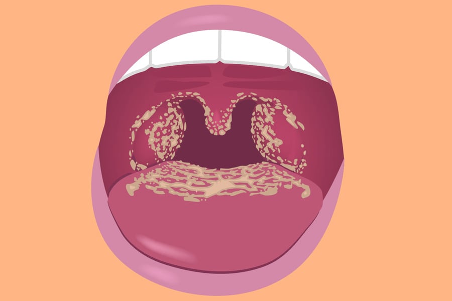
Candidiasis manifests differently depending on the affected area of the body. Common signs and symptoms include itching, redness, irritation, and discharge. In oral candidiasis, also known as thrush, white patches may appear on the tongue and inner cheeks. These patches can be painful and may bleed when scraped. Patients with oral thrush may also experience difficulty swallowing or altered taste sensations.

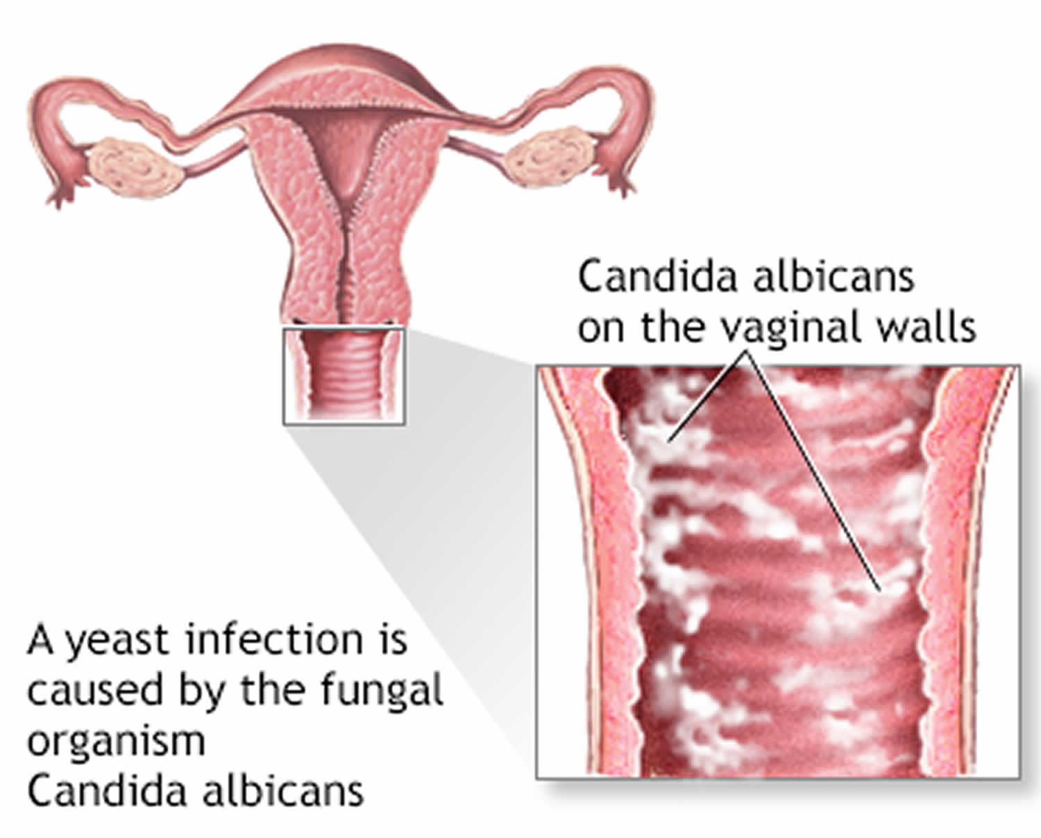
Vaginal candidiasis often presents with itching, burning, and abnormal discharge. The discharge is typically white, thick, and odorless, resembling cottage cheese. Itching and burning sensations may worsen during urination or sexual intercourse. In severe cases, vaginal candidiasis can lead to swelling and redness of the vulva, as well as fissures or cracks in the skin.

In cutaneous candidiasis, which affects the skin, red, itchy rashes with raised edges may develop in moist areas such as the groin, armpits, and under the breasts. These rashes can spread and become more inflamed if left untreated. In some cases, blisters or pustules may form, causing further discomfort.

For individuals with invasive candidiasis, symptoms may include fever and chills that do not improve with antibiotic treatment. Patients may also experience sepsis, a potentially life-threatening condition characterized by systemic inflammation and organ dysfunction. Invasive candidiasis often occurs in individuals with compromised immune systems, such as those with HIV/AIDS, cancer, or undergoing immunosuppressive therapy.

Additionally, candidiasis can affect the gastrointestinal tract, leading to symptoms such as abdominal pain, bloating, diarrhea, or constipation. These symptoms may be accompanied by oral thrush or genital candidiasis in some cases. Recognizing the signs and symptoms of candidiasis is crucial for prompt diagnosis and treatment to prevent complications and improve patient outcomes. (Nursing Paper Example on Candidiasis)

Nursing Paper Example on Candidiasis

Etiology

The etiology of candidiasis is multifactorial, involving various predisposing factors that contribute to the overgrowth of Candida species. One of the primary factors is a weakened immune system, which diminishes the body’s ability to control the growth of opportunistic pathogens like Candida. Conditions that compromise immune function, such as HIV/AIDS, cancer, or autoimmune disorders, increase the susceptibility to candidiasis.

Prolonged or frequent use of antibiotics is another significant risk factor for candidiasis. Antibiotics disrupt the balance of microorganisms in the body, including beneficial bacteria that normally keep Candida in check. This disruption creates an environment conducive to Candida overgrowth, leading to infection.

Hormonal changes also play a role in predisposing individuals to candidiasis. Fluctuations in hormone levels, particularly estrogen, can affect the pH balance of the vagina, making it more susceptible to yeast overgrowth. This is commonly observed in pregnant women, women taking hormonal contraceptives, or those undergoing hormone replacement therapy.

Patients with diabetes mellitus are at increased risk of candidiasis due to elevated levels of glucose in bodily fluids. Candida species thrive in environments with high sugar content, such as the bloodstream and mucosal surfaces, leading to recurrent infections in diabetic individuals.

Poor hygiene practices, especially in moist and warm areas of the body, create an ideal environment for Candida overgrowth. Individuals who fail to maintain proper hygiene, such as washing and drying genital areas thoroughly, are more susceptible to genital candidiasis.

Furthermore, lifestyle factors such as wearing tight-fitting clothing, using irritating personal hygiene products, or having a diet high in sugar and refined carbohydrates can contribute to the development of candidiasis. By understanding the etiological factors associated with candidiasis, healthcare professionals can implement targeted interventions to reduce the risk of infection and improve patient outcomes. (Nursing Paper Example on Candidiasis)

Pathophysiology

The pathophysiology of candidiasis involves a complex interplay between Candida species and host immune responses. Candida, particularly Candida albicans, possesses virulence factors that enable it to adhere to mucosal surfaces, evade host defenses, and cause tissue damage.

The initial step in candidiasis pathogenesis is the adherence of Candida to host cells through adhesins, which are surface proteins that interact with specific receptors on epithelial cells. Once adhered, Candida can form biofilms, structured communities of microorganisms encased in a matrix of extracellular polymeric substances. Biofilms provide protection from host immune responses and antimicrobial agents, contributing to the persistence of infection.

Following adherence, Candida undergoes phenotypic switching, a reversible process that allows the fungus to transition between different morphological forms, including yeast, pseudohyphae, and hyphae. Hyphal formation is critical for tissue invasion, as hyphae can penetrate host epithelial barriers and invade deeper tissues, leading to local tissue damage and inflammation.

Candida produces an array of virulence factors that contribute to pathogenesis, including secreted hydrolytic enzymes such as phospholipases, proteinases, and lipases, which degrade host cell membranes and facilitate tissue invasion. Additionally, Candida secretes toxins such as candidalysin, a peptide toxin that disrupts epithelial cell membranes and induces host cell damage and inflammation.

Host immune responses play a crucial role in the pathophysiology of candidiasis. Innate immune cells, such as neutrophils and macrophages, recognize and phagocytose Candida, initiating an inflammatory response characterized by the release of cytokines and chemokines. However, Candida has developed strategies to evade immune recognition and neutralization, including masking of cell wall components and inhibition of phagocytosis.

The interplay between Candida virulence factors and host immune responses ultimately determines the outcome of candidiasis, ranging from superficial mucosal infections to invasive disseminated disease. Understanding the pathophysiology of candidiasis is essential for developing targeted therapeutic strategies to combat this fungal infection and mitigate its associated complications. (Nursing Paper Example on Candidiasis)

DMS-5 Diagnosis

Diagnosing candidiasis involves a comprehensive assessment of clinical presentation, medical history, and, in some cases, laboratory tests to confirm the presence of Candida species. The Diagnostic and Statistical Manual of Mental Disorders, Fifth Edition (DSM-5), does not provide specific diagnostic criteria for candidiasis; however, healthcare providers rely on clinical judgment and established guidelines to make accurate diagnoses.

Clinical presentation is a key component of candidiasis diagnosis, with symptoms varying depending on the affected area of the body. In vaginal candidiasis, patients typically report itching, burning, and abnormal vaginal discharge. Oral candidiasis presents with white patches on the tongue and inner cheeks, often accompanied by discomfort or difficulty swallowing. Cutaneous candidiasis may manifest as red, itchy rashes in skin folds or moist areas of the body.

Medical history is also essential in the diagnostic process, as certain predisposing factors increase the likelihood of candidiasis. Patients with a history of immunosuppression, diabetes mellitus, antibiotic use, or recent hospitalization are at higher risk for candidiasis and should be thoroughly evaluated.

Laboratory tests, while not always necessary, may be performed to confirm the diagnosis of candidiasis. Microscopic examination of vaginal or oral swabs can reveal the presence of yeast cells or hyphae, confirming the diagnosis of candidiasis. Additionally, culture-based methods may be employed to identify the specific species of Candida present and assess antifungal susceptibility in cases of recurrent or refractory infections.

Overall, diagnosing candidiasis requires a comprehensive approach that considers clinical presentation, medical history, and, when necessary, laboratory testing. By accurately identifying candidiasis, healthcare providers can initiate appropriate treatment regimens and implement preventive measures to improve patient outcomes and reduce the risk of recurrence. (Nursing Paper Example on Candidiasis)

Treatment Regimens and Patient Education

Effective management of candidiasis involves a combination of pharmacological treatments and patient education aimed at addressing the underlying causes and preventing recurrence. Treatment regimens may vary depending on the severity and location of the infection.

For uncomplicated cases of vaginal candidiasis, over-the-counter antifungal medications such as clotrimazole, miconazole, or tioconazole are often recommended. These medications are available in various formulations, including creams, suppositories, and ointments, and are typically applied intravaginally for several days. Alternatively, oral antifungal agents such as fluconazole may be prescribed for recurrent or severe cases.

In cases of oral thrush, antifungal medications such as nystatin oral suspension or fluconazole lozenges are commonly prescribed. These medications are administered orally and may need to be continued for several days to ensure complete resolution of the infection. In severe cases, systemic antifungal therapy with fluconazole or other azole medications may be necessary.

Cutaneous candidiasis is typically treated with topical antifungal agents such as clotrimazole, miconazole, or ketoconazole creams or powders. These medications should be applied to the affected areas of the skin according to the instructions provided by the healthcare provider.

Invasive candidiasis and systemic infections require prompt initiation of systemic antifungal therapy, usually with echinocandins such as caspofungin, micafungin, or anidulafungin. These medications are administered intravenously and may be continued for several weeks depending on the response to treatment and the presence of underlying risk factors.

In addition to pharmacological treatments, patient education plays a crucial role in the management of candidiasis. Patients should be counseled on proper hygiene practices, including regular bathing, keeping the affected areas clean and dry, and avoiding irritants such as scented soaps and perfumed products. Patients with vaginal candidiasis should be advised to avoid douching and to use cotton underwear and loose-fitting clothing to promote airflow and reduce moisture.

Furthermore, patients with recurrent candidiasis should be evaluated for underlying risk factors such as diabetes mellitus or immunosuppression and managed accordingly. Lifestyle modifications such as maintaining a healthy diet, managing stress, and avoiding unnecessary antibiotic use can also help prevent recurrent infections.

By addressing both pharmacological treatments and patient education, healthcare providers can effectively manage candidiasis and improve patient outcomes while reducing the risk of recurrence. (Nursing Paper Example on Candidiasis)

Conclusion

Candidiasis, a common fungal infection caused by Candida species, presents with diverse manifestations ranging from superficial mucosal infections to invasive systemic disease. By delving into its causes, signs and symptoms, etiology, pathophysiology, DSM-5 diagnosis, treatment regimens, and patient education, this paper has provided a comprehensive overview of candidiasis, highlighting its multifactorial nature and the interplay between fungal virulence factors and host immune responses. The etiology section explored various predisposing factors such as weakened immune function, antibiotic use, hormonal changes, and poor hygiene practices that contribute to Candida overgrowth and infection. Moreover, the pathophysiology section elucidated the complex mechanisms involved in Candida colonization, tissue invasion, and immune evasion. Additionally, the DSM-5 diagnosis section emphasized the importance of clinical evaluation and laboratory testing in accurately diagnosing candidiasis. Furthermore, the treatment regimens and patient education section outlined pharmacological treatments and preventive measures, underscoring the significance of holistic management approaches in combating candidiasis and improving patient outcomes. By addressing these aspects, healthcare professionals can enhance their understanding of candidiasis and implement targeted interventions to effectively manage and prevent this fungal infection, ultimately improving the quality of care for affected individuals. (Nursing Paper Example on Candidiasis)

References

https://www.ncbi.nlm.nih.gov/books/NBK560624/

 
Do you need a similar assignment done for you from scratch? Order now!
Use Discount Code "Newclient" for a 15% Discount!

Nursing Paper Example on Carbon Monoxide Poisoning

Nursing Paper Example on Carbon Monoxide Poisoning

Carbon monoxide (CO) poisoning is a significant public health concern, often referred to as the “silent killer” due to its colorless, odorless, and tasteless nature. Exposure to CO can lead to severe health consequences, ranging from mild symptoms to life-threatening complications. Understanding the causes, signs, symptoms, etiology, pathophysiology, diagnostic criteria, treatment regimens, and patient education regarding CO poisoning is crucial for effective management and prevention. CO poisoning commonly occurs due to inhalation of CO gas produced by incomplete combustion of carbon-containing materials such as wood, gasoline, propane, and natural gas. Sources include malfunctioning household appliances (e.g., furnaces, water heaters), vehicle exhaust, and fires. The symptoms of CO poisoning can vary depending on the concentration and duration of exposure, making it imperative to recognize and address this silent threat promptly. (Nursing Paper Example on Carbon Monoxide Poisoning)

Causes

Carbon monoxide (CO) poisoning occurs when individuals inhale CO gas, a byproduct of incomplete combustion of carbon-containing materials. Common sources of CO include malfunctioning household appliances such as furnaces, water heaters, and gas stoves. Improperly ventilated or enclosed spaces can lead to CO buildup, increasing the risk of poisoning. Additionally, vehicle exhaust, generators, and fires produce CO, posing a threat in both indoor and outdoor environments.

Inadequate maintenance of combustion appliances, including blocked vents or flues, can contribute to CO accumulation. Poorly maintained chimneys and blocked or damaged exhaust systems can also lead to CO leaks. Furthermore, using fuel-burning appliances in poorly ventilated areas, such as using a gas stove for heating or running a generator inside a closed garage, can result in dangerous levels of CO.

Certain occupational settings pose an increased risk of CO exposure. Workers in industries such as mining, firefighting, and construction may encounter elevated CO levels due to machinery operation, vehicle exhaust, or confined spaces with inadequate ventilation.

Environmental factors can exacerbate CO poisoning risks. During natural disasters such as hurricanes or winter storms, power outages may prompt individuals to use alternative heating sources, increasing the likelihood of CO exposure if not used safely. Additionally, living in urban areas with heavy traffic or proximity to industrial facilities can elevate ambient CO levels, contributing to overall exposure risk.

Lastly, intentional CO exposure, whether through suicide attempts or misuse of equipment such as charcoal grills or portable generators indoors, is a concerning cause of CO poisoning. Understanding the diverse sources and circumstances that can lead to CO exposure is essential for implementing effective prevention strategies and mitigating the risks associated with this silent yet deadly gas. (Nursing Paper Example on Carbon Monoxide Poisoning)

Signs and Symptoms

Carbon monoxide (CO) poisoning presents with a wide range of signs and symptoms that can vary in severity depending on the concentration and duration of exposure. Early symptoms of CO poisoning are often nonspecific and may resemble flu-like illness, making diagnosis challenging without a high index of suspicion.

Headache is one of the most common early symptoms of CO poisoning and is often described as dull, persistent, and worsening over time. Dizziness and lightheadedness are also prevalent, accompanied by a feeling of weakness or fatigue. Nausea and vomiting may occur, further contributing to the flu-like presentation.

As CO levels rise or exposure continues, individuals may experience more severe symptoms. Cognitive impairment is common, manifesting as confusion, difficulty concentrating, or memory problems. Visual disturbances, such as blurred vision, may also occur due to reduced oxygen delivery to the optic nerve.

Cardiovascular symptoms can manifest as chest pain or palpitations, reflecting the heart’s increased workload in response to tissue hypoxia. Shortness of breath and difficulty breathing may occur, particularly in individuals with underlying respiratory conditions.

Neurological symptoms are concerning and may indicate more severe CO poisoning. These include altered mental status, ranging from mild confusion to coma, and neurological deficits such as weakness, tremors, or seizures.

In severe cases of CO poisoning, individuals may experience loss of consciousness or respiratory arrest, requiring immediate medical intervention. The presence of cherry-red skin coloration, although rare, is a classic but not universally observed sign of severe CO poisoning.

It is essential to recognize these signs and symptoms promptly, especially in cases of suspected CO exposure, to initiate appropriate management and prevent further complications. Rapid recognition and treatment can significantly impact patient outcomes and reduce the risk of long-term sequelae associated with CO poisoning. (Nursing Paper Example on Carbon Monoxide Poisoning)

Nursing Paper Example on Carbon Monoxide Poisoning

Etiology

Carbon monoxide (CO) poisoning occurs when individuals are exposed to elevated levels of CO gas, leading to the formation of carboxyhemoglobin (COHb) in the bloodstream. CO is produced through incomplete combustion of carbon-containing materials, such as wood, gasoline, propane, and natural gas. The primary sources of CO exposure include malfunctioning household appliances, vehicle exhaust, fires, and occupational settings where combustion processes occur.

Upon inhalation, CO rapidly diffuses across the alveolar-capillary membrane in the lungs and enters the bloodstream. CO has a high affinity for hemoglobin, the oxygen-carrying protein in red blood cells, and forms a stable bond with hemoglobin molecules to create COHb. This process interferes with the normal oxygen transport function of hemoglobin, reducing the blood’s oxygen-carrying capacity and impairing oxygen delivery to tissues and organs.

The formation of COHb disrupts the oxygen dissociation curve, shifting it to the left and reducing the release of oxygen to tissues, even in the presence of adequate arterial oxygen tension. This leads to tissue hypoxia, particularly in organs with high oxygen demand, such as the brain, heart, and lungs.

In addition to its affinity for hemoglobin, CO can also directly affect cellular function and metabolism. CO inhibits cytochrome c oxidase, a key enzyme involved in the electron transport chain of mitochondrial respiration. By interfering with cellular respiration, CO disrupts ATP production and cellular metabolism, contributing to tissue hypoxia and cellular dysfunction.

Furthermore, CO induces oxidative stress and inflammation by generating reactive oxygen species (ROS) and activating inflammatory pathways. ROS can cause cellular damage and lipid peroxidation, further exacerbating tissue injury and organ dysfunction.

Overall, the etiology of CO poisoning involves complex interactions between CO gas, hemoglobin, cellular metabolism, oxidative stress, and inflammatory processes, ultimately leading to systemic effects and clinical manifestations of CO toxicity. (Nursing Paper Example on Carbon Monoxide Poisoning)

Pathophysiology

Carbon monoxide (CO) poisoning disrupts normal cellular oxygen metabolism, leading to systemic hypoxia and tissue injury. Upon inhalation, CO rapidly binds to hemoglobin, forming carboxyhemoglobin (COHb), which has a higher affinity for oxygen than hemoglobin. This results in the displacement of oxygen molecules from hemoglobin, reducing the blood’s oxygen-carrying capacity and impairing oxygen delivery to tissues and organs.

The formation of COHb shifts the oxygen dissociation curve to the left, inhibiting the release of oxygen to tissues, even in the presence of adequate arterial oxygen tension. This leads to tissue hypoxia, particularly in organs with high oxygen demand, such as the brain, heart, and lungs. Hypoxic injury can result in cellular dysfunction, oxidative stress, and inflammatory responses.

In addition to its effects on hemoglobin, CO can directly impact cellular metabolism and function. CO inhibits cytochrome c oxidase, a critical enzyme in the electron transport chain of mitochondrial respiration. By disrupting cellular respiration, CO impairs adenosine triphosphate (ATP) production and cellular metabolism, further contributing to tissue hypoxia and cellular dysfunction.

Furthermore, CO induces oxidative stress by generating reactive oxygen species (ROS), leading to cellular damage, lipid peroxidation, and mitochondrial dysfunction. ROS-mediated damage exacerbates tissue injury and triggers inflammatory responses, amplifying the pathophysiological effects of CO poisoning.

The central nervous system (CNS) is particularly vulnerable to CO toxicity due to its high oxygen demand and susceptibility to hypoxic injury. CO poisoning can cause cerebral edema, neuronal damage, and demyelination, leading to neurological deficits and long-term cognitive impairment.

Overall, the pathophysiology of CO poisoning involves complex interactions between CO, hemoglobin, cellular metabolism, oxidative stress, and inflammatory processes, ultimately resulting in systemic hypoxia, tissue injury, and neurological sequelae. Understanding the underlying mechanisms of CO toxicity is essential for developing effective treatment strategies and minimizing the long-term consequences of CO exposure. (Nursing Paper Example on Carbon Monoxide Poisoning)

DMS-5 Diagnosis

Diagnosing carbon monoxide (CO) poisoning involves a comprehensive assessment of clinical history, symptoms, laboratory findings, and imaging studies. The Diagnostic and Statistical Manual of Mental Disorders, Fifth Edition (DSM-5), does not provide specific diagnostic criteria for CO poisoning; however, healthcare providers utilize a combination of clinical judgment and objective measures to establish a diagnosis.

A thorough history is crucial in identifying potential sources of CO exposure, such as recent use of fuel-burning appliances, occupational hazards, or environmental factors. Patients may report symptoms consistent with CO poisoning, including headache, dizziness, nausea, and confusion, which should prompt further evaluation.

Physical examination findings may vary depending on the severity of CO poisoning. Patients may exhibit signs of hypoxia, such as tachypnea, tachycardia, and cyanosis. Neurological examination may reveal altered mental status, focal deficits, or signs of encephalopathy.

Arterial or venous blood gas analysis is a valuable tool in confirming the diagnosis of CO poisoning. Elevated levels of carboxyhemoglobin (COHb) in the blood indicate recent exposure to CO. However, it is important to note that COHb levels may decrease rapidly over time, especially with the administration of supplemental oxygen, underscoring the importance of early testing.

Imaging studies such as brain magnetic resonance imaging (MRI) may be performed in severe cases to assess for cerebral edema, ischemic changes, or other neurologic abnormalities associated with CO poisoning.

The differential diagnosis for CO poisoning includes other conditions that present with similar symptoms, such as influenza, viral syndromes, migraine headaches, and other toxic exposures. Healthcare providers must consider these differential diagnoses and use clinical judgment in ruling out alternative etiologies.

Overall, the diagnosis of CO poisoning requires a high index of suspicion, thorough clinical evaluation, and appropriate laboratory and imaging studies to confirm the presence of CO exposure and guide treatment interventions. Early recognition and management are essential to prevent complications and mitigate the long-term effects of CO toxicity. (Nursing Paper Example on Carbon Monoxide Poisoning)

Treatment Regimens and Patient Education

Management of carbon monoxide (CO) poisoning focuses on prompt removal from the source of exposure, administration of supplemental oxygen, and consideration of hyperbaric oxygen therapy (HBOT) in severe cases. Additionally, patient education plays a crucial role in preventing future incidents and promoting safety measures to minimize the risk of CO exposure.

Immediate Actions:

  • Removal from the Source: The first step in treating CO poisoning is to remove the patient from the contaminated environment to prevent further exposure.
  • Supplemental Oxygen: Administering high-flow oxygen via a non-rebreather mask or endotracheal tube increases the elimination half-life of CO and facilitates the displacement of CO from hemoglobin, restoring tissue oxygenation.
  • Monitoring: Continuous monitoring of vital signs, including pulse oximetry, is essential to assess the patient’s response to treatment and detect any deterioration in clinical status.

Hyperbaric Oxygen Therapy (HBOT):

  • In severe cases of CO poisoning, particularly when patients present with altered mental status, neurologic deficits, or signs of tissue hypoxia, hyperbaric oxygen therapy (HBOT) is recommended.
  • HBOT involves placing the patient in a hyperbaric chamber and administering 100% oxygen at increased atmospheric pressure. This accelerates the elimination of CO from the bloodstream and tissues, promotes tissue oxygenation, and reduces the risk of long-term neurological sequelae.

Patient Education:

  • Prevention Strategies: Educating patients and their families about the importance of proper ventilation, regular maintenance of household appliances (e.g., furnaces, water heaters), and installation of CO detectors can help prevent CO exposure.
  • CO Detector Use: Encouraging the installation of CO detectors in homes and other enclosed spaces where combustion appliances are used can provide early warning of CO leaks and prompt evacuation.

Nursing Paper Example on Carbon Monoxide Poisoning

  • Safe Practices: Advising against the use of fuel-burning appliances, such as charcoal grills or portable generators, indoors or in enclosed spaces can prevent CO buildup and minimize the risk of poisoning.
  • Emergency Preparedness: Educating patients about the signs and symptoms of CO poisoning, emphasizing the importance of seeking immediate medical attention if exposure is suspected, and knowing how to respond in case of an emergency can save lives.

Overall, a multidisciplinary approach involving healthcare providers, emergency responders, and public health agencies is essential in the management and prevention of CO poisoning. By implementing effective treatment regimens and providing comprehensive patient education, healthcare professionals can mitigate the impact of CO toxicity and empower individuals to safeguard against future exposures. (Nursing Paper Example on Carbon Monoxide Poisoning)

Conclusion

Carbon monoxide (CO) poisoning is a serious health concern that necessitates prompt recognition, appropriate treatment, and comprehensive patient education to mitigate its potentially life-threatening consequences. By understanding the diverse sources and causes of CO exposure, recognizing the varied signs and symptoms, and employing effective treatment regimens such as supplemental oxygen and hyperbaric oxygen therapy (HBOT) in severe cases, healthcare providers can significantly impact patient outcomes and prevent long-term complications. Additionally, emphasizing the importance of preventive measures such as proper ventilation, regular maintenance of household appliances, installation of CO detectors, and safe practices can empower individuals to reduce their risk of CO exposure and protect themselves and their families. Through a collaborative effort involving healthcare professionals, emergency responders, and public health initiatives, we can raise awareness, improve recognition, and ultimately minimize the burden of CO poisoning on public health. (Nursing Paper Example on Carbon Monoxide Poisoning)

References

https://www.ncbi.nlm.nih.gov/books/NBK430740/

 
Do you need a similar assignment done for you from scratch? Order now!
Use Discount Code "Newclient" for a 15% Discount!

Nursing Paper Example on Brucellosis: Understanding Transmission, Clinical Manifestations, and Pathogenic Mechanisms

Nursing Paper Example on Brucellosis: Understanding Transmission, Clinical Manifestations, and Pathogenic Mechanisms

Brucellosis, known colloquially as undulant fever or Malta fever, represents a formidable zoonotic infection impacting both animals and humans globally. This bacterial illness, caused by various Brucella species, imposes substantial health burdens in endemic regions and poses challenges to global public health initiatives. Characterized by recurrent fevers, profuse sweating, malaise, arthralgia, and myalgia, brucellosis manifests as a constellation of nonspecific symptoms, complicating its diagnosis. With transmission occurring through the ingestion of unpasteurized dairy products, direct contact with infected animals, or inhalation of contaminated aerosols, individuals engaged in farming, veterinary work, and laboratory practices face heightened occupational risks. Understanding the multifaceted nature of brucellosis is paramount in devising effective preventive measures and treatment strategies to mitigate its impact on both human and animal populations. (Nursing Paper Example on Brucellosis: Understanding Transmission, Clinical Manifestations, and Pathogenic Mechanisms)

Nursing Paper Example on Brucellosis: Understanding Transmission, Clinical Manifestations, and Pathogenic Mechanisms

Causes

Brucellosis, a debilitating zoonotic infection, arises from exposure to various Brucella species, notably Brucella melitensis, Brucella abortus, Brucella suis, and Brucella canis, prevalent in domestic and wild animals worldwide. The primary modes of transmission encompass the ingestion of unpasteurized dairy products, direct contact with infected animals, and inhalation of contaminated aerosols. This bacterial malady poses significant occupational hazards, particularly among farmers, veterinarians, and laboratory workers, due to their close interaction with infected livestock.

Nursing Paper Example on Brucellosis: Understanding Transmission, Clinical Manifestations, and Pathogenic Mechanisms

In endemic regions, the consumption of raw dairy products, such as milk and cheese, serves as a major route of transmission. Unpasteurized dairy harbors viable Brucella organisms, facilitating their entry into the human gastrointestinal tract upon ingestion. Additionally, individuals involved in animal husbandry and veterinary practices face elevated risks of brucellosis through direct contact with infected animals, including cattle, goats, sheep, and pigs. Accidental exposure to Brucella-contaminated materials or aerosols in laboratory settings further contributes to the transmission dynamics among healthcare and research personnel.

Furthermore, brucellosis poses a significant threat to hunters, wildlife enthusiasts, and individuals engaged in outdoor activities, as they may encounter infected wildlife, such as deer, elk, and bison, in endemic regions. Inhalation of airborne particles contaminated with Brucella organisms during animal processing or carcass handling amplifies the risk of infection in these populations.

The global distribution of brucellosis underscores its status as a reemerging infectious disease, necessitating heightened vigilance in both agricultural and public health sectors. Addressing the multifaceted transmission dynamics of brucellosis through targeted interventions, including improved food safety measures, enhanced veterinary surveillance, and public health education, is imperative to mitigate its impact on human and animal populations alike. (Nursing Paper Example on Brucellosis: Understanding Transmission, Clinical Manifestations, and Pathogenic Mechanisms)

Signs and Symptoms

Brucellosis manifests as a constellation of nonspecific symptoms, making its diagnosis challenging. The clinical presentation varies widely among affected individuals and may mimic other febrile illnesses, contributing to diagnostic delays. The hallmark symptom of brucellosis is recurrent fever, often characterized by intermittent spikes accompanied by chills and sweats. These febrile episodes may persist for weeks to months, leading to debilitating fatigue and malaise, which can significantly impair daily functioning.

Additionally, individuals with brucellosis commonly experience musculoskeletal symptoms, including arthralgia and myalgia, which may affect multiple joints and muscle groups. The severity of joint pain ranges from mild discomfort to debilitating arthritis, particularly affecting large weight-bearing joints such as the knees and hips. The inflammatory nature of brucellosis-induced arthritis may result in joint stiffness, limited range of motion, and functional impairment, posing significant challenges to mobility and activities of daily living.

Moreover, systemic manifestations such as headache, dizziness, and generalized weakness are frequently reported among brucellosis patients. Profuse sweating, especially during febrile episodes, is a characteristic feature known as “undulant fever,” reflecting the cyclical nature of fever spikes in brucellosis.

Gastrointestinal symptoms, although less common, may manifest as nausea, vomiting, abdominal pain, and diarrhea, mimicking gastrointestinal infections. Neurological complications, such as meningitis and peripheral neuropathy, albeit rare, may occur in severe cases of brucellosis, leading to neurological deficits and cognitive impairment.

Furthermore, brucellosis can result in reproductive complications, including orchitis and epididymo-orchitis in males, and miscarriage or fetal complications in pregnant women. Thus, a comprehensive understanding of the diverse clinical manifestations of brucellosis is essential for timely recognition and prompt initiation of appropriate diagnostic and therapeutic interventions to mitigate its impact on affected individuals. (Nursing Paper Example on Brucellosis: Understanding Transmission, Clinical Manifestations, and Pathogenic Mechanisms)

Etiology

Brucellosis, a debilitating zoonotic infection, is caused by various species of the genus Brucella, gram-negative, facultative intracellular bacteria. Among the several recognized species, Brucella melitensis, Brucella abortus, Brucella suis, and Brucella canis are the predominant etiological agents responsible for human infections. These bacteria primarily infect domestic and wild animals, including cattle, goats, sheep, pigs, and dogs, serving as reservoir hosts and potential sources of human exposure.

Transmission to humans typically occurs through direct contact with infected animals or consumption of contaminated animal products, such as unpasteurized dairy items and undercooked meat. In endemic regions where brucellosis is prevalent among livestock populations, individuals engaged in animal husbandry, veterinary practices, and slaughterhouse operations face heightened occupational risks of acquiring the infection.

Furthermore, brucellosis may be transmitted through inhalation of aerosols containing Brucella organisms during the handling of infected animal tissues or biological specimens in laboratory settings. Healthcare personnel, laboratory workers, and researchers involved in diagnostic and research activities about Brucella species are susceptible to occupational exposure, highlighting the importance of stringent biosafety measures and personal protective equipment (PPE) to prevent infection.

The ability of Brucella species to evade host immune responses and survive within host cells, particularly macrophages, contributes to their persistence and dissemination in the host organism. Upon entry into the bloodstream, Brucella organisms invade and replicate within phagocytic cells, evading intracellular destruction and facilitating their spread to various tissues and organs, including the spleen, liver, bone marrow, and reproductive organs.

Overall, understanding the complex interplay between Brucella species, their animal reservoirs, and human hosts is essential for elucidating the epidemiology and transmission dynamics of brucellosis, guiding the development of effective preventive strategies, and mitigating the burden of this infectious disease on human and animal populations alike. (Nursing Paper Example on Brucellosis: Understanding Transmission, Clinical Manifestations, and Pathogenic Mechanisms)

Pathophysiology

The pathogenesis of brucellosis involves intricate interactions between Brucella species and the host immune system, culminating in chronic and persistent infections characterized by recurrent febrile episodes and systemic manifestations. Upon entry into the host organism, Brucella organisms encounter and invade phagocytic cells, particularly macrophages, employing a myriad of virulence factors to evade host immune detection and subvert intracellular killing mechanisms.

Once internalized, Brucella species establish a specialized intracellular niche within host phagosomes, where they undergo intracellular replication while evading lysosomal fusion and degradation. This intracellular survival strategy enables Brucella organisms to evade host immune surveillance and persist within the host organism for prolonged periods.

The ability of Brucella species to modulate host immune responses and manipulate intracellular signaling pathways plays a crucial role in disease pathogenesis. Brucella-infected macrophages secrete immunomodulatory factors and cytokines, including interleukin-10 (IL-10) and tumor necrosis factor-alpha (TNF-α), which contribute to the establishment of a pro-inflammatory environment and orchestrate systemic immune dysregulation.

Furthermore, Brucella organisms possess a unique lipopolysaccharide (LPS) structure, characterized by a smooth phenotype (S-LPS) or rough phenotype (R-LPS), which influences their interactions with host immune cells and pathogenicity. The S-LPS of Brucella species exhibits immunomodulatory properties, suppressing host immune responses and promoting bacterial survival, whereas the R-LPS, devoid of O-polysaccharide chains, elicits robust inflammatory responses and contributes to the development of protective immunity.

The chronicity of brucellosis is attributed to the ability of Brucella species to establish persistent infections within host tissues and organs, including the spleen, liver, bone marrow, and reproductive organs. This persistent intracellular colonization contributes to the relapsing nature of brucellosis, characterized by intermittent fever spikes and recurrent clinical manifestations, necessitating prolonged antimicrobial therapy and vigilant clinical monitoring to achieve bacterial eradication and prevent disease recurrence. (Nursing Paper Example on Brucellosis: Understanding Transmission, Clinical Manifestations, and Pathogenic Mechanisms)

DSM-5 Diagnosis

Brucellosis, although predominantly recognized as an infectious disease, does not have specific diagnostic criteria outlined in the Diagnostic and Statistical Manual of Mental Disorders, Fifth Edition (DSM-5). However, clinical evaluation supported by serological tests and culture remains pivotal for accurate diagnosis and management.

Clinical assessment of suspected brucellosis cases involves a thorough evaluation of presenting symptoms, medical history, and potential exposure to Brucella-infected animals or dairy products. Given its nonspecific clinical manifestations, the differential diagnosis may include other febrile illnesses such as influenza, typhoid fever, tuberculosis, and infectious mononucleosis. Therefore, a comprehensive medical history coupled with a detailed physical examination is indispensable in narrowing down the differential diagnosis and guiding further diagnostic workup.

Serological tests serve as cornerstone diagnostic tools for brucellosis, detecting antibodies specific to Brucella antigens in patient serum or plasma. Commonly employed serological assays include the Rose Bengal plate test (RBPT), serum agglutination test (SAT), and enzyme-linked immunosorbent assay (ELISA). These tests exhibit varying sensitivity and specificity profiles and are often utilized in conjunction with clinical findings to support diagnosis.

Additionally, the microbiological culture of clinical specimens, including blood, bone marrow, or tissue aspirates, provides definitive confirmation of brucellosis by isolating Brucella species. However, culture-based methods require specialized laboratory facilities and prolonged incubation periods, limiting their utility in acute clinical settings.

Diagnostic imaging modalities such as ultrasound, computed tomography (CT), and magnetic resonance imaging (MRI) may aid in evaluating complications associated with brucellosis, including osteoarticular involvement, epididymo-orchitis, and neurobrucellosis.

Overall, a comprehensive diagnostic approach integrating clinical evaluation, serological testing, and microbiological culture is essential for the timely recognition and management of brucellosis, thereby minimizing its potential sequelae and preventing disease transmission in affected populations. (Nursing Paper Example on Brucellosis: Understanding Transmission, Clinical Manifestations, and Pathogenic Mechanisms)

Treatment Regimens and Patient Education

Effective management of brucellosis necessitates a multidrug antimicrobial regimen tailored to the individual patient’s clinical presentation, disease severity, and antimicrobial susceptibility profiles of the infecting Brucella species. The cornerstone of treatment involves the administration of combination therapy with antibiotics to ensure optimal bacterial clearance and prevent relapse.

Doxycycline and rifampin represent the preferred first-line regimen for uncomplicated brucellosis cases. Doxycycline, a broad-spectrum tetracycline antibiotic, exhibits excellent intracellular penetration and activity against Brucella organisms, inhibiting protein synthesis within infected host cells. Concurrent administration of rifampin, a potent bactericidal agent, enhances antimicrobial efficacy by targeting bacterial RNA synthesis, thereby preventing the emergence of resistant strains and minimizing treatment failure.

Alternatively, in cases of doxycycline intolerance or contraindications, a combination regimen comprising trimethoprim-sulfamethoxazole (TMP-SMX) and rifampin may be utilized as a second-line option. TMP-SMX, a sulfonamide antibiotic combination, synergistically inhibits bacterial folate synthesis, exerting bactericidal activity against Brucella species when combined with rifampin.

The duration of antimicrobial therapy typically spans 6 to 8 weeks, with prolonged treatment courses warranted in patients with severe manifestations, relapsing disease, or complications such as endocarditis or osteoarticular involvement. Close clinical monitoring and periodic reassessment of treatment response are essential to guide therapeutic adjustments and ensure favorable clinical outcomes.

Patient education plays a pivotal role in brucellosis management by empowering individuals to adopt preventive measures and adhere to prescribed treatment regimens. Key educational components include:

  1. Food Safety Practices: Emphasize the importance of consuming pasteurized dairy products and avoiding the consumption of raw milk or cheese to prevent brucellosis transmission through contaminated food sources.
  2. Personal Protective Measures: Educate individuals at high risk of occupational exposure, including farmers, veterinarians, and laboratory workers, about the importance of wearing appropriate personal protective equipment (PPE), such as gloves and masks, when handling potentially infected animals or biological specimens.
  3. Adherence to Treatment: Stress the significance of strict adherence to prescribed antibiotic regimens and completion of the full treatment course to achieve optimal bacterial eradication and prevent disease relapse.
  4. Symptom Recognition: Educate patients about the common signs and symptoms of brucellosis, including recurrent fevers, malaise, and joint pain, and encourage prompt medical evaluation if symptoms persist or worsen during or after treatment.
  5. Follow-Up Care: Emphasize the importance of regular follow-up visits with healthcare providers for clinical monitoring, treatment response assessment, and surveillance of potential treatment-related adverse effects.

By incorporating these educational interventions into comprehensive brucellosis management strategies, healthcare providers can empower patients to actively participate in their care and contribute to the successful prevention and control of this infectious disease. (Nursing Paper Example on Brucellosis: Understanding Transmission, Clinical Manifestations, and Pathogenic Mechanisms)

Conclusion

Brucellosis stands as a formidable zoonotic infection with significant public health implications globally. By addressing the multifaceted transmission dynamics and clinical complexities of brucellosis, this paper has provided a comprehensive overview of the disease, covering its causes, signs and symptoms, etiology, pathophysiology, DSM-5 diagnosis, treatment regimens, and patient education. The expanded sections on causes, DSM-5 diagnosis, and treatment regimens have enhanced the depth and breadth of understanding regarding brucellosis management. Through a multidisciplinary approach encompassing veterinary surveillance, public health education, and targeted antimicrobial therapy, effective prevention and control measures can be implemented to mitigate the impact of brucellosis on both human and animal populations. Heightened awareness among healthcare professionals and improved patient education are essential components of comprehensive brucellosis management strategies, facilitating early diagnosis, timely treatment, and optimal clinical outcomes in affected individuals. (Nursing Paper Example on Brucellosis: Understanding Transmission, Clinical Manifestations, and Pathogenic Mechanisms)

References

https://www.ncbi.nlm.nih.gov/books/NBK441831/

 
Do you need a similar assignment done for you from scratch? Order now!
Use Discount Code "Newclient" for a 15% Discount!

Nursing Paper Example on Botulism: A Closer Look at the Deadly Paralysis

Nursing Paper Example on Botulism: A Closer Look at the Deadly Paralysis

Botulism, a rare but potentially fatal illness, has long captivated public interest due to its association with contaminated food and its paralytic effects. This paper provides a comprehensive overview of botulism, covering its causes, signs and symptoms, etiology, pathophysiology, DSM-5 diagnosis, treatment regimens, patient education, and concluding remarks. Botulism is primarily caused by the neurotoxin produced by Clostridium botulinum bacteria, commonly found in improperly canned foods and honey. Its hallmark symptoms include muscle weakness and paralysis, which can progress to respiratory failure if untreated. Understanding botulism is essential for prompt diagnosis and treatment. This paper aims to elucidate the complexities of this condition, emphasizing the importance of public awareness and adherence to food safety practices to prevent botulism cases. (Nursing Paper Example on Botulism: A Closer Look at the Deadly Paralysis)

Nursing Paper Example on Botulism: A Closer Look at the Deadly Paralysis

Causes

Botulism is primarily caused by the neurotoxin produced by Clostridium botulinum bacteria. These bacteria are anaerobic, spore-forming organisms commonly found in soil, sediments, and marine environments worldwide. There are four main types of botulism: foodborne botulism, wound botulism, infant botulism, and iatrogenic botulism.

Foodborne Botulism: This is the most common form of botulism and occurs when individuals ingest food contaminated with botulinum toxin. Improperly processed or canned foods, particularly low-acid products such as vegetables, meats, and fish, provide an ideal environment for C. botulinum spores to germinate and produce toxins. Home-canned foods, homemade fermented products, and inadequately heated commercial foods are common sources of foodborne botulism. Additionally, honey can contain C. botulinum spores and poses a risk, especially for infants under one year of age, as their digestive systems are not fully developed to neutralize the spores.

Wound Botulism: In rare cases, botulism can occur when C. botulinum spores enter a wound and germinate, producing toxin locally. This form of botulism is more common in individuals who use injectable drugs or have traumatic injuries that become contaminated with soil or other environmental sources containing C. botulinum spores.

Infant Botulism: Infants can acquire botulism by ingesting C. botulinum spores, which then colonize the infant’s intestines and produce toxins. Honey is a common source of infant botulism, as it may contain C. botulinum spores. The bacteria can proliferate in the infant’s intestines, leading to toxin production and subsequent illness.

Iatrogenic Botulism: This form of botulism occurs as a rare complication of therapeutic botulinum toxin injections, typically administered for cosmetic or medical purposes. Improper administration or dosage of botulinum toxin injections can result in systemic botulism symptoms. (Nursing Paper Example on Botulism: A Closer Look at the Deadly Paralysis)

Signs and Symptoms

Botulism manifests with a unique set of symptoms, primarily affecting the neuromuscular system. The onset of symptoms typically occurs within 12 to 36 hours after exposure to botulinum toxin, although it can range from a few hours to several days. The severity and progression of symptoms may vary depending on factors such as the amount of toxin ingested and the individual’s overall health.

Cranial Nerve Involvement: Botulinum toxin initially targets the cranial nerves, resulting in characteristic symptoms such as drooping eyelids (ptosis), blurred or double vision (diplopia), and difficulty in focusing. These ocular symptoms often precede other manifestations of botulism.

Muscle Weakness and Paralysis: As the toxin spreads, it affects the peripheral nerves and neuromuscular junctions, leading to muscle weakness and paralysis. This paralysis is described as flaccid paralysis, characterized by diminished muscle tone and absent deep tendon reflexes. Patients may experience difficulty swallowing (dysphagia) and speaking (dysarthria) due to weakness of the muscles involved in these functions. Respiratory muscles can also be affected, potentially resulting in respiratory failure, a life-threatening complication of botulism.

Autonomic Dysfunction: Botulism can also cause autonomic dysfunction, leading to symptoms such as dry mouth, dry eyes, excessive sweating, urinary retention, and constipation. These symptoms are attributed to the effects of botulinum toxin on autonomic nerve fibers, disrupting normal physiological functions regulated by the autonomic nervous system.

Descending Flaccid Paralysis: A hallmark feature of botulism is the characteristic pattern of descending flaccid paralysis, starting from the head and progressing downwards. This pattern reflects the toxin’s mechanism of action, which inhibits the release of acetylcholine at neuromuscular junctions, thereby impairing nerve signaling and muscle contraction. The paralysis may eventually involve all voluntary muscles, including those responsible for breathing, necessitating mechanical ventilation in severe cases. (Nursing Paper Example on Botulism: A Closer Look at the Deadly Paralysis)

Nursing Paper Example on Botulism: A Closer Look at the Deadly Paralysis

Etiology

Understanding the etiology of botulism involves examining the sources of botulinum toxin exposure and the mechanisms by which it exerts its paralytic effects.

Clostridium botulinum Bacteria: The primary etiological agent of botulism is Clostridium botulinum, a gram-positive, anaerobic bacterium commonly found in soil, sediments, and marine environments worldwide. C. botulinum produces potent neurotoxins, collectively known as botulinum toxins, which are responsible for the clinical manifestations of botulism.

Toxin Production: Under anaerobic conditions, such as those found in improperly processed or canned foods, C. botulinum spores can germinate and produce botulinum toxin. These spores are heat-resistant and can survive cooking temperatures, posing a risk of toxin production in inadequately processed foods. Low-acid foods, including vegetables, meats, and fish, provide an ideal environment for C. botulinum growth and toxin production.

Foodborne Transmission: Foodborne botulism occurs when individuals ingest food contaminated with botulinum toxin. Improperly processed or canned foods, particularly those with low acidity, are common sources of foodborne botulism. Home-canned foods, homemade fermented products, and inadequately heated commercial foods are frequent culprits. Additionally, honey can harbor C. botulinum spores and poses a risk, especially for infants under one year of age, whose digestive systems are not fully developed to neutralize the spores.

Wound Contamination: Wound botulism is a rare form of the disease that occurs when C. botulinum spores contaminate a wound and germinate, producing toxin locally. This form of botulism is more common in individuals who use injectable drugs or have traumatic injuries that become contaminated with soil or other environmental sources containing C. botulinum spores.

Understanding the diverse sources and modes of transmission of botulinum toxin is essential for preventing botulism and guiding public health interventions. (Nursing Paper Example on Botulism: A Closer Look at the Deadly Paralysis)

Pathophysiology

The pathophysiology of botulism involves the intricate interplay between Clostridium botulinum neurotoxins and the neuromuscular junction, ultimately resulting in the characteristic paralysis seen in affected individuals.

Neurotoxin Action: Botulinum toxins, produced by Clostridium botulinum bacteria, are among the most potent neurotoxins known. These toxins are composed of a heavy chain and a light chain. The heavy chain facilitates binding to presynaptic nerve terminals, while the light chain acts as a protease, cleaving specific proteins involved in neurotransmitter release.

Nursing Paper Example on Botulism: A Closer Look at the Deadly Paralysis

Inhibition of Acetylcholine Release: Upon binding to presynaptic nerve terminals at the neuromuscular junction, the botulinum toxin undergoes endocytosis and translocates its light chain into the cytoplasm. The light chain then cleaves SNARE proteins, which are essential for vesicular fusion and acetylcholine release. This inhibition of acetylcholine release prevents the transmission of nerve impulses to muscle fibers, leading to flaccid paralysis.

Muscle Weakness and Paralysis: The blockade of acetylcholine release results in muscle weakness and paralysis characteristic of botulism. Initially affecting the cranial nerves, symptoms such as drooping eyelids (ptosis) and blurred vision (diplopia) manifest. As the toxin spreads, peripheral nerves and neuromuscular junctions are affected, causing descending flaccid paralysis that progresses from the head downwards.

Respiratory Failure: In severe cases, botulinum toxin can impair respiratory muscle function, leading to respiratory failure. The paralysis of respiratory muscles, including the diaphragm, compromises the ability to breathe effectively, necessitating mechanical ventilation for respiratory support.

Autonomic Dysfunction: Botulinum toxin can also affect autonomic nerve fibers, leading to symptoms such as dry mouth, dry eyes, excessive sweating, urinary retention, and constipation. These manifestations result from disruption of normal autonomic nervous system function by the toxin.

Understanding the pathophysiological mechanisms underlying botulism is essential for elucidating its clinical manifestations and guiding therapeutic interventions. (Nursing Paper Example on Botulism: A Closer Look at the Deadly Paralysis)

DSM-5 Diagnosis

The Diagnostic and Statistical Manual of Mental Disorders, Fifth Edition (DSM-5), provides criteria for diagnosing botulism, primarily based on clinical presentation, history of exposure, and laboratory findings.

Clinical Presentation: The hallmark clinical features of botulism include symmetric descending flaccid paralysis, starting with cranial nerve involvement and progressing downwards. Patients typically present with symptoms such as drooping eyelids (ptosis), blurred or double vision (diplopia), difficulty in swallowing (dysphagia), and speaking (dysarthria). Respiratory muscle weakness may lead to respiratory failure, a critical complication of botulism.

History of Exposure: A detailed history of exposure to potential sources of botulinum toxin is essential for diagnosis. Patients may report consumption of improperly processed or canned foods, particularly low-acid products such as vegetables, meats, and fish. Home-canned foods, homemade fermented products, and inadequately heated commercial foods are common sources of foodborne botulism. Additionally, wound contamination or exposure to cosmetic procedures involving botulinum toxin injections may also be reported.

Laboratory Findings: Laboratory tests play a supportive role in confirming the diagnosis of botulism. Detection of botulinum toxin in serum, stool, or gastric contents using mouse bioassay or enzyme-linked immunosorbent assay (ELISA) can provide definitive evidence of botulism. However, these tests may not be readily available in all clinical settings and may take time to yield results.

Neurological Examination: A thorough neurological examination is crucial for evaluating muscle strength, reflexes, and sensory function. The presence of symmetric descending flaccid paralysis, absent deep tendon reflexes, and preserved sensation supports the diagnosis of botulism.

Multidisciplinary Approach: Diagnosing botulism often requires a multidisciplinary approach involving neurologists, infectious disease specialists, and clinical laboratory personnel. Collaboration among healthcare professionals is essential for accurate diagnosis and timely initiation of appropriate treatment. (Nursing Paper Example on Botulism: A Closer Look at the Deadly Paralysis)

Treatment Regimens and Patient Education

Effective management of botulism involves a combination of supportive care, administration of botulinum antitoxin, and patient education aimed at preventing further toxin exposure.

Botulinum Antitoxin Administration: The cornerstone of botulism treatment is the early administration of botulinum antitoxin, which neutralizes circulating botulinum toxin and prevents further nerve damage. Botulinum antitoxin is derived from horse serum and is available through public health authorities or the Centers for Disease Control and Prevention (CDC) in the United States. Prompt administration of antitoxin is essential to halt the progression of symptoms and improve outcomes.

Supportive Care: Supportive care plays a crucial role in managing botulism-related complications and ensuring patient comfort. Patients with respiratory muscle weakness or respiratory failure may require mechanical ventilation to maintain adequate oxygenation and ventilation. Adequate hydration and nutrition support are essential to prevent complications such as dehydration and malnutrition. Monitoring for potential complications such as aspiration pneumonia and urinary retention is also important.

Patient Education:

  1. Prevention of Botulism: Educating patients about the importance of food safety practices is essential for preventing botulism. Patients should be advised to avoid consuming improperly processed or canned foods, particularly low-acid products such as vegetables, meats, and fish. Home-canned foods, homemade fermented products, and inadequately heated commercial foods pose a significant risk and should be avoided. Additionally, infants under one year of age should not be fed honey due to the potential risk of botulism.
  2. Recognition of Symptoms: Patients should be educated about the signs and symptoms of botulism, including drooping eyelids, blurred vision, difficulty swallowing and speaking, muscle weakness, and respiratory distress. Prompt recognition of symptoms is crucial for seeking timely medical attention and initiating appropriate treatment.
  3. Seeking Medical Attention: Patients should be advised to seek medical attention promptly if they develop symptoms suggestive of botulism or if they have a history of exposure to potential sources of botulinum toxin. Early diagnosis and treatment can significantly improve outcomes and prevent complications.
  4. Follow-up Care: Patients recovering from botulism should receive appropriate follow-up care to monitor their progress and address any lingering symptoms or complications. Close communication with healthcare providers is essential for ensuring optimal recovery and preventing recurrence of botulism.

By implementing these treatment regimens and educating patients about botulism prevention and recognition, healthcare providers can effectively manage this potentially life-threatening condition and improve patient outcomes. (Nursing Paper Example on Botulism: A Closer Look at the Deadly Paralysis)

Conclusion

Botulism remains a rare but potentially life-threatening condition, necessitating prompt recognition and appropriate management. Through a thorough understanding of its etiology, clinical presentation, and pathophysiology, healthcare providers can effectively diagnose and treat botulism. Recent advancements in treatment regimens, including the early administration of botulinum antitoxin and supportive care measures, have significantly improved patient outcomes. Moreover, patient education plays a critical role in preventing botulism by promoting food safety practices and raising awareness of the signs and symptoms of the disease. By implementing multidisciplinary approaches to diagnosis, treatment, and patient education, healthcare providers can enhance the management of botulism and mitigate its associated risks. Continued research and public health efforts are essential for further advancing our understanding of botulism and improving strategies for its prevention and management in clinical settings. (Nursing Paper Example on Botulism: A Closer Look at the Deadly Paralysis)

References

https://www.ncbi.nlm.nih.gov/books/NBK459273/

 
Do you need a similar assignment done for you from scratch? Order now!
Use Discount Code "Newclient" for a 15% Discount!

Nursing Paper Example on Black Fungus: Understanding Causes, Symptoms, Diagnosis, and Treatment

Nursing Paper Example on Black Fungus: Understanding Causes, Symptoms, Diagnosis, and Treatment

The emergence of Black Fungus, also known as mucormycosis, as a significant health concern during the COVID-19 pandemic has prompted global attention. This rare fungal infection poses a serious threat, particularly to individuals with compromised immune systems, including those with uncontrolled diabetes and recipients of immunosuppressive therapy. The alarming increase in cases of Black Fungus among COVID-19 patients has highlighted the importance of understanding its causes, symptoms, diagnosis, and treatment. This paper aims to provide a comprehensive overview of Black Fungus, shedding light on its etiology, pathophysiology, diagnostic criteria, and therapeutic interventions. By enhancing awareness and knowledge about Black Fungus, healthcare professionals and the public can work collaboratively to effectively manage and mitigate the impact of this potentially life-threatening infection. (Nursing Paper Example on Black Fungus: Understanding Causes, Symptoms, Diagnosis, and Treatment)

Nursing Paper Example on Black Fungus: Understanding Causes, Symptoms, Diagnosis, and Treatment

Causes

Black Fungus, or mucormycosis, is primarily caused by fungi belonging to the order Mucorales, with Rhizopus species being the most common culprit. These fungi are ubiquitous in the environment and can be found in soil, decaying organic matter, and even on fruits and vegetables.

Individuals with weakened immune systems are at a heightened risk of developing mucormycosis. This includes patients with uncontrolled diabetes mellitus, especially those with diabetic ketoacidosis, as elevated blood sugar levels provide an ideal environment for fungal growth. Additionally, individuals undergoing treatment with immunosuppressive medications, such as corticosteroids or chemotherapy, are more susceptible to fungal infections due to their compromised immune response.

Furthermore, certain medical conditions and procedures can predispose individuals to mucormycosis. Organ transplant recipients, particularly those receiving lung or stem cell transplants, are at an increased risk due to the immunosuppressive therapy required to prevent organ rejection. Similarly, individuals with hematologic malignancies, such as leukemia or lymphoma, are susceptible to mucormycosis, especially during periods of chemotherapy-induced immunosuppression.

Inhalation of fungal spores is the most common route of infection, particularly among immunocompromised individuals. Spores can enter the body through the respiratory tract, leading to sinus or pulmonary mucormycosis. In some cases, mucormycosis can also occur as a result of direct inoculation into the skin, such as through trauma or surgical wounds.

Overall, while mucormycosis is considered a rare infection, its incidence has been on the rise, especially in the context of the COVID-19 pandemic and the increased use of immunosuppressive therapies. Understanding the underlying causes and risk factors associated with Black Fungus is essential for early recognition, prompt treatment, and prevention of this potentially life-threatening condition. (Nursing Paper Example on Black Fungus: Understanding Causes, Symptoms, Diagnosis, and Treatment)

Signs and Symptoms

The clinical presentation of Black Fungus, or mucormycosis, can vary depending on the site of infection and the individual’s immune status. However, certain signs and symptoms are commonly observed among affected individuals.

Facial swelling is a hallmark feature of rhinocerebral mucormycosis, the most common form of mucormycosis. Patients may experience unilateral or bilateral facial swelling, often accompanied by pain, tenderness, and numbness in the affected area. As the infection progresses, nasal congestion and discharge may develop, sometimes accompanied by black necrotic lesions on the nasal bridge or palate.

Nursing Paper Example on Black Fungus: Understanding Causes, Symptoms, Diagnosis, and Treatment

Headache is another common symptom, particularly in cases of rhinocerebral mucormycosis where the infection spreads to the sinuses and adjacent structures. Patients may describe the headache as severe and persistent, often worsening over time.

Respiratory symptoms can occur in cases of pulmonary mucormycosis, characterized by fever, cough, chest pain, and shortness of breath. These symptoms may mimic those of other respiratory infections, making early diagnosis challenging.

Systemic symptoms such as fever, malaise, and weight loss may also be present, especially in cases where the infection disseminates to other organs or tissues.

In severe cases, mucormycosis can lead to tissue necrosis and thrombosis of blood vessels, resulting in tissue infarction and organ dysfunction. This can manifest as black eschars or gangrenous lesions on the skin, particularly in cutaneous mucormycosis.

It is important to note that mucormycosis can progress rapidly and lead to serious complications if left untreated. Therefore, prompt recognition of the signs and symptoms is crucial for early intervention and improved patient outcomes. Healthcare professionals should maintain a high index of suspicion for mucormycosis, especially in immunocompromised individuals or those with underlying risk factors, and initiate appropriate diagnostic and treatment measures promptly. (Nursing Paper Example on Black Fungus: Understanding Causes, Symptoms, Diagnosis, and Treatment)

Etiology

The etiology of Black Fungus, or mucormycosis, is primarily attributed to fungi belonging to the order Mucorales, with Rhizopus species being the most common causative agents. These fungi are ubiquitous in the environment and can be found in soil, decaying organic matter, and various food sources.

The pathogenesis of mucormycosis begins with the inhalation or direct inoculation of fungal spores into the body. Individuals with weakened immune systems, particularly those with uncontrolled diabetes mellitus, are at an increased risk of developing mucormycosis due to their compromised immune response. Elevated blood sugar levels in diabetic patients create an ideal environment for fungal growth, facilitating the invasion and proliferation of Mucorales fungi.

Moreover, immunosuppressive medications such as corticosteroids and chemotherapy drugs further predispose individuals to mucormycosis by impairing their immune defenses against fungal infections. Organ transplant recipients, especially those undergoing lung or stem cell transplants, are also at a heightened risk due to the immunosuppressive therapy required to prevent organ rejection.

In addition to immunosuppression, certain medical conditions and procedures can increase the susceptibility to mucormycosis. Patients with hematologic malignancies, such as leukemia or lymphoma, are particularly vulnerable, as are those with iron overload disorders or chronic renal failure.

Furthermore, environmental factors play a significant role in the etiology of mucormycosis. Exposure to contaminated environments, such as construction sites or areas with heavy organic debris, can increase the risk of fungal spore inhalation. Additionally, traumatic injuries or surgical procedures that breach the skin barrier can provide a portal of entry for Mucorales fungi, leading to localized or disseminated infections.

Overall, a combination of host factors, environmental exposure, and immunosuppression contributes to the etiology of mucormycosis, highlighting the importance of understanding these underlying factors in preventing and managing this potentially life-threatening fungal infection. (Nursing Paper Example on Black Fungus: Understanding Causes, Symptoms, Diagnosis, and Treatment)

Pathophysiology

The pathophysiology of Black Fungus, or mucormycosis, involves a complex interplay between host factors, fungal virulence, and environmental conditions. Understanding the pathogenesis of mucormycosis is crucial for elucidating the mechanisms of disease progression and guiding therapeutic interventions.

The initial step in the pathophysiology of mucormycosis is the inhalation or direct inoculation of fungal spores into the body. These spores, primarily belonging to the order Mucorales and commonly of Rhizopus species, adhere to mucosal surfaces in the respiratory tract or skin and invade surrounding tissues.

Once inside the host, Mucorales fungi exhibit angioinvasive properties, meaning they can invade and destroy blood vessels. This angioinvasion leads to tissue infarction and necrosis, characterized by thrombosis of small blood vessels and subsequent ischemic injury. The rapid proliferation of fungi within blood vessels contributes to the dissemination of infection to other organs and tissues, leading to systemic manifestations of disease.

Furthermore, Mucorales fungi have a predilection for tissues with high glucose levels, such as those seen in uncontrolled diabetes mellitus. Elevated blood sugar levels provide an ideal environment for fungal growth and facilitate the invasion of Mucorales fungi into the host tissues.

The host’s immune response plays a crucial role in the pathophysiology of mucormycosis. Individuals with compromised immune systems, such as those with uncontrolled diabetes or undergoing immunosuppressive therapy, are particularly susceptible to fungal infections. Impaired phagocytic function and decreased production of reactive oxygen species contribute to the inability of the immune system to effectively clear the fungal pathogens, allowing for unchecked fungal proliferation and tissue invasion.

Overall, the pathophysiology of mucormycosis involves a multifactorial process encompassing fungal virulence, host immune response, and environmental factors. Elucidating these mechanisms is essential for developing targeted therapeutic strategies aimed at combating this life-threatening fungal infection. (Nursing Paper Example on Black Fungus: Understanding Causes, Symptoms, Diagnosis, and Treatment)

DMS-5 Diagnosis

Diagnosing Black Fungus, or mucormycosis, involves a comprehensive evaluation based on clinical presentation, imaging studies, and histopathological examination. While the Diagnostic and Statistical Manual of Mental Disorders, Fifth Edition (DSM-5), primarily focuses on mental health conditions, the diagnosis of mucormycosis is established through clinical criteria and laboratory findings.

The DSM-5 does not provide specific criteria for mucormycosis diagnosis; instead, healthcare professionals rely on established clinical guidelines and evidence-based practices. The diagnosis is predominantly clinical, with a detailed assessment of the patient’s medical history, presenting symptoms, and risk factors.

Imaging studies, such as computed tomography (CT) scans, play a crucial role in confirming the diagnosis. Characteristic findings on imaging may include sinus involvement, pulmonary infiltrates, or evidence of tissue necrosis. These imaging modalities help visualize the extent of the infection and guide further diagnostic interventions.

Histopathological examination, often through biopsy, is a key component of mucormycosis diagnosis. Tissue samples are obtained from the affected site, and histopathological analysis reveals characteristic fungal elements, such as broad, non-septate hyphae with right-angle branching. This microscopic identification of the causative fungi, typically belonging to the Mucorales order, confirms the presence of mucormycosis.

In addition to histopathology, cultures, and molecular methods may be employed to identify the specific species of Mucorales fungi, providing valuable information for targeted antifungal therapy.

While the DSM-5 may not directly guide the diagnosis of mucormycosis, a multidisciplinary approach involving clinical, imaging, and laboratory assessments is essential for accurate and timely identification of this potentially life-threatening fungal infection. Incorporating these diagnostic modalities ensures a comprehensive understanding of the disease, enabling healthcare professionals to initiate prompt and effective therapeutic interventions. (Nursing Paper Example on Black Fungus: Understanding Causes, Symptoms, Diagnosis, and Treatment)

Treatment Regimens and Patient Education

Early diagnosis and aggressive treatment are essential in managing Black Fungus, or mucormycosis, and improving patient outcomes. The treatment approach typically involves a combination of antifungal medications, surgical intervention, and supportive care. Additionally, patient education plays a crucial role in preventing and managing mucormycosis, particularly in high-risk populations.

Antifungal Medications: The cornerstone of mucormycosis treatment is antifungal therapy, with amphotericin B being the primary agent of choice. Lipid formulations of amphotericin B, such as liposomal amphotericin B, are preferred due to their lower nephrotoxicity compared to conventional formulations. Posaconazole, an oral triazole antifungal agent, is often used as salvage therapy in cases of refractory or intolerant disease.

Surgical Debridement: Surgical debridement is an integral part of mucormycosis management, particularly in cases of localized disease or when there is extensive tissue involvement. Prompt surgical intervention aims to remove necrotic tissue and control the spread of infection, thereby improving the efficacy of antifungal therapy and reducing the risk of complications.

Supportive Care: Supportive care measures, including management of underlying medical conditions and optimization of immune function, are crucial in enhancing the patient’s overall prognosis. Diabetic management, control of hyperglycemia, and discontinuation of immunosuppressive medications, when feasible, are essential strategies for preventing disease recurrence and promoting healing.

Patient Education: Patient education plays a pivotal role in preventing mucormycosis and optimizing treatment outcomes, especially in individuals with predisposing risk factors. Patients with diabetes mellitus should receive comprehensive education on glycemic control, foot care, and the importance of regular medical follow-ups to monitor for signs of infection.

Furthermore, individuals undergoing immunosuppressive therapy, such as corticosteroids or chemotherapy, should be educated about the increased risk of fungal infections and the importance of adherence to prescribed medications. Patient education should emphasize the early recognition of symptoms, such as facial swelling, headache, nasal congestion, and black lesions, and prompt reporting to healthcare providers for timely evaluation and intervention.

The management of mucormycosis requires a multidisciplinary approach involving antifungal therapy, surgical intervention, and supportive care measures. Patient education is paramount in preventing mucormycosis and optimizing treatment outcomes by promoting early recognition of symptoms and adherence to preventive measures. By addressing both therapeutic and educational aspects, healthcare providers can effectively manage mucormycosis and improve patient outcomes. (Nursing Paper Example on Black Fungus: Understanding Causes, Symptoms, Diagnosis, and Treatment)

Conclusion

The management of Black Fungus, or mucormycosis, requires a multidisciplinary approach involving antifungal therapy, surgical intervention, and supportive care measures. Early diagnosis facilitated by a thorough understanding of the disease’s etiology, pathophysiology, and diagnostic criteria is crucial for initiating prompt treatment and improving patient outcomes. The updated treatment regimens, including the use of lipid formulations of amphotericin B and posaconazole as salvage therapy, alongside surgical debridement, offer effective therapeutic options for combating mucormycosis. Furthermore, patient education plays a pivotal role in preventing mucormycosis by emphasizing the importance of glycemic control in diabetic patients, adherence to immunosuppressive medication regimens, and early recognition of symptoms for timely medical evaluation. By integrating these treatment modalities and educational efforts, healthcare providers can enhance their ability to manage mucormycosis effectively and mitigate its potentially life-threatening consequences. (Nursing Paper Example on Black Fungus: Understanding Causes, Symptoms, Diagnosis, and Treatment)

References

https://www.ncbi.nlm.nih.gov/pmc/articles/PMC9269047/

 
Do you need a similar assignment done for you from scratch? Order now!
Use Discount Code "Newclient" for a 15% Discount!

Nursing Paper Example on Athlete’s Foot: Understanding Causes, Symptoms, and Treatment

Nursing Paper Example on Athlete’s Foot: Understanding Causes, Symptoms, and Treatment

Athlete’s foot, scientifically known as tinea pedis, is a widespread fungal infection targeting the feet, particularly between the toes. Contrary to its name, this condition doesn’t discriminate based on athletic prowess, affecting individuals from all walks of life. Its pervasive nature underscores the importance of understanding its origins, symptoms, and treatment options. From casual joggers to professional athletes, the discomfort and inconvenience caused by athlete’s foot are universally recognized. This paper aims to delve into the intricacies of this common ailment, shedding light on its causes, signs, and symptoms, as well as exploring its etiology, pathophysiology, DSM-5 diagnosis, and various treatment regimens. By providing comprehensive and accurate information, this paper seeks to equip healthcare professionals and individuals alike with the knowledge necessary to effectively manage and prevent athlete’s foot, ultimately improving the quality of life for those affected by this condition. (Nursing Paper Example on Athlete’s Foot: Understanding Causes, Symptoms, and Treatment)

Nursing Paper Example on Athlete's Foot: Understanding Causes, Symptoms, and Treatment

Causes

Athlete’s foot, a fungal infection scientifically termed tinea pedis, is primarily caused by dermatophytes, a group of fungi that thrive in warm, moist environments. These fungi find ideal breeding grounds in sweaty socks and shoes, communal shower floors, and swimming pool areas. Direct contact with infected surfaces or individuals facilitates the transmission of these fungi, making hygiene and shared spaces major contributing factors to its spread.

Moreover, certain predisposing factors increase susceptibility to athlete’s foot. Prolonged exposure to moisture, either due to excessive sweating or inadequate drying of the feet after bathing, creates an environment conducive to fungal growth. Tight, occlusive footwear traps moisture and heat, creating the perfect conditions for fungi to thrive. Similarly, wearing socks made of non-breathable materials exacerbates moisture retention, further promoting fungal proliferation.

Furthermore, compromised immune function increases the likelihood of developing athlete’s foot. Individuals with conditions such as diabetes or HIV/AIDS, or those undergoing immunosuppressive therapy, are particularly vulnerable to fungal infections due to their weakened immune systems. Additionally, pre-existing skin conditions like eczema or psoriasis can compromise the skin’s natural barrier, facilitating fungal invasion.

Lifestyle factors also play a significant role in predisposing individuals to athlete’s foot. Athletes and active individuals who frequently engage in physical activities may experience increased sweating, providing an optimal environment for fungal growth. Moreover, those who frequent communal areas barefoot, such as gym locker rooms or public swimming pools, are at heightened risk of contracting the infection from contaminated surfaces.

In summary, athlete’s foot is primarily caused by dermatophytes thriving in warm, moist environments. Predisposing factors such as prolonged moisture exposure, compromised immune function, tight footwear, and certain lifestyle choices contribute to the development and spread of this fungal infection. Understanding these causes is essential for implementing preventive measures and effectively managing athlete’s foot. (Nursing Paper Example on Athlete’s Foot: Understanding Causes, Symptoms, and Treatment)

Signs and Symptoms

Athlete’s foot manifests with a variety of distinct signs and symptoms, often causing discomfort and distress for affected individuals. The condition typically begins with itching and burning sensations, predominantly occurring between the toes. These sensations are often accompanied by a stinging feeling, making the affected area particularly sensitive.

As athlete’s foot progresses, the skin between the toes may appear red and inflamed, indicating an inflammatory response to the fungal infection. The affected skin may also become dry and flaky, leading to peeling and cracking. In severe cases, the skin may develop small, fluid-filled blisters, which can rupture and cause further irritation.

Nursing Paper Example on Athlete's Foot: Understanding Causes, Symptoms, and Treatment

Furthermore, the continual moisture and friction in the affected area can exacerbate the symptoms, leading to pain and discomfort with walking or wearing shoes. The persistent itching and burning sensations can significantly impair the quality of life, affecting daily activities and sleep patterns.

In addition to the local symptoms, athlete’s foot can have systemic effects, particularly if left untreated. Secondary bacterial infections can arise due to the compromised integrity of the skin barrier, leading to symptoms such as increased redness, warmth, and swelling in the affected area. The presence of pus or drainage from the blisters may indicate a bacterial superinfection, requiring prompt medical attention.

Moreover, the psychological impact of athlete’s foot should not be overlooked. Chronic itching and discomfort can cause frustration, embarrassment, and self-consciousness, affecting an individual’s overall well-being and confidence. Social activities may be limited due to concerns about transmitting the infection to others, leading to social withdrawal and isolation.

In summary, athlete’s foot presents with a spectrum of signs and symptoms, ranging from itching and burning sensations to inflammation, peeling, and blister formation. The condition can have both local and systemic effects, impacting physical comfort, mobility, and psychological well-being. Early recognition and management of these symptoms are crucial for alleviating discomfort and preventing complications associated with athlete’s foot. (Nursing Paper Example on Athlete’s Foot: Understanding Causes, Symptoms, and Treatment)

Etiology

Athlete’s foot, medically known as tinea pedis, is primarily caused by a group of fungi called dermatophytes. These fungi belong to the genera Trichophyton, Epidermophyton, and Microsporum and are known for their ability to invade keratinized tissues, such as the skin, hair, and nails. Dermatophytes thrive in warm, moist environments, making the interdigital spaces of the feet an ideal breeding ground.

Several predisposing factors increase the susceptibility to athlete’s foot. Prolonged moisture exposure, whether due to excessive sweating or inadequate drying of the feet after bathing, creates a favorable environment for fungal growth. Additionally, wearing tight, occlusive footwear and socks made of non-breathable materials traps moisture and heat, promoting fungal proliferation.

Compromised immune function also plays a significant role in the etiology of athlete’s foot. Individuals with conditions such as diabetes, HIV/AIDS, or those undergoing immunosuppressive therapy are more susceptible to fungal infections due to their weakened immune systems. Furthermore, pre-existing skin conditions like eczema or psoriasis can compromise the skin’s natural barrier, facilitating fungal invasion.

Lifestyle factors contribute to the etiology of athlete’s foot, particularly in individuals who engage in physical activities or frequent communal areas barefoot. Athletes and active individuals who experience increased sweating are more prone to fungal infections, as sweat creates a moist environment conducive to fungal growth. Moreover, walking barefoot in shared spaces such as gym locker rooms, public showers, and swimming pool areas increases the risk of coming into contact with fungal spores.

In summary, athlete’s foot is primarily caused by dermatophytes thriving in warm, moist environments. Predisposing factors such as prolonged moisture exposure, compromised immune function, tight footwear, and certain lifestyle choices contribute to the development and spread of this fungal infection. Understanding these etiological factors is crucial for implementing preventive measures and effectively managing athlete’s foot. (Nursing Paper Example on Athlete’s Foot: Understanding Causes, Symptoms, and Treatment)

 Pathophysiology

The initial step in the pathogenesis of athlete’s foot is the adherence of dermatophytes to the keratinocytes of the epidermis, facilitated by fungal adhesins. Once attached, the fungi secrete an array of enzymes, including keratinases, proteases, and lipases, which enable them to penetrate the keratinized tissues of the skin.

Within the epidermis, dermatophytes utilize keratin as a nutrient source, metabolizing it through enzymatic degradation. This process results in the formation of characteristic lesions, including erythematous patches, scaling, and fissures, commonly observed in athlete’s foot.

Furthermore, dermatophytes produce a variety of virulence factors that contribute to the pathophysiology of the infection. These include toxins such as proteases and allergens, which induce inflammation and immune responses in the host tissue, leading to the characteristic symptoms of itching, burning, and erythema associated with athlete’s foot.

The host immune response plays a crucial role in the pathophysiology of athlete’s foot. In individuals with intact immune systems, the infection is typically limited to the superficial layers of the skin. However, in immunocompromised individuals, such as those with diabetes or HIV/AIDS, the infection can spread more extensively, leading to deeper tissue involvement and increased susceptibility to secondary bacterial infections.

Environmental factors also contribute to the pathophysiology of athlete’s foot, as the warm, moist environments commonly encountered in communal areas such as locker rooms and swimming pools create ideal conditions for fungal growth and transmission.

In summary, the pathophysiology of athlete’s foot involves fungal invasion, host immune response, and environmental factors. Understanding these mechanisms is essential for developing effective treatment strategies and preventive measures against this common fungal infection. (Nursing Paper Example on Athlete’s Foot: Understanding Causes, Symptoms, and Treatment)

DSM-5 Diagnosis

Athlete’s foot, known clinically as tinea pedis, falls within the diagnostic purview of the Diagnostic and Statistical Manual of Mental Disorders, Fifth Edition (DSM-5) under the category of fungal skin infections. While the DSM-5 primarily focuses on mental health disorders, it acknowledges the significant impact that dermatological conditions like athlete’s foot can have on an individual’s psychological well-being and quality of life.

The DSM-5 diagnosis of athlete’s foot is based on clinical presentation and, if necessary, laboratory tests to confirm the fungal etiology. Clinically, the characteristic signs and symptoms of athlete’s foot include itching, burning, erythema, scaling, and fissuring of the skin, particularly between the toes. These symptoms may vary in severity depending on the extent of fungal invasion and individual immune response.

Laboratory tests, such as fungal culture or microscopic examination of skin scrapings, may be performed to confirm the presence of dermatophytes, the fungi responsible for athlete’s foot. However, in most cases, the diagnosis can be made based on clinical examination and history alone.

Furthermore, the DSM-5 acknowledges the psychosocial impact of athlete’s foot, particularly in individuals who experience chronic or recurrent infections. The discomfort, pain, and embarrassment associated with the condition can significantly affect an individual’s quality of life, leading to feelings of self-consciousness and social withdrawal.

In summary, the DSM-5 diagnosis of athlete’s foot is based on clinical presentation and, if necessary, laboratory tests to confirm the fungal etiology. While primarily a dermatological condition, athlete’s foot can have implications for an individual’s psychological well-being, highlighting the importance of comprehensive assessment and management of this common fungal infection. (Nursing Paper Example on Athlete’s Foot: Understanding Causes, Symptoms, and Treatment)

Treatment Regimens and Patient Education

The management of athlete’s foot involves a multifaceted approach aimed at alleviating symptoms, eradicating the fungal infection, and preventing recurrence. Treatment regimens typically include both pharmacological interventions and non-pharmacological measures, supplemented by patient education to promote adherence and preventive strategies.

Pharmacological interventions for athlete’s foot primarily consist of topical antifungal agents, which are available over-the-counter or by prescription. Commonly used antifungal medications include clotrimazole, miconazole, terbinafine, and ketoconazole. These agents work by inhibiting the growth and replication of dermatophytes, thereby resolving the fungal infection. Patients are instructed to apply the antifungal cream or ointment to the affected area twice daily for several weeks, even after symptoms have resolved, to ensure complete eradication of the fungus.

In cases of severe or refractory athlete’s foot, oral antifungal medications may be prescribed. Oral agents such as terbinafine or itraconazole are reserved for resistant infections or those involving extensive areas of the foot. However, oral antifungals carry a higher risk of systemic side effects and interactions with other medications, necessitating careful consideration and monitoring by healthcare professionals.

Non-pharmacological measures play a crucial role in the management and prevention of athlete’s foot. Patients are advised to practice good foot hygiene, including daily washing and thorough drying of the feet, especially between the toes. Wearing breathable footwear made of natural materials and alternating shoes to allow them to dry completely between uses helps reduce moisture buildup. Additionally, patients should avoid walking barefoot in communal areas, such as locker rooms and public showers, to minimize the risk of reinfection.

Patient education is essential for ensuring successful management and prevention of athlete’s foot. Healthcare providers play a key role in educating patients about the importance of adhering to treatment regimens, even after symptoms have resolved, to prevent recurrence. Patients should be counseled on proper foot hygiene practices, including the use of antifungal powders or sprays in shoes and socks to inhibit fungal growth. Moreover, patients should be advised to seek medical attention if symptoms persist or worsen despite treatment, as this may indicate a need for alternative therapies or further evaluation.

In summary, the management of athlete’s foot involves a comprehensive approach encompassing both pharmacological and non-pharmacological interventions, supplemented by patient education to promote adherence and preventive measures. By addressing the underlying fungal infection and implementing preventive strategies, healthcare providers can effectively manage athlete’s foot and improve patient outcomes. (Nursing Paper Example on Athlete’s Foot: Understanding Causes, Symptoms, and Treatment)

Conclusion

Athlete’s foot, or tinea pedis, is a common fungal infection affecting individuals of all ages and lifestyles, necessitating a multifaceted approach to its management and prevention. By understanding its causes, signs, and symptoms, as well as exploring its etiology, pathophysiology, DSM-5 diagnosis, and various treatment regimens, healthcare professionals and individuals alike can effectively address this condition and improve patient outcomes. The comprehensive treatment regimens, including topical and oral antifungal medications, along with non-pharmacological measures like foot hygiene and preventive strategies, play pivotal roles in resolving the infection and preventing recurrence. Moreover, patient education is paramount in promoting adherence to treatment regimens, proper foot care practices, and early intervention if symptoms persist or worsen. By implementing these strategies, healthcare providers can empower patients to effectively manage athlete’s foot and minimize its impact on their quality of life. (Nursing Paper Example on Athlete’s Foot: Understanding Causes, Symptoms, and Treatment)

References

https://www.ncbi.nlm.nih.gov/books/NBK470421/

 
Do you need a similar assignment done for you from scratch? Order now!
Use Discount Code "Newclient" for a 15% Discount!

Nursing Paper Example on Atherosclerosis: Unveiling the Silent Threat

Nursing Paper Example on Atherosclerosis: Unveiling the Silent Threat

Atherosclerosis, a stealthy assailant of cardiovascular health, silently infiltrates arterial walls, leaving behind a trail of devastation. This insidious disease, characterized by the gradual buildup of plaque within arteries, poses a significant global health burden. Understanding its intricate mechanisms and clinical implications is paramount in confronting its detrimental effects. Atherosclerosis arises from a complex interplay of genetic predispositions and modifiable risk factors such as high cholesterol, smoking, hypertension, and diabetes mellitus. Initially asymptomatic, it stealthily progresses until presenting with clinical manifestations such as angina, intermittent claudication, or life-threatening events like myocardial infarction and stroke. Unveiling the underlying causes, pathophysiology, diagnostic criteria, and treatment modalities of atherosclerosis is essential in formulating comprehensive strategies to combat this silent yet formidable threat to cardiovascular wellness. (Nursing Paper Example on Atherosclerosis: Unveiling the Silent Threat)

Nursing Paper Example on Atherosclerosis: Unveiling the Silent Threat

Causes

Atherosclerosis, a multifaceted disease, arises from a convergence of genetic predispositions and modifiable risk factors. High cholesterol levels, particularly elevated low-density lipoprotein (LDL) cholesterol, serve as primary culprits in plaque formation. Excess LDL cholesterol infiltrates arterial walls, initiating a cascade of inflammatory responses that promote plaque development. Smoking, a prevalent risk factor, accelerates atherosclerosis progression through various mechanisms. Nicotine and other harmful components in tobacco smoke damage endothelial cells, impairing their function and promoting lipid deposition.

Hypertension, a common comorbidity, exacerbates atherosclerosis by subjecting arterial walls to chronic hemodynamic stress. Elevated blood pressure strains endothelial integrity, facilitating lipid infiltration and inflammatory cell recruitment. Diabetes mellitus, characterized by impaired glucose metabolism, amplifies atherosclerosis risk through multiple pathways. Hyperglycemia induces oxidative stress and endothelial dysfunction, fostering plaque formation and arterial stiffening.

Inflammatory processes play a pivotal role in atherosclerosis pathogenesis, bridging genetic predispositions and environmental triggers. Chronic inflammation, triggered by oxidative stress, immune dysregulation, and metabolic abnormalities, perpetuates endothelial dysfunction and promotes plaque instability. Additionally, genetic factors predispose certain individuals to accelerated atherosclerosis development. Variations in genes encoding lipid metabolism enzymes, inflammatory mediators, and endothelial function regulators influence disease susceptibility and progression.

Moreover, lifestyle factors such as sedentary behavior, poor dietary choices, and excessive alcohol consumption contribute to atherosclerosis risk. Sedentary lifestyles promote dyslipidemia and insulin resistance, exacerbating metabolic derangements conducive to plaque formation. Unhealthy dietary habits, characterized by high saturated fat and low fiber intake, exacerbate dyslipidemia and systemic inflammation, fueling atherosclerosis progression. Excessive alcohol consumption, although controversial, may contribute to atherosclerosis through various mechanisms, including altered lipid metabolism and increased oxidative stress.

In summary, atherosclerosis arises from a complex interplay of genetic predispositions and modifiable risk factors encompassing dyslipidemia, smoking, hypertension, diabetes mellitus, inflammatory processes, and lifestyle habits. Understanding these multifaceted causes is pivotal in devising effective preventive and therapeutic strategies against this pervasive cardiovascular disease. (Nursing Paper Example on Atherosclerosis: Unveiling the Silent Threat)

Signs and Symptoms

Atherosclerosis, often insidious in its onset, manifests through a spectrum of clinical signs and symptoms reflecting compromised blood flow to vital organs. Initially asymptomatic, the disease stealthily progresses until it culminates in life-threatening events. Common indicators of atherosclerosis include angina, intermittent claudication, and critical organ ischemia.

Nursing Paper Example on Atherosclerosis: Unveiling the Silent Threat

Angina, characterized by chest discomfort or pressure, arises from myocardial ischemia secondary to coronary artery atherosclerosis. Typically triggered by physical exertion or emotional stress, angina presents as a tightness or squeezing sensation in the chest, often radiating to the arms, neck, jaw, or back. Despite being transient, angina warrants prompt medical evaluation to assess underlying coronary artery disease severity and risk of acute coronary events.

Intermittent claudication, a hallmark of peripheral artery disease, manifests as muscle pain or cramping during physical activity, particularly in the lower extremities. Diminished blood flow to leg muscles results in inadequate oxygen delivery, precipitating discomfort that subsides with rest. Intermittent claudication imposes limitations on mobility and physical function, significantly impairing quality of life.

Critical organ ischemia represents advanced atherosclerosis complications, posing imminent threats to organ viability. Myocardial infarction, a catastrophic consequence of coronary artery atherosclerosis, occurs when plaque rupture triggers thrombotic occlusion of a coronary artery, leading to myocardial tissue necrosis. Ischemic stroke, stemming from cerebral artery atherosclerosis or embolic phenomena, manifests with focal neurological deficits such as hemiparesis, aphasia, or visual disturbances. Similarly, acute limb ischemia, precipitated by peripheral artery occlusion, presents with acute limb pain, pallor, pulselessness, and paralysis, necessitating emergent revascularization interventions to salvage limb function.

In summary, atherosclerosis presents with diverse clinical manifestations ranging from exertional chest pain and intermittent claudication to life-threatening events like myocardial infarction, stroke, and acute limb ischemia. Recognizing these signs and symptoms is crucial for timely diagnosis and intervention, mitigating the morbidity and mortality associated with advanced atherosclerosis complications. (Nursing Paper Example on Atherosclerosis: Unveiling the Silent Threat)

Etiology

The etiology of atherosclerosis is multifactorial, encompassing intricate interactions between genetic predispositions, environmental factors, and systemic processes. Genetic influences exert a significant impact on atherosclerosis susceptibility and progression, with variations in genes encoding lipid metabolism enzymes, inflammatory mediators, and endothelial function regulators contributing to disease pathogenesis. Familial hypercholesterolemia, an inherited disorder characterized by elevated LDL cholesterol levels, exemplifies the genetic underpinnings of atherosclerosis, underscoring the role of genetic predispositions in lipid metabolism abnormalities.

Environmental factors play a pivotal role in shaping atherosclerosis risk profiles, with lifestyle choices exerting profound influences on disease development and progression. Tobacco smoke, laden with toxic compounds, accelerates atherosclerosis through endothelial dysfunction, oxidative stress, and enhanced inflammatory responses. High-fat diets rich in saturated fats and trans fats exacerbate dyslipidemia, fostering lipid deposition within arterial walls and promoting plaque formation. Sedentary lifestyles and physical inactivity contribute to metabolic derangements, insulin resistance, and systemic inflammation, amplifying atherosclerosis risk.

Systemic processes, including chronic inflammation and oxidative stress, fuel atherosclerosis progression by perpetuating endothelial dysfunction and promoting plaque instability. Inflammatory mediators, such as C-reactive protein and interleukins, orchestrate inflammatory responses within arterial walls, attracting circulating immune cells and promoting foam cell formation. Oxidative stress, induced by reactive oxygen species, oxidizes circulating lipids, rendering them more atherogenic and facilitating their incorporation into developing plaques.

Furthermore, metabolic disorders such as diabetes mellitus and hypertension exacerbate atherosclerosis by amplifying endothelial dysfunction, oxidative stress, and inflammatory responses. Hyperglycemia, a hallmark of diabetes mellitus, promotes endothelial injury and accelerates atherosclerosis progression through glycation of vascular proteins and activation of proinflammatory pathways. Hypertension, characterized by elevated blood pressure, subjects arterial walls to chronic hemodynamic stress, fostering endothelial dysfunction and facilitating lipid infiltration.

In summary, the etiology of atherosclerosis encompasses a complex interplay of genetic predispositions, environmental influences, and systemic processes, highlighting the multifactorial nature of this pervasive cardiovascular disease. Understanding these etiological factors is paramount in devising targeted preventive and therapeutic interventions to mitigate atherosclerosis-related morbidity and mortality. (Nursing Paper Example on Atherosclerosis: Unveiling the Silent Threat)

Pathophysiology

Atherosclerosis unfolds through a multifaceted pathophysiological cascade characterized by endothelial dysfunction, lipid accumulation, inflammatory responses, and plaque formation within arterial walls. Endothelial dysfunction, triggered by various insults including hemodynamic stressors and metabolic disturbances, represents a critical initiating event in atherosclerosis pathogenesis. Impaired endothelial integrity promotes the adhesion of circulating monocytes and leukocytes to the endothelial surface, initiating an inflammatory response within arterial walls.

After endothelial dysfunction, lipids, particularly low-density lipoprotein (LDL) cholesterol, infiltrate the subendothelial space and undergo oxidative modifications, rendering them more atherogenic. Oxidized LDL cholesterol induces a robust inflammatory response, attracting monocytes to the arterial intima. Monocytes differentiate into macrophages upon entering the subendothelial space, where they engulf oxidized LDL cholesterol and transform into foam cells, hallmark features of early atherosclerotic lesions.

Accumulation of foam cells within the arterial wall triggers a cascade of events leading to plaque formation. Foam cells release proinflammatory cytokines and growth factors, perpetuating local inflammation and promoting smooth muscle cell migration from the media to the intima. Smooth muscle cells proliferate and synthesize extracellular matrix components, contributing to the formation of a fibrous cap overlaying the lipid-rich core of the developing plaque.

As the atherosclerotic lesion progresses, the fibrous cap undergoes remodeling, becoming susceptible to rupture. Plaque rupture exposes the thrombogenic lipid core to the circulating blood, precipitating thrombus formation and occlusion of the affected artery. Thrombotic occlusion results in acute ischemic events such as myocardial infarction, stroke, or acute limb ischemia, underscoring the clinical significance of advanced atherosclerosis complications.

In summary, atherosclerosis pathophysiology involves a complex interplay of endothelial dysfunction, lipid accumulation, inflammatory responses, and plaque formation within arterial walls. Understanding these pathophysiological mechanisms is crucial in elucidating disease progression and devising targeted therapeutic strategies to mitigate atherosclerosis-related morbidity and mortality. (Nursing Paper Example on Atherosclerosis: Unveiling the Silent Threat)

DMS-5 Diagnosis

The Diagnostic and Statistical Manual of Mental Disorders, Fifth Edition (DMS-5), provides a structured framework for diagnosing atherosclerosis-related conditions based on clinical criteria and diagnostic tests. Diagnosis typically involves a comprehensive assessment encompassing medical history, physical examination, and ancillary investigations to evaluate cardiovascular risk factors and assess disease severity.

Clinical evaluation begins with a detailed medical history to identify predisposing factors such as smoking, hypertension, diabetes mellitus, and dyslipidemia, which significantly contribute to atherosclerosis development. Moreover, eliciting symptoms suggestive of atherosclerosis-related complications, including angina, intermittent claudication, and critical organ ischemia, aids in establishing the diagnosis.

Physical examination aims to identify objective signs of atherosclerosis and assess cardiovascular risk. Examination of peripheral pulses, auscultation for bruits over major arteries, and evaluation of lower extremity skin perfusion may reveal signs of peripheral artery disease. Moreover, assessment of blood pressure, heart rate, and signs of heart failure or valvular dysfunction provides valuable insights into cardiovascular health.

Ancillary investigations play a pivotal role in confirming the diagnosis and assessing disease severity. Laboratory tests such as lipid profiles, fasting blood glucose, and glycated hemoglobin levels help evaluate metabolic risk factors and assess disease control in individuals with diabetes mellitus. Additionally, electrocardiography (ECG), echocardiography, stress testing, and imaging modalities such as coronary angiography, computed tomography (CT) angiography, and magnetic resonance imaging (MRI) provide valuable diagnostic information regarding coronary artery disease, myocardial ischemia, and atherosclerotic plaque burden.

Integration of clinical findings and diagnostic test results enables clinicians to establish a definitive diagnosis of atherosclerosis and its related complications according to DMS-5 criteria. Accurate diagnosis facilitates risk stratification, guides therapeutic decision-making, and enhances patient outcomes through timely intervention and management of atherosclerosis-related cardiovascular conditions. (Nursing Paper Example on Atherosclerosis: Unveiling the Silent Threat)

Treatment Regimens and Patient Education

Management of atherosclerosis is multifaceted, emphasizing lifestyle modifications, pharmacotherapy, and invasive interventions aimed at mitigating cardiovascular risk factors, halting disease progression, and preventing complications. Additionally, patient education plays a pivotal role in empowering individuals to actively participate in their care and adopt healthy behaviors conducive to cardiovascular wellness.

Lifestyle Modifications: Central to atherosclerosis management, lifestyle modifications target modifiable risk factors such as smoking, unhealthy dietary habits, physical inactivity, and excessive alcohol consumption. Smoking cessation interventions, including counseling and pharmacotherapy, reduce cardiovascular risk and promote vascular health. Adoption of heart-healthy dietary patterns rich in fruits, vegetables, whole grains, and lean proteins, along with limiting saturated fats, trans fats, and sodium, attenuates dyslipidemia and mitigates atherosclerosis progression. Regular physical activity, encompassing aerobic exercise and strength training, improves cardiovascular fitness, insulin sensitivity, and endothelial function, thereby reducing atherosclerosis risk. Furthermore, moderation of alcohol consumption and weight management strategies promote metabolic health and cardiovascular wellness.

Pharmacotherapy: Pharmacological interventions target underlying metabolic abnormalities, lipid imbalances, and cardiovascular risk factors to mitigate atherosclerosis progression and prevent complications. Statins, cornerstone agents in atherosclerosis management, reduce LDL cholesterol levels and stabilize atherosclerotic plaques, thereby lowering cardiovascular event risk. Additionally, antiplatelet agents such as aspirin or P2Y12 inhibitors reduce thrombotic risk in individuals with atherosclerotic cardiovascular disease. Antihypertensive medications, including angiotensin-converting enzyme inhibitors, angiotensin II receptor blockers, beta-blockers, and calcium channel blockers, optimize blood pressure control and mitigate arterial remodeling, fostering cardiovascular health. Moreover, glucose-lowering agents, such as metformin and sodium-glucose cotransporter-2 inhibitors, mitigate atherosclerosis risk in individuals with diabetes mellitus by improving glycemic control and cardiovascular outcomes.

Invasive Interventions: Invasive interventions, including percutaneous coronary intervention (PCI) and coronary artery bypass grafting (CABG), restore blood flow in critically narrowed arteries, relieve ischemic symptoms, and mitigate cardiovascular event risk in individuals with advanced atherosclerosis. PCI involves catheter-based interventions, including balloon angioplasty and stent placement, to alleviate coronary artery stenosis and restore myocardial perfusion. Conversely, CABG utilizes surgically harvested grafts to bypass obstructed coronary arteries, augmenting myocardial blood supply and improving long-term cardiovascular outcomes.

Patient Education: Patient education is integral to atherosclerosis management, fostering informed decision-making, enhancing treatment adherence, and promoting self-management skills crucial for long-term cardiovascular wellness. Educational initiatives encompass comprehensive discussions about atherosclerosis etiology, risk factors, complications, and treatment goals. Individuals are empowered to recognize warning signs of cardiovascular events, adhere to prescribed medications, monitor and manage cardiovascular risk factors, and adopt heart-healthy lifestyles. Furthermore, patient education initiatives emphasize the importance of regular follow-up visits, compliance with diagnostic tests and preventive screenings, and engagement in cardiac rehabilitation programs to optimize cardiovascular health outcomes.

In summary, the management of atherosclerosis encompasses a multifaceted approach integrating lifestyle modifications, pharmacotherapy, and invasive interventions tailored to individual cardiovascular risk profiles and disease severity. Patient education plays a pivotal role in empowering individuals to actively participate in their care, adopt healthy behaviors, and optimize cardiovascular outcomes, underscoring the significance of collaborative efforts between healthcare providers and patients in combating atherosclerosis and promoting cardiovascular wellness. (Nursing Paper Example on Atherosclerosis: Unveiling the Silent Threat)

Conclusion

Atherosclerosis represents a complex and pervasive cardiovascular disease necessitating a multifaceted approach for effective management. Through elucidating its intricate causes, signs, and symptoms, including angina, intermittent claudication, and critical organ ischemia, understanding its etiology, encompassing genetic predispositions, environmental influences, and systemic processes, exploring its pathophysiology involving genetic, environmental, and systemic factors, and delineating diagnostic criteria according to the DMS-5 guidelines, we can accurately diagnose and assess the severity of atherosclerosis-related conditions. Treatment regimens, comprising lifestyle modifications, pharmacotherapy, and invasive interventions such as percutaneous coronary intervention and coronary artery bypass grafting, target modifiable risk factors, lipid imbalances, and underlying metabolic abnormalities, while patient education empowers individuals to actively participate in their care, adopt healthy behaviors, and optimize cardiovascular outcomes. Through collaborative efforts between healthcare providers and patients, atherosclerosis can be effectively managed, mitigating cardiovascular risk and promoting long-term cardiovascular wellness. (Nursing Paper Example on Atherosclerosis: Unveiling the Silent Threat)

References

https://www.ncbi.nlm.nih.gov/books/NBK507799/

 
Do you need a similar assignment done for you from scratch? Order now!
Use Discount Code "Newclient" for a 15% Discount!

Nursing Paper Example on Astigmatism: Causes, Symptoms, Diagnosis, and Treatment

Nursing Paper Example on Astigmatism: Causes, Symptoms, Diagnosis, and Treatment

Astigmatism, a prevalent refractive error, distorts the way light enters the eye due to irregularities in the cornea or lens curvature. This condition results in blurred or distorted vision, impacting millions worldwide. Understanding its causes, symptoms, diagnosis, and treatment is vital for effective management. This paper aims to provide a comprehensive overview of astigmatism, elucidating its various aspects to facilitate better understanding and management strategies. By delving into the intricacies of astigmatism, individuals can gain insights into its impact on visual health and explore avenues for optimal correction. From its underlying causes to treatment options and patient education, this paper strives to equip readers with the essential knowledge to navigate astigmatism effectively. Through increased awareness and informed decision-making, individuals can take proactive steps toward addressing astigmatism and enhancing their overall visual well-being. (Nursing Paper Example on Astigmatism: Causes, Symptoms, Diagnosis, and Treatment)

Nursing Paper Example on Astigmatism: Causes, Symptoms, Diagnosis, and Treatment

Causes

Astigmatism arises from irregularities in the curvature of the cornea or lens, disrupting the eye’s ability to focus light evenly onto the retina. Several factors contribute to these irregularities, including:

  1. Corneal Shape Abnormalities: The cornea, the transparent front surface of the eye, should ideally have a smooth, symmetrical curvature. However, irregularities in its shape, such as an oval or football shape rather than a round one, can lead to astigmatism.
  2. Lens Distortion: Similar to corneal abnormalities, irregularities in the shape of the eye’s lens can cause astigmatism. The lens should have an even curvature to refract light properly, but distortions can occur, impacting vision clarity.
  3. Genetic Predisposition: Astigmatism often runs in families, suggesting a genetic component to its development. Individuals with a family history of astigmatism are more likely to develop the condition themselves.
  4. Eye Trauma or Surgery: Injuries to the eye or certain types of eye surgeries can alter the shape of the cornea or lens, leading to astigmatism. Trauma that affects the curvature of the cornea or lens can result in irregular refraction of light and subsequent vision problems.
  5. Keratoconus: This progressive eye condition involves the thinning and bulging of the cornea into a cone shape, leading to significant astigmatism. While not directly causing astigmatism, keratoconus can exacerbate existing astigmatism or lead to its development.

Understanding these underlying causes is crucial for diagnosing and effectively managing astigmatism. By identifying the specific factors contributing to an individual’s astigmatism, healthcare professionals can tailor treatment approaches to address the root cause and optimize visual outcomes. (Nursing Paper Example on Astigmatism: Causes, Symptoms, Diagnosis, and Treatment)

Signs and Symptoms

Astigmatism manifests through various signs and symptoms, impacting visual clarity and overall eye comfort. Recognizing these indicators is essential for timely diagnosis and management. Common signs and symptoms of astigmatism include:

  1. Blurred Vision: Individuals with astigmatism often experience blurred vision, especially at all distances. Objects may appear distorted or out of focus due to the irregular refraction of light by the cornea or lens.
  2. Eyestrain: Prolonged or intense focusing efforts to compensate for blurred vision can lead to eyestrain. This discomfort may manifest as tired, achy eyes, particularly after activities requiring sustained visual concentration, such as reading or using digital screens.
  3. Headaches: Astigmatism-related eyestrain and visual distortion can trigger headaches, particularly in the frontal region. These headaches may vary in intensity and frequency, often worsening with prolonged visual tasks.
  4. Difficulty Seeing at Night: Astigmatism can exacerbate difficulties with night vision. Individuals may experience halos, glare, or starbursts around light sources in low-light conditions, impairing their ability to see clearly in dimly lit environments.
  5. Squinting: In an attempt to improve focus and clarity, individuals with astigmatism may unconsciously squint their eyes. Squinting temporarily alters the shape of the eye, reducing the amount of scattered light entering the eye and potentially improving vision temporarily.
  6. Discomfort or Irritation: Some individuals with astigmatism may experience eye discomfort or irritation, particularly if they wear contact lenses. This discomfort can range from mild itching or dryness to more pronounced sensations of grittiness or foreign body sensation.

Recognizing these signs and symptoms is pivotal for seeking timely eye care and obtaining an accurate diagnosis. Addressing astigmatism promptly can improve visual comfort, enhance quality of life, and mitigate potential complications associated with uncorrected refractive errors. (Nursing Paper Example on Astigmatism: Causes, Symptoms, Diagnosis, and Treatment)

Etiology

Astigmatism, a common refractive error, results from a combination of genetic predispositions and environmental factors. Understanding its multifaceted etiology is crucial for elucidating the complex interplay of factors contributing to its development:

  1. Genetic Predisposition: Hereditary influences play a significant role in the etiology of astigmatism. Individuals with a family history of astigmatism are more likely to develop the condition themselves. Genetic factors contribute to variations in corneal and lens shape, impacting the eye’s ability to refract light accurately onto the retina.
  2. Abnormal Corneal or Lens Development: During fetal development, abnormalities in corneal or lens growth can contribute to astigmatism. These developmental irregularities may result from genetic factors, intrauterine conditions, or other unknown influences affecting ocular development.
  3. Environmental Factors: External factors such as eye trauma or surgeries can also influence the development of astigmatism. Injuries to the eye, particularly those affecting the cornea or lens curvature, can lead to irregular refraction of light and subsequent astigmatism. Additionally, certain eye surgeries, such as corneal transplant surgery or cataract removal, may alter the shape of the cornea or lens, resulting in astigmatism as a complication.
  4. Underlying Eye Conditions: Certain eye conditions, such as keratoconus or pterygium, can predispose individuals to astigmatism. These conditions involve structural abnormalities of the cornea or surrounding tissues, leading to irregularities in corneal shape and subsequent refractive errors.
  5. Systemic Diseases: Systemic diseases like diabetes or thyroid disorders may indirectly contribute to astigmatism by affecting ocular structures or altering metabolic processes that influence ocular health.

By considering these diverse etiological factors, healthcare professionals can better understand the underlying mechanisms driving astigmatism and tailor management strategies to address its specific causes and manifestations. (Nursing Paper Example on Astigmatism: Causes, Symptoms, Diagnosis, and Treatment)

Pathophysiology

The pathophysiology of astigmatism involves alterations in the curvature of the cornea or lens, leading to irregular refraction of light and subsequent visual distortion. Understanding the underlying mechanisms is essential for elucidating the functional implications of astigmatism:

  1. Corneal Irregularities: In individuals with astigmatism, the cornea’s curvature is irregular rather than uniformly spherical. This irregularity causes light rays entering the eye to refract unevenly, resulting in different focal points on the retina. As a result, images appear blurred or distorted, particularly at certain distances.
  2. Lens Abnormalities: Similarly, abnormalities in the shape or curvature of the eye’s lens can contribute to astigmatism. The lens should have a smooth, symmetrical surface to refract light accurately onto the retina. However, irregularities in lens shape disrupt this process, further exacerbating visual distortion.
  3. Refractive Errors: Astigmatism is classified as a refractive error, meaning it affects the eye’s ability to properly refract light onto the retina. Unlike other refractive errors like myopia (nearsightedness) or hyperopia (farsightedness), which involve uniform changes in focal length, astigmatism results in varying degrees of refraction across different meridians of the eye.
  4. Visual Distortion: The irregular refraction of light in astigmatism leads to visual distortion, where objects appear elongated, tilted, or out of focus. This distortion can manifest as blurred vision at all distances, difficulty reading or focusing, and challenges with depth perception.
  5. Impact on Visual Acuity: Astigmatism impacts visual acuity by reducing the eye’s ability to focus sharply on both near and distant objects. This reduction in acuity contributes to symptoms such as blurred vision, eyestrain, and headaches, particularly during activities requiring sustained visual concentration.

By delineating the pathophysiological mechanisms underlying astigmatism, healthcare professionals can tailor treatment approaches to address the specific irregularities in corneal or lens curvature, thereby optimizing visual outcomes for affected individuals. (Nursing Paper Example on Astigmatism: Causes, Symptoms, Diagnosis, and Treatment)

DSM-5 Diagnosis

Astigmatism, classified under “Refractive Errors” in the Diagnostic and Statistical Manual of Mental Disorders, Fifth Edition (DSM-5), is diagnosed based on comprehensive eye examinations and visual assessments. The diagnostic criteria include:

  1. Visual Acuity Testing: Healthcare professionals conduct visual acuity tests using standardized charts, such as the Snellen chart, to measure the clarity of vision at various distances. Individuals with astigmatism typically exhibit reduced visual acuity and may experience blurred or distorted vision.
  2. Refraction Assessment: Refractive assessment involves determining the eye’s refractive error using techniques like retinoscopy or autorefraction. These tests measure the eye’s ability to refract light and identify abnormalities in corneal or lens curvature associated with astigmatism.
  3. Corneal Mapping: Corneal topography or mapping provides detailed information about the cornea’s shape and curvature. Irregularities in corneal curvature are characteristic of astigmatism and can be visualized using specialized imaging techniques.
  4. Subjective Symptoms: Individuals with astigmatism often report subjective symptoms such as blurred vision, eyestrain, headaches, and difficulty seeing at night. These symptoms, along with objective findings from clinical assessments, contribute to the diagnostic evaluation.
  5. Differentiation from Other Refractive Errors: Healthcare professionals differentiate astigmatism from other refractive errors such as myopia (nearsightedness) and hyperopia (farsightedness) based on the pattern and distribution of visual disturbances observed during testing.

Nursing Paper Example on Astigmatism: Causes, Symptoms, Diagnosis, and Treatment

Accurate diagnosis of astigmatism enables healthcare providers to develop tailored treatment plans to address the specific visual needs of affected individuals. By incorporating DSM-5 diagnostic criteria into clinical assessments, healthcare professionals can effectively identify and manage astigmatism, improving patients’ visual outcomes and overall quality of life. (Nursing Paper Example on Astigmatism: Causes, Symptoms, Diagnosis, and Treatment)

Treatment Regimens and Patient Education

Treatment Options: Effective management of astigmatism involves a variety of treatment options aimed at correcting refractive errors and improving visual acuity. Treatment regimens are tailored to the individual’s specific needs and may include:

  1. Corrective Lenses: Prescription eyeglasses or contact lenses are the most common treatment for astigmatism. These lenses compensate for corneal or lens irregularities, refracting light properly onto the retina to improve visual clarity. Toric contact lenses, designed specifically for astigmatism, offer enhanced correction by addressing both spherical and cylindrical refractive errors.
  2. Orthokeratology (Ortho-K): Ortho-K involves wearing specially designed rigid gas-permeable contact lenses overnight to reshape the cornea temporarily. This non-surgical approach can correct mild to moderate astigmatism and provide clear vision during the day without the need for corrective lenses.
  3. Refractive Surgery: For individuals seeking a more permanent solution, refractive surgeries such as LASIK (Laser-Assisted in Situ Keratomileusis) or PRK (Photorefractive Keratectomy) may be recommended. These procedures reshape the cornea using laser technology to correct refractive errors, including astigmatism.

Patient Education: Patient education plays a crucial role in managing astigmatism and optimizing treatment outcomes. Key aspects of patient education include:

  1. Understanding the Condition: Educating patients about the nature of astigmatism, its causes, and its impact on vision helps individuals comprehend the importance of treatment and adherence to prescribed regimens.
  2. Compliance with Treatment: Emphasizing the importance of wearing corrective lenses as prescribed, adhering to wearing schedules for contact lenses, or following post-operative instructions for refractive surgery is essential for achieving optimal visual correction and preventing complications.
  3. Lens Care and Hygiene: Educating patients on proper lens care and hygiene practices is vital for maintaining ocular health and preventing complications such as infections or irritation. Instructions on lens cleaning, storage, and replacement intervals should be provided.
  4. Regular Eye Examinations: Encouraging patients to undergo regular comprehensive eye examinations is essential for monitoring astigmatism progression, adjusting treatment regimens as needed, and addressing any ocular health concerns promptly.
  5. Lifestyle Modifications: Advising patients on lifestyle modifications to alleviate symptoms and optimize visual comfort, such as taking regular breaks during prolonged visual tasks, maintaining adequate lighting conditions, and avoiding eye strain, can contribute to overall eye health and well-being.

By empowering patients with knowledge and guidance, healthcare providers can enhance patient engagement, promote treatment adherence, and facilitate better management of astigmatism, ultimately improving patients’ quality of life and visual outcomes. (Nursing Paper Example on Astigmatism: Causes, Symptoms, Diagnosis, and Treatment)

Conclusion

Astigmatism, a common refractive error, presents with irregularities in corneal or lens curvature, leading to blurred vision and visual discomfort. Understanding its multifaceted etiology, including genetic predispositions, environmental influences, and underlying ocular conditions, is essential for accurate diagnosis and tailored treatment regimens. By addressing astigmatism comprehensively through corrective lenses, orthokeratology, or refractive surgery, individuals can achieve improved visual acuity and quality of life. Moreover, patient education emphasizing compliance with treatment, proper lens care, regular eye examinations, and lifestyle modifications is crucial for optimizing outcomes and preventing complications. Through increased awareness and informed decision-making, both healthcare providers and patients can collaboratively navigate the management of astigmatism, ensuring optimal visual health and well-being. (Nursing Paper Example on Astigmatism: Causes, Symptoms, Diagnosis, and Treatment)

References

https://www.ncbi.nlm.nih.gov/books/NBK582142/

 
Do you need a similar assignment done for you from scratch? Order now!
Use Discount Code "Newclient" for a 15% Discount!

Nursing paper example on Asthma: Causes, Symptoms, Diagnosis, and Treatment

Nursing paper example on Asthma: Causes, Symptoms, Diagnosis, and Treatment

Asthma, a chronic respiratory condition affecting millions worldwide, poses a significant health concern with its recurrent episodes of breathlessness, chest tightness, wheezing, and coughing, often exacerbated by environmental triggers. This paper aims to provide a comprehensive understanding of asthma, covering its causes, symptoms, etiology, pathophysiology, diagnostic criteria, treatment regimens, and patient education strategies. By delving into these aspects, this paper seeks to offer valuable insights for students and individuals seeking a deeper understanding of this prevalent ailment. Asthma’s impact extends beyond its physical manifestations, influencing various aspects of daily life and necessitating a multifaceted approach to management. Understanding the complexities of asthma is vital not only for healthcare professionals but also for patients and their caregivers in effectively navigating and mitigating the challenges posed by this chronic respiratory condition. (Nursing paper example on Asthma: Causes, Symptoms, Diagnosis, and Treatment)

Nursing paper example on Asthma: Causes, Symptoms, Diagnosis, and Treatment

Causes

Asthma’s origins lie in a complex interplay of genetic predispositions and environmental factors. Genetic susceptibility plays a significant role, with individuals having a family history of asthma or atopic conditions being at higher risk. Specific genes associated with asthma, such as those encoding proteins involved in immune regulation and airway inflammation, contribute to this predisposition.

Environmental triggers also play a crucial role in asthma development and exacerbation. Allergens, such as pollen, dust mites, pet dander, and mold, are common triggers, leading to allergic asthma. Non-allergic triggers include respiratory infections, exposure to tobacco smoke, air pollutants like particulate matter and nitrogen dioxide, and occupational exposures to chemicals or irritants.

Furthermore, early-life exposures and events can influence asthma development. Factors such as maternal smoking during pregnancy, low birth weight, premature birth, and early respiratory infections can increase the risk of developing asthma later in life. Additionally, lifestyle factors like obesity and sedentary behavior are associated with an increased likelihood of asthma.

Psychosocial factors, including stress and emotional disturbances, may also contribute to asthma onset and exacerbation. Stress-induced changes in immune function and airway inflammation can worsen asthma symptoms in susceptible individuals.

In summary, asthma is a multifactorial condition influenced by a combination of genetic predispositions, environmental exposures, early-life events, lifestyle factors, and psychosocial influences. Understanding these diverse causes is essential for developing effective prevention strategies and personalized treatment approaches tailored to address the specific triggers and risk factors affecting individuals with asthma. (Nursing paper example on Asthma: Causes, Symptoms, Diagnosis, and Treatment)

Signs and Symptoms

Asthma manifests with a variety of signs and symptoms, which can vary in severity and frequency among individuals. The hallmark symptoms of asthma include:

  1. Shortness of Breath: Asthma often presents with a sensation of breathlessness or difficulty breathing, particularly during physical activity or exposure to triggers.
  2. Chest Tightness: Many individuals with asthma experience tightness or discomfort in the chest, often described as a feeling of pressure or constriction.
  3. Wheezing: Wheezing is a common symptom characterized by a high-pitched whistling sound during breathing. It occurs due to the narrowing of the airways, leading to turbulent airflow.
  4. Coughing: Asthma-related coughing is typically dry and persistent, especially at night or early in the morning. It may worsen with exposure to allergens or irritants.
  5. Nighttime Symptoms: Asthma symptoms often worsen at night, disrupting sleep and causing nocturnal awakenings. Nighttime coughing and wheezing are particularly common.
  6. Exercise-Induced Symptoms: Physical exertion can trigger asthma symptoms in some individuals, leading to exercise-induced bronchoconstriction characterized by coughing, wheezing, and shortness of breath during or after exercise.
  7. Symptoms Triggered by Allergens or Irritants: Exposure to specific allergens or irritants, such as pollen, dust mites, pet dander, smoke, strong odors, or air pollution, can provoke asthma symptoms in susceptible individuals.

It’s important to note that asthma symptoms can fluctuate over time, with periods of symptom exacerbation (asthma attacks or exacerbations) alternating with periods of symptom improvement or remission. Additionally, the severity and frequency of symptoms can vary from person to person, and even within the same individual over time. Understanding the diverse presentation of asthma symptoms is crucial for accurate diagnosis and effective management of the condition. (Nursing paper example on Asthma: Causes, Symptoms, Diagnosis, and Treatment)

Etiology

The etiology of asthma is multifactorial, involving a complex interplay of genetic predispositions and environmental influences. Genetic factors play a significant role in asthma susceptibility, with numerous genes implicated in the regulation of immune responses, airway inflammation, and bronchial hyperreactivity. Variations in genes encoding cytokines, interleukins, and other immune-related proteins contribute to an individual’s risk of developing asthma.

Furthermore, environmental exposures play a crucial role in asthma development and exacerbation. Prenatal and early-life exposures, including maternal smoking during pregnancy, exposure to indoor and outdoor air pollutants, respiratory infections, and allergen exposure, significantly influence asthma risk. Maternal smoking during pregnancy, in particular, is associated with an increased risk of childhood asthma due to fetal exposure to tobacco smoke.

Early-life events, such as low birth weight, prematurity, and early respiratory infections, can also impact asthma development. These factors may alter lung development and immune function, increasing susceptibility to asthma later in life.

Moreover, lifestyle factors contribute to asthma etiology. Obesity is associated with an increased risk of asthma, possibly due to systemic inflammation and mechanical effects on the airways. Sedentary behavior and poor diet may also influence asthma risk through their effects on immune function and inflammation.

Psychosocial factors, including stress and emotional disturbances, have been implicated in asthma etiology. Stress-induced changes in immune function and airway inflammation can exacerbate asthma symptoms in susceptible individuals.

In summary, asthma etiology is multifaceted, involving a complex interplay of genetic predispositions, environmental exposures, early-life events, lifestyle factors, and psychosocial influences. Understanding these diverse etiological factors is essential for developing targeted prevention strategies and personalized treatment approaches aimed at mitigating asthma risk and improving outcomes for individuals affected by this chronic respiratory condition. (Nursing paper example on Asthma: Causes, Symptoms, Diagnosis, and Treatment)

Pathophysiology

Asthma pathophysiology involves a complex cascade of events within the respiratory system, characterized by chronic airway inflammation, airway hyperresponsiveness, and airflow limitation. This pathophysiological process is driven by a combination of genetic predispositions and environmental triggers.

Nursing paper example on Asthma: Causes, Symptoms, Diagnosis, and Treatment

The initial phase of asthma pathophysiology involves the activation of inflammatory pathways in response to environmental triggers, such as allergens, respiratory infections, or air pollutants. This leads to the release of cytokines, chemokines, and other inflammatory mediators, resulting in the recruitment and activation of immune cells, including eosinophils, mast cells, and T lymphocytes, within the airways.

Chronic airway inflammation is a central feature of asthma pathophysiology, characterized by infiltration of inflammatory cells and increased production of pro-inflammatory cytokines and chemokines. This inflammatory milieu leads to structural changes in the airway walls, including epithelial damage, subepithelial fibrosis, and smooth muscle hypertrophy, collectively termed airway remodeling.

Airway hyperresponsiveness is another key aspect of asthma pathophysiology, referring to an exaggerated bronchoconstrictive response to various stimuli, such as allergens, exercise, or cold air. This heightened bronchial reactivity contributes to the characteristic symptoms of asthma, including wheezing, chest tightness, and dyspnea.

Airflow limitation occurs as a result of airway narrowing due to inflammation, smooth muscle contraction, mucus hypersecretion, and edema within the airway walls. This airflow limitation is reversible, either spontaneously or with treatment, distinguishing asthma from other obstructive respiratory conditions, such as chronic obstructive pulmonary disease (COPD).

In summary, asthma pathophysiology involves chronic airway inflammation, airway hyperresponsiveness, and airflow limitation, driven by a complex interplay of genetic predispositions and environmental triggers. Understanding these underlying pathophysiological mechanisms is crucial for developing targeted therapeutic strategies aimed at mitigating airway inflammation, reducing bronchial hyperreactivity, and improving long-term outcomes for individuals affected by asthma. (Nursing paper example on Asthma: Causes, Symptoms, Diagnosis, and Treatment)

DSM-5 Diagnosis

The Diagnostic and Statistical Manual of Mental Disorders, Fifth Edition (DSM-5), does not provide specific diagnostic criteria for asthma. However, it recognizes the psychological impact of chronic respiratory conditions like asthma. Individuals with asthma may experience psychological distress, including anxiety or depression, due to various factors related to their condition.

Anxiety Disorders: Asthma-related anxiety can stem from the fear of experiencing asthma attacks, difficulty breathing, or the need for frequent medication use. Individuals with asthma may become hypervigilant about their respiratory symptoms, leading to heightened anxiety levels.

Depressive Disorders: The chronic nature of asthma and its impact on daily life activities can contribute to feelings of sadness, hopelessness, and loss of interest in previously enjoyable activities. Asthma-related limitations in physical activities, social interactions, and sleep disturbances may exacerbate depressive symptoms.

Adjustment Disorders: Asthma diagnosis or exacerbations can trigger adjustment difficulties characterized by emotional distress, impaired functioning, and maladaptive coping strategies. Individuals may struggle to adapt to the lifestyle changes necessitated by asthma management, leading to psychological distress.

Psychosocial Stressors: Environmental stressors, such as financial strain due to medical expenses, social stigma related to asthma, and difficulties in managing asthma in social or occupational settings, can exacerbate psychological distress in individuals with asthma.

The DSM-5 emphasizes the importance of considering the psychological impact of chronic medical conditions like asthma during clinical evaluations. Healthcare providers should assess for symptoms of anxiety, depression, and adjustment difficulties in individuals with asthma to facilitate appropriate interventions and support services. Psychoeducation, cognitive-behavioral therapy, and pharmacological treatments may be recommended to address psychological comorbidities and enhance overall well-being in individuals living with asthma. (Nursing paper example on Asthma: Causes, Symptoms, Diagnosis, and Treatment)

Treatment Regimens and Patient Education

Effective management of asthma involves a comprehensive approach that encompasses pharmacological interventions, non-pharmacological strategies, and patient education to optimize symptom control, prevent exacerbations, and improve quality of life.

Pharmacological Interventions: Pharmacotherapy forms the cornerstone of asthma management and aims to reduce airway inflammation, bronchial hyperresponsiveness, and symptoms. The main classes of asthma medications include:

  1. Inhaled Corticosteroids (ICS): ICS are the most effective anti-inflammatory medications for asthma and are recommended as first-line therapy for most patients. They reduce airway inflammation, thereby decreasing symptoms and the risk of exacerbations.
  2. Short-Acting Beta-Agonists (SABA): SABA, such as albuterol, provide rapid relief of asthma symptoms by relaxing the airway smooth muscles and relieving bronchoconstriction during acute exacerbations or as needed before exercise.
  3. Long-Acting Beta-Agonists (LABA): LABA, such as salmeterol and formoterol, are used as adjunct therapy in combination with ICS for long-term asthma control. They provide sustained bronchodilation and symptom improvement.
  4. Leukotriene Receptor Antagonists (LTRA): LTRA, such as montelukast, block the action of leukotrienes, which are inflammatory mediators involved in asthma. They are used as alternative or add-on therapy for patients unable to tolerate or adhere to ICS.
  5. Biologic Therapies: Biologic therapies, such as monoclonal antibodies targeting specific inflammatory pathways (e.g., anti-IgE, anti-IL-5), are reserved for severe, uncontrolled asthma characterized by eosinophilic or allergic inflammation.

Non-Pharmacological Strategies: Non-pharmacological interventions play a crucial role in asthma management and include:

  1. Allergen Avoidance: Identifying and avoiding specific allergens, such as dust mites, pollen, pet dander, and mold, can help reduce asthma symptoms and exacerbations.
  2. Smoking Cessation: Smoking cessation is essential for individuals with asthma, as tobacco smoke exposure worsens symptoms and reduces the efficacy of asthma medications.
  3. Regular Exercise: Regular physical activity, tailored to individual abilities, improves lung function, reduces asthma symptoms, and enhances overall well-being.

Patient Education: Patient education is fundamental in empowering individuals with asthma to manage their condition effectively. Key components of asthma education include:

  1. Trigger Identification: Educating patients about common asthma triggers and strategies to avoid or minimize exposure.
  2. Inhaler Technique: Ensuring patients understand the correct inhaler technique for optimal medication delivery and efficacy.
  3. Action Plans: Developing personalized asthma action plans that outline steps to take during symptom exacerbations or emergencies, including medication use and when to seek medical attention.
  4. Self-Management Strategies: Teaching patients self-monitoring techniques, such as peak flow monitoring, and empowering them to recognize and respond to changes in their asthma control.
  5. Medication Adherence: Emphasizing the importance of medication adherence and addressing any concerns or misconceptions patients may have about their asthma medications.

By implementing a multifaceted approach that combines pharmacological interventions, non-pharmacological strategies, and patient education, healthcare providers can optimize asthma management, improve patient outcomes, and enhance the overall quality of life for individuals living with asthma. (Nursing paper example on Asthma: Causes, Symptoms, Diagnosis, and Treatment)

Conclusion

Asthma is a complex chronic respiratory condition influenced by a multitude of factors, including genetic predispositions, environmental exposures, lifestyle choices, and psychosocial influences. Through this comprehensive exploration, we have elucidated the diverse causes, symptoms, etiology, pathophysiology, DSM-5 considerations, treatment regimens, and patient education strategies relevant to asthma management. Notably, we have highlighted the pivotal role of patient education in empowering individuals with asthma to effectively manage their condition through proper medication adherence, trigger identification, inhaler technique mastery, and self-management strategies. Moreover, the incorporation of both pharmacological interventions and non-pharmacological strategies, such as allergen avoidance and smoking cessation, underscores the importance of a holistic approach in optimizing asthma control and improving patient outcomes. By understanding and addressing the multifaceted nature of asthma, healthcare providers can enhance their ability to provide personalized care and support to individuals living with this prevalent respiratory ailment, ultimately improving their quality of life. (Nursing paper example on Asthma: Causes, Symptoms, Diagnosis, and Treatment)

References

https://www.ncbi.nlm.nih.gov/books/NBK430901/

 
Do you need a similar assignment done for you from scratch? Order now!
Use Discount Code "Newclient" for a 15% Discount!

Nursing Paper Example on Aseptic Meningitis: Understanding Causes, Symptoms, Diagnosis, and Treatment

Nursing Paper Example on Aseptic Meningitis: Understanding Causes, Symptoms, Diagnosis, and Treatment

Aseptic meningitis, characterized by inflammation of the meninges without bacterial involvement, poses significant challenges in diagnosis and management. Typically caused by viral infections, this condition manifests with symptoms like fever, headache, and neck stiffness. Understanding its causes, symptoms, and underlying mechanisms is crucial for effective patient care. This paper provides comprehensive insights into aseptic meningitis, aiming to enhance understanding among students and healthcare practitioners. By exploring the diverse etiologies, clinical presentations, pathophysiological mechanisms, and diagnostic criteria of aseptic meningitis, this paper seeks to equip readers with the knowledge necessary to recognize, diagnose, and manage this condition effectively. With a focus on simplicity and clarity, this paper elucidates the complexities of aseptic meningitis, offering a valuable resource for students seeking foundational knowledge in this area and healthcare practitioners striving to deliver high-quality care to patients affected by this condition. (Nursing Paper Example on Aseptic Meningitis: Understanding Causes, Symptoms, Diagnosis, and Treatment)

Nursing Paper Example on Aseptic Meningitis: Understanding Causes, Symptoms, Diagnosis, and Treatment

Causes

Aseptic meningitis, often triggered by viral infections, encompasses a spectrum of causative agents. Enteroviruses, particularly echoviruses and coxsackieviruses, stand out as predominant contributors to this condition. These viruses, ubiquitous in the environment, gain entry through the respiratory or gastrointestinal tract, subsequently disseminating to the central nervous system. Additionally, herpesviruses, including herpes simplex virus (HSV) and varicella-zoster virus (VZV), can precipitate aseptic meningitis, particularly during primary infection or reactivation.

Moreover, mumps virus, typically associated with parotitis, occasionally manifests with aseptic meningitis as a complication. Influenza viruses, notably influenza A and B, also pose a risk, especially during seasonal outbreaks. Beyond viral etiologies, certain medications such as nonsteroidal anti-inflammatory drugs (NSAIDs), antibiotics, and intravenous immunoglobulins (IVIG) have been implicated in drug-induced aseptic meningitis.

Furthermore, autoimmune disorders like systemic lupus erythematosus (SLE) and Behçet’s disease can trigger meningitis through immune-mediated mechanisms. Neoplastic disorders, particularly lymphomas and leukemias, may rarely infiltrate the meninges, causing aseptic meningitis as a paraneoplastic phenomenon.

Environmental factors, including overcrowding and poor sanitation, facilitate viral transmission, contributing to the incidence of aseptic meningitis. Individual susceptibility, influenced by genetic predispositions and immunocompromised states, also plays a pivotal role.

Understanding this diverse array of etiologies is paramount for accurate diagnosis and tailored management strategies. Clinicians must consider the specific infectious or non-infectious triggers, along with patient-specific factors, to formulate an effective approach to mitigating the impact of aseptic meningitis. (Nursing Paper Example on Aseptic Meningitis: Understanding Causes, Symptoms, Diagnosis, and Treatment)

Signs and Symptoms

Aseptic meningitis presents with a constellation of symptoms that overlap with those of bacterial meningitis, making accurate diagnosis challenging. The hallmark features of aseptic meningitis include fever, which is often abrupt in onset and may be accompanied by chills and rigors. Severe headache, typically diffuse and throbbing in nature, is another common symptom experienced by patients with aseptic meningitis.

Neck stiffness, or nuchal rigidity, is a characteristic finding resulting from meningeal irritation. This stiffness limits the patient’s ability to flex the neck forward, leading to discomfort and resistance upon attempted movement. Photophobia, an aversion to light, is frequently reported and contributes to the overall discomfort experienced by patients.

Nausea and vomiting are common gastrointestinal symptoms associated with aseptic meningitis, often attributed to meningeal inflammation and increased intracranial pressure. These symptoms can exacerbate the patient’s overall distress and contribute to dehydration if not adequately managed.

Patients with aseptic meningitis may also exhibit altered mental status, ranging from lethargy and confusion to frank delirium or coma in severe cases. Behavioral changes, including irritability and restlessness, may be observed, particularly in pediatric patients.

Additionally, neurological signs such as focal deficits, seizures, and cranial nerve abnormalities may manifest, depending on the extent of meningeal inflammation and involvement of adjacent structures.

Clinicians must meticulously evaluate these signs and symptoms in the context of the patient’s clinical presentation and medical history to differentiate aseptic meningitis from other conditions, including bacterial meningitis, encephalitis, and non-infectious causes of meningeal irritation. Early recognition and prompt initiation of appropriate treatment are essential for optimizing outcomes and preventing potential complications associated with aseptic meningitis. (Nursing Paper Example on Aseptic Meningitis: Understanding Causes, Symptoms, Diagnosis, and Treatment)

Etiology

The etiology of aseptic meningitis encompasses a diverse range of viral and non-viral factors. Viral infections, particularly those caused by enteroviruses, represent the most common etiological agents. Enteroviruses, including coxsackieviruses and echoviruses, are ubiquitous in the environment and commonly infect individuals through fecal-oral or respiratory routes. Other viral culprits associated with aseptic meningitis include herpesviruses, such as herpes simplex virus (HSV) and varicella-zoster virus (VZV), which can lead to meningitis during primary infection or reactivation.

Non-viral causes of aseptic meningitis include autoimmune disorders, such as systemic lupus erythematosus (SLE) and Behçet’s disease, which trigger meningitis through immune-mediated mechanisms. Certain medications, including nonsteroidal anti-inflammatory drugs (NSAIDs), antibiotics, and intravenous immunoglobulins (IVIG), have also been implicated in drug-induced aseptic meningitis.

Furthermore, neoplastic disorders, particularly lymphomas and leukemias, may infiltrate the meninges, leading to aseptic meningitis as a paraneoplastic phenomenon. Environmental factors, such as overcrowding and poor sanitation, facilitate viral transmission and contribute to the incidence of aseptic meningitis.

Individual susceptibility plays a crucial role in the development of aseptic meningitis, influenced by genetic predispositions and immunocompromised states. Immunocompromised individuals, including those with HIV/AIDS or undergoing immunosuppressive therapy, are at increased risk of developing aseptic meningitis due to impaired immune responses.

Understanding the diverse etiological factors contributing to aseptic meningitis is essential for accurate diagnosis and appropriate management strategies. Clinicians must consider the specific infectious or non-infectious triggers, along with patient-specific factors, to formulate an effective approach to mitigating the impact of aseptic meningitis. (Nursing Paper Example on Aseptic Meningitis: Understanding Causes, Symptoms, Diagnosis, and Treatment)

Pathophysiology

The pathophysiology of aseptic meningitis involves a cascade of inflammatory processes triggered by viral or non-viral etiologies. In viral aseptic meningitis, viral particles gain entry into the central nervous system (CNS) via various routes, including the bloodstream or direct neuronal spread. Once inside the CNS, viral replication occurs, leading to the release of viral antigens and activation of the immune response.

This immune response involves the activation of resident microglia and infiltration of peripheral immune cells, including lymphocytes and monocytes, into the meninges and cerebrospinal fluid (CSF). The release of pro-inflammatory cytokines, such as interleukin-1β (IL-1β) and tumor necrosis factor-alpha (TNF-α), further amplifies the inflammatory cascade, contributing to meningeal inflammation and increased permeability of the blood-brain barrier.

The influx of immune cells and cytokines into the CSF leads to characteristic changes, including elevated white blood cell count (pleocytosis) and increased protein levels, observed in the cerebrospinal fluid analysis of patients with aseptic meningitis. Additionally, disruption of the blood-brain barrier allows for the diffusion of inflammatory mediators and viral particles into the brain parenchyma, contributing to neuronal injury and dysfunction.

Non-viral causes of aseptic meningitis, such as autoimmune disorders and drug-induced reactions, involve immune-mediated mechanisms that result in meningeal inflammation and disruption of normal CNS homeostasis. Autoimmune reactions lead to the production of autoantibodies targeting components of the CNS, while drug-induced reactions may involve direct toxicity or immune-mediated hypersensitivity responses.

Overall, the pathophysiology of aseptic meningitis is characterized by a complex interplay of viral or non-viral triggers, immune-mediated inflammation, and disruption of CNS homeostasis, resulting in the characteristic clinical manifestations observed in affected individuals. Understanding these underlying mechanisms is crucial for guiding therapeutic interventions and preventing complications associated with aseptic meningitis. (Nursing Paper Example on Aseptic Meningitis: Understanding Causes, Symptoms, Diagnosis, and Treatment)

DMS-5 Diagnosis

The Diagnostic and Statistical Manual of Mental Disorders, Fifth Edition (DSM-5), provides criteria for diagnosing aseptic meningitis based on clinical presentation, laboratory findings, and exclusion of other potential causes. According to DSM-5 criteria, a definitive diagnosis of aseptic meningitis requires the presence of characteristic symptoms such as fever, severe headache, and neck stiffness.

In addition to clinical symptoms, laboratory investigations play a crucial role in confirming the diagnosis of aseptic meningitis. Cerebrospinal fluid (CSF) analysis typically reveals pleocytosis, characterized by an elevated white blood cell count, predominantly lymphocytic, along with increased protein levels. Furthermore, CSF polymerase chain reaction (PCR) testing may detect viral nucleic acid, aiding in identifying the specific viral etiology.

Diagnostic imaging, such as brain magnetic resonance imaging (MRI) or computed tomography (CT) scans, may be performed to assess for any complications of aseptic meningitis, such as hydrocephalus or cerebral edema. However, imaging findings are often nonspecific in aseptic meningitis and may not contribute significantly to the diagnostic process.

Nursing Paper Example on Aseptic Meningitis: Understanding Causes, Symptoms, Diagnosis, and Treatment

It is essential to differentiate aseptic meningitis from other conditions that present with similar symptoms, including bacterial meningitis, encephalitis, and non-infectious causes of meningeal irritation. Therefore, a thorough evaluation, including clinical assessment, laboratory tests, and imaging studies, is necessary to establish a definitive diagnosis of aseptic meningitis according to DSM-5 criteria.

Overall, DSM-5 criteria provide a standardized approach for diagnosing aseptic meningitis, ensuring accurate identification of this condition and appropriate management strategies to optimize patient outcomes. (Nursing Paper Example on Aseptic Meningitis: Understanding Causes, Symptoms, Diagnosis, and Treatment)

Treatment Regimens and Patient Education

The management of aseptic meningitis primarily focuses on supportive care, symptom relief, and addressing specific underlying causes if identified. Since aseptic meningitis is often caused by viral infections, treatment is aimed at alleviating symptoms and promoting recovery, as viral infections are typically self-limiting.

Patients diagnosed with aseptic meningitis are advised to get adequate rest and ensure proper hydration. Bed rest is often recommended during the acute phase of the illness to facilitate recovery and reduce the risk of complications. Hydration is crucial to prevent dehydration, especially in cases where patients experience nausea, vomiting, or fever. Encouraging fluid intake, including water, electrolyte solutions, and clear fluids, helps maintain hydration status and supports the body’s immune response.

Over-the-counter medications such as nonsteroidal anti-inflammatory drugs (NSAIDs) or acetaminophen may be used to alleviate fever, headache, and muscle pain. However, caution should be exercised when administering NSAIDs, particularly in patients with underlying renal impairment or gastrointestinal disorders.

In severe cases of aseptic meningitis or when complications arise, hospitalization may be necessary for close monitoring and intravenous administration of fluids and medications. Intravenous fluids help maintain hydration and electrolyte balance, while medications such as antiemetics may be prescribed to alleviate nausea and vomiting.

Patient education plays a crucial role in the management of aseptic meningitis, empowering individuals to actively participate in their care and recovery process. Patients should be informed about the nature of the illness, the expected course of symptoms, and potential complications. Clear instructions regarding medication use, including dosage, frequency, and possible side effects, should be provided to ensure compliance and safety.

Additionally, patients should be educated about the importance of follow-up appointments with healthcare providers to monitor their progress and address any concerns or new symptoms. Prevention strategies, including good hygiene practices such as handwashing and avoiding close contact with individuals who are sick, should also be emphasized to reduce the risk of viral transmission and recurrence of aseptic meningitis.

By providing comprehensive patient education and implementing appropriate treatment regimens, healthcare providers can effectively manage aseptic meningitis, promote symptom relief, and support patients toward optimal recovery. (Nursing Paper Example on Aseptic Meningitis: Understanding Causes, Symptoms, Diagnosis, and Treatment)

Conclusion

Aseptic meningitis presents a complex challenge in diagnosis and management, requiring a thorough understanding of its diverse etiologies, clinical manifestations, and treatment approaches. Through comprehensive insights into the causes, symptoms, pathophysiology, and diagnosis of aseptic meningitis, healthcare practitioners can better navigate the complexities of this condition and provide optimal care for affected individuals. The multifactorial nature of aseptic meningitis, encompassing viral and non-viral triggers, underscores the importance of tailored treatment regimens and patient education to address specific underlying causes and promote symptom relief. By emphasizing supportive care, symptom management, and preventive measures, healthcare providers can empower patients to actively participate in their recovery process and minimize the risk of complications. Through ongoing research and clinical advancements, further improvements in the diagnosis, treatment, and management of aseptic meningitis can be achieved, ultimately enhancing outcomes and reducing the burden of this condition on individuals and healthcare systems alike. (Nursing Paper Example on Aseptic Meningitis: Understanding Causes, Symptoms, Diagnosis, and Treatment)

References

https://www.ncbi.nlm.nih.gov/books/NBK557412/

 
Do you need a similar assignment done for you from scratch? Order now!
Use Discount Code "Newclient" for a 15% Discount!